. 2008 Sep 23;105(38):14545-50.
doi: 10.1073/pnas.0807298105. Epub 2008 Sep 17.

Structural determinants for membrane association and dynamic organization of the hepatitis C virus NS3-4A complex

Affiliations

Structural determinants for membrane association and dynamic organization of the hepatitis C virus NS3-4A complex

Volker Brass et al. Proc Natl Acad Sci U S A. .

Abstract

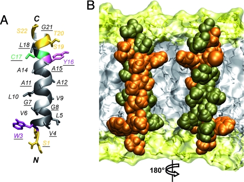
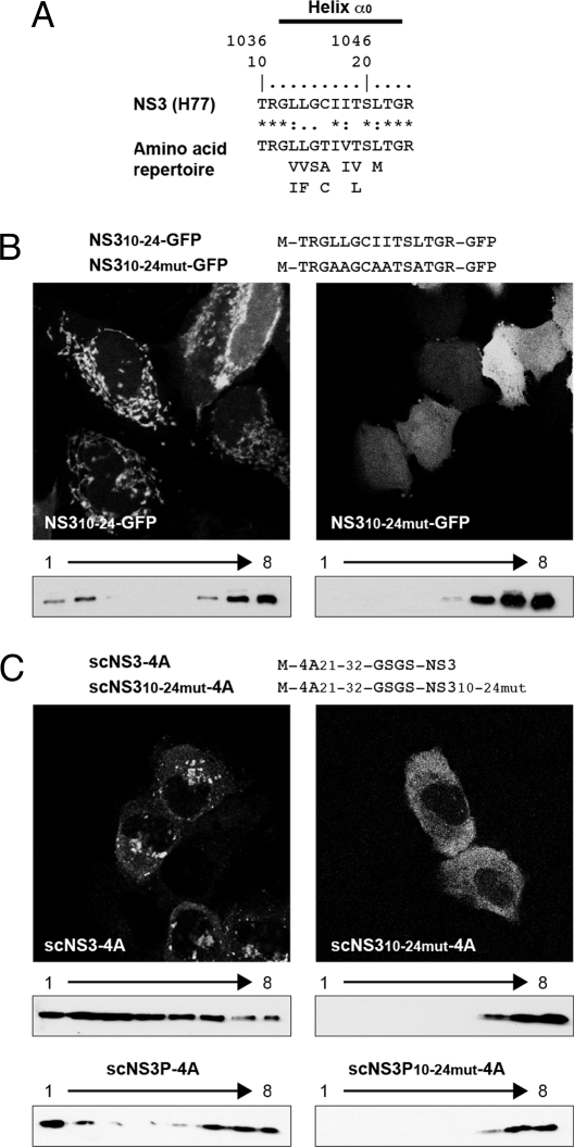
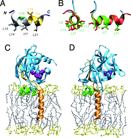
Hepatitis C virus (HCV) NS3-4A is a membrane-associated multifunctional protein harboring serine protease and RNA helicase activities. It is an essential component of the HCV replication complex and a prime target for antiviral intervention. Here, we show that membrane association and structural organization of HCV NS3-4A are ensured in a cooperative manner by two membrane-binding determinants. We demonstrate that the N-terminal 21 amino acids of NS4A form a transmembrane alpha-helix that may be involved in intramembrane protein-protein interactions important for the assembly of a functional replication complex. In addition, we demonstrate that amphipathic helix alpha(0), formed by NS3 residues 12-23, serves as a second essential determinant for membrane association of NS3-4A, allowing proper positioning of the serine protease active site on the membrane. These results allowed us to propose a dynamic model for the membrane association, processing, and structural organization of NS3-4A on the membrane. This model has implications for the functional architecture of the HCV replication complex, proteolytic targeting of host factors, and drug design.


Conflict of interest statement

The authors declare no conflict of interest.

Figures

Fig. 1.
Fig. 1.
Identification of the membrane segment of NS4A. (A) Amino acid sequence analyses. The NS4A sequence from the HCV H77 consensus clone (GenBank accession no. AF009606) is shown at the top. Amino acids are numbered with respect to NS4A and the HCV polyprotein (top row). The amino acid repertoire of 26 reference sequences representative of all major HCV genotypes and subtypes (http://euhcvdb.ibcp.fr; ref. 21) is given below, with the observed amino acids listed in decreasing order of frequency and the similarity index according to ClustalW convention (asterisk, invariant; colon, highly similar; dot, similar). (B) Subcellular localization of NS4A-GFP fusion constructs. U-2 OS cells transiently transfected with pCMVNS4A-GFP (NS4A) or C- and N-terminal NS4A deletion constructs fused to GFP (see SI Text for details) were examined by fluorescence microscopy. (C) Membrane topology of NS4A. Lysates of U-2 OS cells transfected with pCMVgt-NS4A-GFP or pCMVgtmut-NS4A-GFP (see SI Text for details) were digested with endoglycosidase H (Endo H) or N-glycosidase F (N-glyc F), followed by 15% SDS/PAGE and immunoblot with monoclonal antibody JL-8 against GFP (Clontech). The position of glycosylated gt-NS4A-GFP is indicated by an asterisk.
Fig. 2.
Fig. 2.
Structure of the NS4A transmembrane segment. (A) Ribbon representation of the best representative structure of NS4A[1-22]* selected from the final set of 26 calculated NMR structures (BMRB entry 15580). Residue side chains are shown as sticks and are colored on the basis of the chemical properties of their side chains (hydrophobic, dark gray; polar, yellow). Gly are light gray. Tyr, Trp, and Cys are violet, magenta, and green, respectively. Fully conserved residues are underlined. (B) Amino acid van der Waals representation and tentative position of NS4A amino acids 1–22 within a phospholipid bilayer. Fully and less conserved residues are colored orange and bronze, respectively. Orientation of the left structure is the same as in image A. Note that the majority of fully conserved residues are located on one side of the transmembrane α-helix. The membrane is represented as a simulated model of a 1-palmitoyl-2-oleoyl-sn-glycero-3-phosphocholine (POPC) bilayer (http://moose.bio.ucalgary.ca). Polar heads and hydrophobic tails of phospholipids (surface and stick structures) are light yellow and gray, respectively.
Fig. 3.
Fig. 3.
NS3 helix α0 mediates membrane association. (A) Sequence analyses of the NS3 amino acid 10–24 segment. See Fig. 1 legend for details. (B) Membrane association of NS3 helix α0-GFP fusion construct. Constructs NS310–24-GFP or NS310–24mut-GFP (see SI Text for details) were expressed in U-2 OS cells and examined by fluorescence microscopy and membrane flotation analyses. Monoclonal antibody JL-8 against GFP was used for immunoblot. Membranes float to the upper, low-density fractions (left portion of the blots). (C) Membrane association of NS3-4A single-chain constructs. Constructs scNS3–4A, scNS310–24mut-4A, scNS3P-4A, and scNS3P10–24mut-4A (see SI Text for details) were examined by immunofluorescence microscopy and membrane flotation by using monoclonal antibody 1B6 against NS3 (3).
Fig. 4.
Fig. 4.
Structural analyses of NS3 helix α0. (A) Ribbon representation of the best representative structure of NS3[10–24] selected from the final set of 26 calculated NMR structures in 100 mM SDS-d25 (BMRB entry 15582). Residue side chains are colored as in Fig. 2A. (B) Comparison of NS3 amino acid 10–24 structures obtained by NMR (red) and by X-ray crystallography (ref. ; PDB entry 1CU1; green and cyan). The two structures were superimposed from residues 12–23. Left and right images correspond to axial and perpendicular views. (C and D) Model of the membrane-associated NS3 serine protease domain complexed with NS4A (perpendicular and axial views relative to NS3 helix α0). This model was constructed by using the coordinates reported by Yao et al. (ref. ; PDB entry 1CU1) and the structure of NS4A[1-22]* reported in Fig. 2. The NS3 serine protease domain is cyan, with side chain atoms of the catalytic triad (His 57, Asp 81, and Ser 139) highlighted as purple spheres. NS3 helix α0 is green and the five hydrophobic residues are represented by sticks and balls. The N-terminal transmembrane (amino acids 1–20) and central (amino acids 21–32) segments of NS4A are orange and light orange, respectively. The membrane is represented as a simulated model of POPC bilayer (see Fig. 2 legend for details).
Fig. 5.
Fig. 5.
Mechanistic model of the membrane association process of NS3-4A. See Discussion for comments. The cytosolic side of the membrane bilayer is on the top. (1) N-terminal portion of NS3 (amino acids 13–23 highlighted in green) before folding of helix α0 (constructed by using the NS3 serine protease structure without the central segment of NS4A solved by X-ray crystallography (ref. ; PDB entry 1A1Q). (2) Membrane association of uncleaved NS3-NS4A by NS3 helix α0. The NS4A structure is undefined at this step. It is represented as sticks with N-terminal segment 1–20, central segment 21–32, and the beginning of the C-terminal segment (amino acids 33–40) in medium, light, and dark orange, respectively. The NS3 structure was constructed by using: (i) The crystal structure of scNS3–4A (ref. ; PDB entry 1CU1), where the C terminus of the helicase domain (silver) lies within the active site of the serine protease domain (cyan); (ii) The NS3 serine protease structure without the central segment of NS4A solved by NMR (ref. ; PDB entry 1BT7) for the N-terminal β-barrel subdomain; and (iii) The NMR structure of NS3[10–24] comprising helix α0 (this study). Well folded structures (helix α0, C-terminal β-barrel subdomain, and helicase domain) are represented as ribbon diagrams whereas the less stable or unfolded structures are represented as sticks (NS3 segment 1–9 and N-terminal β-barrel subdomain). Side-chain atoms of the catalytic triad (His 57, Asp 81, and Ser 139) are highlighted as purple spheres. (3) Cleavage at the NS3/NS4A site allows membrane insertion of the N-terminal segment of NS4A, resulting in a transmembrane α-helix. (4) Cofolding of the central segment 21–32 of NS4A into the N-terminal β-barrel subdomain stabilizes the structure of the serine protease, which is locked onto the membrane by NS3 helix α0 and the NS4A transmembrane α-helix. Note that the hydrophilic helicase domain would be partially immersed into the membrane in the NS3-NS4A cis-cleavage conformation (5) (model in brackets). (5) Final topology of the NS3-4A complex on the membrane.

References

    1. Moradpour D, Penin F, Rice CM. Replication of hepatitis C virus. Nat Rev Microbiol. 2007;5:453–463. - PubMed
    1. De Francesco R, Steinkühler C. Structure and function of the hepatitis C virus NS3-NS4A serine protease. Curr Top Microbiol Immunol. 2000;242:149–169. - PubMed
    1. Kim JL, et al. Crystal structure of the hepatitis C virus NS3 protease domain complexed with a synthetic NS4A cofactor peptide. Cell. 1996;87:343–355. - PubMed
    1. Yan Y, et al. Complex of NS3 protease and NS4A peptide of BK strain of hepatitis C virus: A 2.2 Å resolution structure in a hexagonal crystal form. Protein Sci. 1998;7:837–847. - PMC - PubMed
    1. Yao N, et al. Molecular views of viral polyprotein processing revealed by the crystal structure of the hepatitis C virus bifunctional protease-helicase. Structure Fold Des. 1999;7:1353–1363. - PubMed

Publication types

MeSH terms

Substances

LinkOut - more resources