. 2008 Sep 23;105(38):14503-8.
doi: 10.1073/pnas.0803998105. Epub 2008 Sep 17.

The Parkinson's disease genes pink1 and parkin promote mitochondrial fission and/or inhibit fusion in Drosophila

Affiliations

The Parkinson's disease genes pink1 and parkin promote mitochondrial fission and/or inhibit fusion in Drosophila

Hansong Deng et al. Proc Natl Acad Sci U S A. .

Abstract

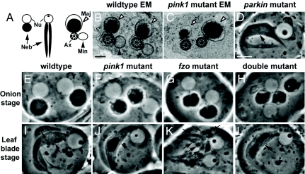
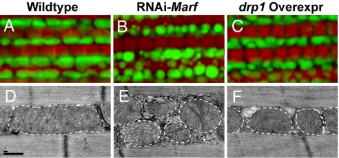
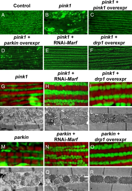
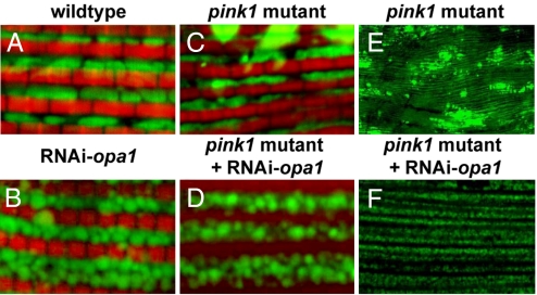
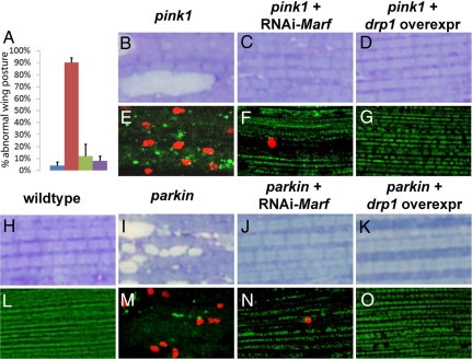
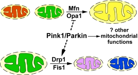
Mutations in PTEN-induced kinase 1 (pink1) or parkin cause autosomal-recessive and some sporadic forms of Parkinson's disease. pink1 acts upstream of parkin in a common genetic pathway to regulate mitochondrial integrity in Drosophila. Mitochondrial morphology is maintained by a dynamic balance between the opposing actions of mitochondrial fusion, controlled by Mitofusin (mfn) and Optic atrophy 1 (opa1), and mitochondrial fission, controlled by drp1. Here, we explore interactions between pink1/parkin and the mitochondrial fusion/fission machinery. Muscle-specific knockdown of the fly homologue of Mfn (Marf) or opa1, or overexpression of drp1, results in significant mitochondrial fragmentation. Mfn-knockdown flies also display altered cristae morphology. Interestingly, knockdown of Mfn or opa1 or overexpression of drp1, rescues the phenotypes of muscle degeneration, cell death, and mitochondrial abnormalities in pink1 or parkin mutants. In the male germline, we also observe genetic interactions between pink1 and the testes-specific mfn homologue fuzzy onion, and between pink1 and drp1. Our data suggest that the pink1/parkin pathway promotes mitochondrial fission and/or inhibits fusion by negatively regulating mfn and opa1 function, and/or positively regulating drp1. However, pink1 and parkin mutant flies show distinct mitochondrial phenotypes from drp1 mutant flies, and flies carrying a heterozygous mutation in drp1 enhance the pink1-null phenotype, resulting in lethality. These results suggest that pink1 and parkin are likely not core components of the drp1-mediated mitochondrial fission machinery. Modification of fusion and fission may represent a novel therapeutic strategy for Parkinson's disease.


Conflict of interest statement

The authors declare no conflict of interest.

Figures

Fig. 1.
Fig. 1.
pink1 and parkin mutants show phenotypes in spermatogenesis suggestive of defects in mitochondrial fission; pink1 genetically interacts with fzo in testes. (A) Schematic of an onion-staged (left) and a leaf-blade-staged (middle) spermatid showing the nucleus (Nu) and nebenkern (Neb, arrow), and a cross-section through a spermatid tail immediately before individualization (right). In these spermatid tails, an axoneme (Ax) is associated with two mitochondrial derivatives, the major (Maj, open arrowhead) and minor (Min, filled arrowhead). (B and C) EM images of WT (B) and pink1-mutant (C) spermatid tails show the presence of only the major mitochondrial derivative, but not the minor derivative in pink1 mutants. (D) A parkin-mutant spermatid during the leaf-blade stage, as with a similarly staged pink1-mutant spermatid (J), contains only one mitochondrial derivative compared with two seen in WT specimens (I). (E–L) Genetic interactions between pink1 and fzo. During the onion stage, pink1 mutants show vacuolations of the nebenkern (F), which are not seen in WT specimens (E). fzo1/Df(3R)P2O mutants have nebenkerns with irregular borders (G). Similar phenotypes are also seen in fzo1/fzo2 and fzo1-homozygous testes (data not shown). Double mutants removing both pink1 and fzo function exhibit nebenkerns that have smooth borders, yet are still vacuolated (H). (I) WT leaf-blade–staged spermatids have two mitochondrial derivatives. During the leaf-blade stage, fzo1/Df(3R)P2O spermatids show fragmented mitochondria (K). Double mutants with pink1 and fzo removed show a single mitochondrial derivative, the pink1 mutant-like phenotype (L). Note that the cell membranes encapsulating spermatids often contain multiple spermatids. This is a result of the disruption of cytoplasmic bridges connecting spermatids during sample preparation, and is not a phenotype. Genotypes: (B, E, and I) w/Y; (C, F, and J) w pink15 (G and K) w/Y; e fzo1/Df(3R)P2O; (H and L) w pink15/Y; e fzo1/Df(3R)P2O; Scale bars: 200 nm in B and C; 10 μm in D–L.
Fig. 2.
Fig. 2.
Muscle-specific knockdown of Marf and overexpression of drp1 results in abnormal mitochondrial morphology. MitoGFP- (green) and phalloidin-labeled (red) muscle (A–C) and EM images (D–F) from 1- to 2-day-old flies. Compared with control (A and D), both Marf knockdown (B and E) and drp1 overexpression (C and F) result in mitochondrial fragmentation, with Marf-knockdown flies also showing cristae irregularities and more severe mitochondrial fragmentation. The borders of mitochondria are marked with white dashed lines. Genotypes: (A and D) FM6/Y; Mef2-Gal4, UAS-mitoGFP/+; (B and E) w; UAS-RNAi-Marf/+; Mef2-Gal4, UAS-mitoGFP/+; (C and F) w; UAS-drp1/+; Mef2-Gal4, UAS-mitoGFP/+. Note that, as controls, Mef2-Gal4, UAS-mitoGFP flies show similar mitochondrial phenotypes in backgrounds of w/Y, FM6/Y, or w/Y; UAS-LacZ. Scale bars: 0.5 μm in D–F.
Fig. 3.
Fig. 3.
Both Marf knockdown and drp1 overexpression suppress mitochondrial phenotypes in pink1- and parkin-mutant muscle. MitoGFP-labeled muscle at low magnification (A–F), mitoGFP/phalloidin double-labeled muscle at higher magnification (G–I and M–O), and EM images (J–L and P–R) from 1- to 2-day-old flies. Both pink1 (B and G) and parkin (M) mutants show weakened mitoGFP labeling and clumps of intense mitoGFP signal. In pink1 mutants, these phenotypes can be completely suppressed by pink1 overexpression (C) and partially rescued by parkin overexpression (D). Moreover, pink1 and parkin phenotypes can also be suppressed by knockdown of Marf (E, H, and N) or by drp1 overexpression (F, I, and O). At the EM level, pink1 and parkin mutants show broken cristae (J and P), which can be suppressed by Marf knockdown (K and Q) or drp1 overexpression (L and R). However, pink1- and parkin-mutant flies with Marf knockdown (K and Q) still show significant mitochondrial fragmentation and cristae abnormalities reminiscent of those seen in Marf-knockdown flies alone (Fig. 2E). The borders of mitochondria are marked with white dashed lines. Genotypes: (A) FM6/Y; (B, G, and J) w pink15f/Y; Mef2-Gal4, UAS-mitoGFP/+; (C) w pink15 f/Y; Mef2-Gal4, UAS-pink1/+; UAS-mitoGFP/+; (D) w pink15 f/Y; UAS-parkin/+; Mef2-Gal4, UAS-mitoGFP/+; (E, H, and K) w pink15 f/Y; Mef2-Gal4, UAS-mitoGFP/UAS-RNAi-Marf; (F, I, and L) w pink15 f/Y; Mef2-Gal4, UAS-mitoGFP/UAS-drp1; (M and P) w/Y; UAS-mitoGFP/+; 24B-Gal4 park25/park25; (N and Q) w/Y; UAS-mitoGFP/UAS-RNAi-Marf; 24B-Gal4 park25/park25; (O and R) w/Y; UAS-mitoGFP/UAS-drp1; 24B-Gal4 park25/park25. Scale bars: 0.5 μm in J–L and P–R.
Fig. 4.
Fig. 4.
Muscle-specific opa1 knockdown results in mitochondrial fragmentation and suppression of mitochondrial defects observed in pink1 mutants. Muscle from 1- to 2-day-old flies labeled with mitoGFP (green) and phalloidin (red) at high magnification (A–D) or labeled with mitoGFP (green) at low magnification. Compared with control (A), opa1 knockdown results in smaller and rounder mitochondria (B), similar to what is observed in Marf knockdown (Fig. 2B). However, we note that the borders of opa1 knockdown mitochondria appear fuzzy, whereas those of Marf knockdown do not. Compared with pink1 mutants alone (C and E), opa1 knockdown in pink1 mutants displays striking rescue of mitochondrial morphology (D and F), Genotypes: (A) FM6/Y; Mef2-Gal4, UAS-mitoGFP/+; (B) w; UAS-RNAi-opa1/+; Mef2-Gal4, UAS-mitoGFP/+; (C and E) w pink15f/Y; Mef2-Gal4, UAS-mitoGFP/+; (D and F) w pink15 f/Y; Mef2-Gal4, UAS-mitoGFP/UAS-RNAi-opa1.
Fig. 5.
Fig. 5.
Marf knockdown or drp1 overexpression in muscle results in functional rescue of pink1- and parkin-mutant phenotypes. (A) Abnormal wing posture seen in pink1 mutants (red) compared with WT specimens (blue) is significantly suppressed by Marf knockdown (green) or drp1 overexpression (purple) in muscle. The y axis denotes the percentile of flies showing “upheld” or “downheld” wings, an indication of muscle degeneration. Toluidine blue (B–D and H–K) or TUNEL/mitoGFP stainings (E–G and L–O) of muscles. Compared with WT specimens (H), muscles from pink1 and parkin mutants show vacuolations indicative of degeneration (B and I). These phenotypes can be suppressed by Marf knockdown (C and J) or drp1 overexpression (D and K). WT muscle does not show any TUNEL-positive cell death (L), whereas pink1 and parkin mutants show prominent TUNEL-positive staining (red; E and M). These phenotypes are suppressed by expression of RNAi-Marf (F and N) or drp1 (G and O). Genotypes are as shown in Fig. 3.
Fig. 6.
Fig. 6.
Interactions of pink1 and parkin with genes regulating mitochondrial fusion and fission. Mitochondrial fusion requires Mfn and Opa1, and mitochondrial fission requires Drp1 and Fis1. Pink1 and Parkin promote fission and/or inhibit fusion, either directly or indirectly (dashed lines). In addition, Pink1 and Parkin are unlikely to be components of the canonical pathways regulating mitochondrial dynamics. Rather, Pink1 and Parkin may regulate other mitochondrial functions that also impact mitochondrial integrity.

References

    1. Dauer W, Przedborski S. Parkinson's disease: Mechanisms and models. Neuron. 2003;39:889–909. - PubMed
    1. Langston JW, et al. Chronic Parkinsonism in humans due to a product of meperidine-analog synthesis. Science. 1983;219:979–980. - PubMed
    1. Sherer TB, Betarbet R, Greenamyre JT. Environment, mitochondria, and Parkinson's disease. Neuroscientist. 2002;8:192–197. - PubMed
    1. Dodson MW, Guo M. Pink1, parkin, DJ-1 and mitochondrial dysfunction in Parkinson's disease. Curr Opin Neurobiol. 2007;17:331–337. - PubMed
    1. Wood-Kaczmar A, Gandhi S, Wood NW. Understanding the molecular causes of Parkinson's disease. Trends Mol Med. 2006;12:521–528. - PubMed

Publication types