Review
. 2008 Oct 1;121(Pt 19):3109-19.
doi: 10.1242/jcs.035683.

The molecular logic of Notch signaling--a structural and biochemical perspective

Affiliations
Review

The molecular logic of Notch signaling--a structural and biochemical perspective

Wendy R Gordon et al. J Cell Sci. .

Abstract

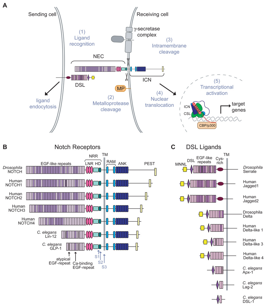
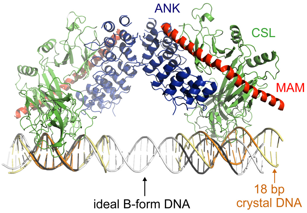
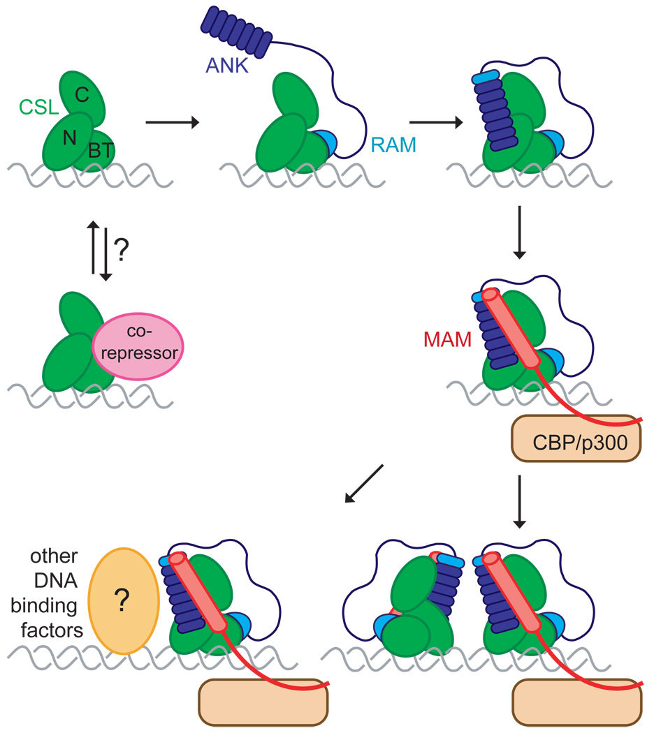
The Notch signaling pathway constitutes an ancient and conserved mechanism for cell-cell communication in metazoan organisms, and has a central role both in development and in adult tissue homeostasis. Here, we summarize structural and biochemical advances that contribute new insights into three central facets of canonical Notch signal transduction: (1) ligand recognition, (2) autoinhibition and the switch from protease resistance to protease sensitivity, and (3) the mechanism of nuclear-complex assembly and the induction of target-gene transcription. These advances set the stage for future mechanistic studies investigating ligand-dependent activation of Notch receptors, and serve as a foundation for the development of mechanism-based inhibitors of signaling in the treatment of cancer and other diseases.


Figures

Figure 1
Figure 1
Notch signaling pathway and domain organization of Notch receptors and DSL ligands. (A) Model for the major events in the Notch signaling pathway. Signals initiated by the engagement of ligand (1) lead to metalloprotease cleavage (MP) at site S2 (2). This proteolytic step allows the cleavage of Notch by the γ-secretase complex at site S3 within the transmembrane domain (3), and release of intracellular notch (ICN) from the membrane (4). ICN translocates to the nucleus where it enters into a transcriptional activation complex with CSL and Mastermind (MAM;5). (B, C) The domain organization of Notch receptors (B) and DSL-family ligands (C) from fly, human and worm.
Figure 2
Figure 2
Structures of NOTCH1 and JAGGED1 ectodomain fragments and models for the Notch ectodomain. (A) NMR structure of EGF-like repeats 11–13 from human NOTCH1 (PDB ID code 1TOZ). Two of 20 calculated structures in the ensemble are shown. The double-headed arrow indicates the range of positions that are occupied by repeat 13 among the 20 calculated structures. Disulfide bonds are colored orange, and hydrophobic residues engaged in interdomain contacts are shown as sticks. Bound calcium ions, placed by homology to other EGF-like-repeat structures, are shown as green spheres. (B, C) Two proposed models for the overall organization of the Notch1 ectodomain. Panel B shows a rod-like, extended model, whereas panel C illustrates one possible compact model. EGF-like repeats (ovals) are shaded dark purple when they contain a consensus calcium-binding site (per the definition used by UNIPROT), and light purple when they do not. (D). Cartoon representation of the X-ray structure for the JAGGED1 polypeptide (PDB code 2VJ2) comprising the DSL domain and EGF-like repeats 1–3. Disulfide bonds are yellow. Residues at the proposed Notch binding surface are rendered as colored sticks.
Figure 3
Figure 3
X-ray structure of the human NOTCH2 NRR in the autoinhibited conformation and models for signal activation. (A) Ribbon representation of the NRR. The LNR modules are colored different shades of pink and purple and the HD domain is colored in two shades of cyan; the light and dark cyan represent residues that are N- and C-terminal, respectively, to the furin cleavage loop. The three bound Ca++ ions are green, the bound Zn++ ion is blue, and the ten disulfide bonds are red. The positions of S1 and S2 cleavage are indicated with red arrows. (B) The LNR-AB linker sterically blocks access to the metalloprotease cleavage site. The hydrophobic pocket in the HD domain that houses the S2 site is rendered in a surface representation, and residues from the LNR-AB linker are in ball-and-stick representation. (C) Model for activation by mechanical force. Endocytosis of bound ligand generates a mechanical force that tugs on the LNR domain (panel 1) disengaging the hydrophobic plug from the hydrophobic pocket containing the S2 site (panel 2), and peeling the LNR repeats away from HD domain (panel 3). Partial or complete relaxation of the HD domain then allows access of metalloprotease to the S2 site, and cleavage of the scissile bond to trigger Notch activation (panel 4). (D) Peeling of the LNR repeats away from the HD domain may not confer sufficient exposure of the S2 site to allow cleavage by metalloprotease. The left panel depicts a hypothetical model for the negative regulatory region upon peeling of the first two LNR repeats away from the HD domain; the right panel shows the structure of the catalytic domain of the metalloprotease TACE (PDB ID code 1BKC). The deep active site cleft is indicated with an arrow. Panels A and B are adapted from reference (Gordon et al., 2007) and reproduced with permission (http://www.nature.com/nsmb/).
Figure 4
Figure 4
Ribbon diagrams representing the structure of human and worm Notch ternary complexes. (A, left) Human complex of the ANK domain of Notch1, CSL and the N-terminal region of Mastermind-like 1 (MAM) bound to an 18 base-pair DNA sequence from the hes1 promoter (PDB code 2F8X). (A, right) Worm complex of the RAMANK region of Lin12, Lag-1, and the N-terminal region of Sel-8 (PDB code 2FO1). The structures illustrate the cooperative binding of MAM to a composite surface that is created at the interface between the Notch ANK domain and CSL. Bottom panels show a 105° rotation around the vertical axis. (B) Superposition of CSL structures showing the difference in loop conformation between CSL-DNA complexes and complexes that include RAM, ANK domains or both. Mouse CSL-DNA (PDB code 3BRG, magenta) and worm CSL-DNA (PDB code 1TTU, green) structures have a “closed” loop. Worm RAM-CSL-DNA (PDB code 3BRF, yellow and PDB code 3BRD, cyan), human ANK-MAM-CSL-DNA (red), and worm RAMANK-MAM-CSL-DNA (blue) complexes have an “open” loop. (C) Superposition of worm (colors) and human (grey) ternary complex structures. Several unique insertions at the N-terminus of RAM and within the ANK domain are found in worm Lin12 but not in other Notch molecules (orange). These features may play a role in the more compact packing of the worm NTC structure when compared to the human NTC structure.
Figure 5
Figure 5
Ribbon diagram of two symmetry-related copies of the human NTC (PDB code 2F8X), revealing the near-linear orientation of the two DNA elements that mimics the inverted repeat of the SPS element. Superposition of the symmetry-related pseudo-dimer (DNA in yellow, with CSL binding sites in orange) on ideal B-form DNA corresponding to 42 base-pairs of the hes1 promoter (grey, with CSL binding sites in black) reveals that the orientation and spacing between the two CSL sites in the crystal approximates, but does not match, the expected spacing and orientation in a natural SPS.
Figure 6
Figure 6
Model for assembly of Notch ternary complexes. A high-affinity interaction between the N-terminal RAM peptide of Notch and the β-trefoil domain (BTD) of CSL is likely to be the first event in the assembly of Notch transcriptional activation complexes. This step allows the lower-affinity ANK domain to bind at its docking site, resulting in ordering of the ankyrin-like N-cap and first repeat of the ANK domain. The interface between the ANK domain and the RHR-N and RHR-C domains of CSL create a composite surface for the binding of MAM, which recruits CBP/p300. Higher-order homotypic assemblies of Notch complexes or heterotypic assemblies with other transcription factors may be required for transcription of specific Notch targets.

References

    1. Acar M, Jafar-Nejad H, Takeuchi H, Rajan A, Ibrani D, Rana NA, Pan H, Haltiwanger RS, Bellen HJ. Rumi Is a CAP10 Domain Glycosyltransferase that Modifies Notch and Is Required for Notch Signaling. Cell. 2008;132:247–258. - PMC - PubMed
    1. Ahimou F, Mok LP, Bardot B, Wesley C. The adhesion force of Notch with Delta and the rate of Notch signaling. J Cell Biol. 2004;167:1217–1229. - PMC - PubMed
    1. Aster JC, Pear WS, Blacklow SC. Notch signaling in leukemia. Annu Rev Pathol. 2008;3:587–613. - PMC - PubMed
    1. Aster JC, Robertson ES, Hasserjian RP, Turner JR, Kieff E, Sklar J. Oncogenic forms of NOTCH1 lacking either the primary binding site for RBP-Jkappa or nuclear localization sequences retain the ability to associate with RBP-Jkappa and activate transcription. J Biol Chem. 1997;272:11336–11343. - PubMed
    1. Bailey AM, Posakony JW. Suppressor of hairless directly activates transcription of enhancer of split complex genes in response to Notch receptor activity. Genes Dev. 1995;9:2609–2622. - PubMed