

The positron emission tomography ligand DAA1106 binds with high affinity to activated microglia in human neurological disorders
- PMID: 18800007
- PMCID: PMC2669281
- DOI: 10.1097/NEN.0b013e318188b204
The positron emission tomography ligand DAA1106 binds with high affinity to activated microglia in human neurological disorders
Abstract
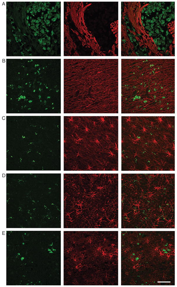
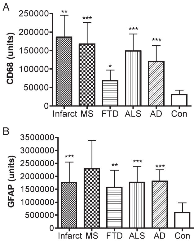
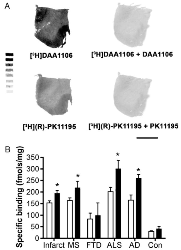
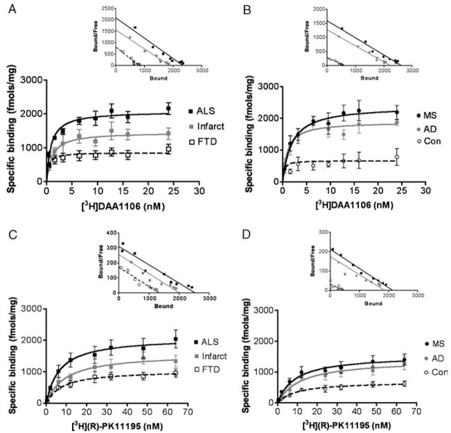
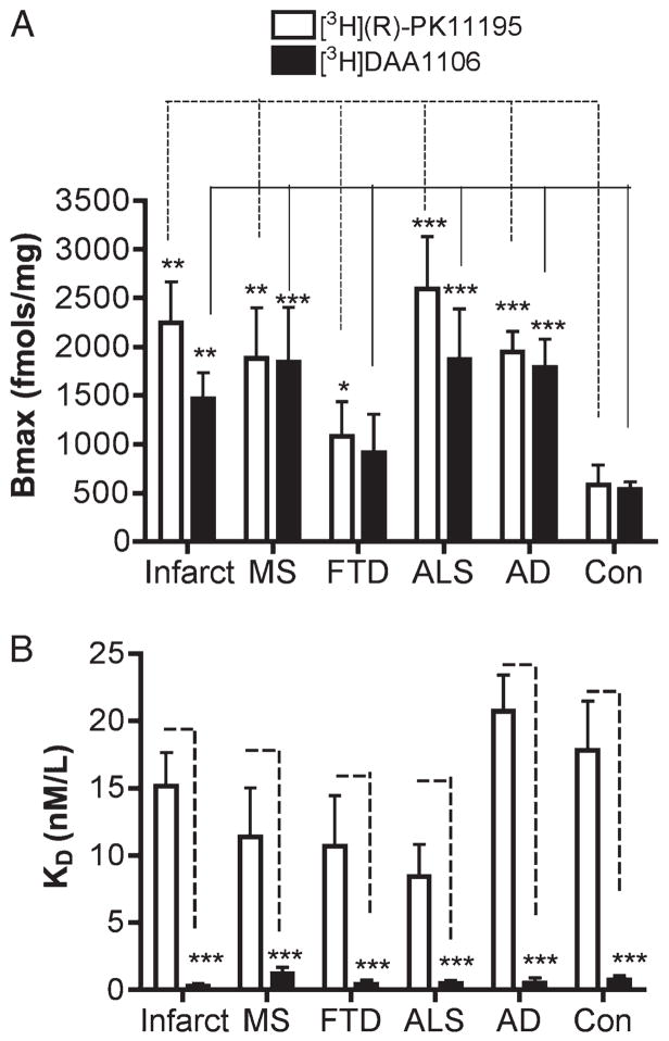
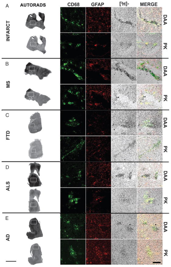
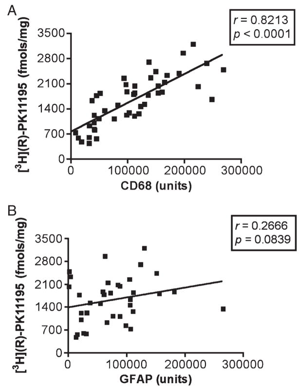
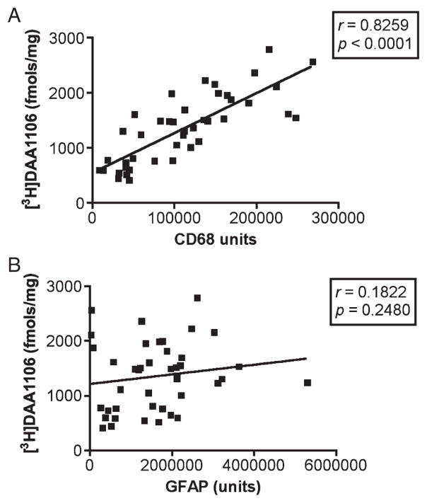
Chronic microglial activation is an important component of many neurological disorders, and imaging activated microglia in vivo will enable the detection and improved treatment of neuroinflammation. 1-(2-chlorphenyl)-N-methyl-N-(1-methylpropyl)-3-isoquinoline-carbox-amide (PK11195), a peripheral benzodiazepine receptor ligand, has been used to image neuroinflammation, but the extent to which PK11195 binding distinguishes activated microglia and reactive astrocytes is unclear. Moreover, PK11195 may lack sufficient sensitivity for detecting mild neuroinflammation. We hypothesized that N-(2,5-dimethoxybenzyl)-N-(4-fluoro-2-phenoxyphenyl) acetamide (DAA1106), a new ligand that binds specifically to peripheral benzodiazepine receptor, binds to activated microglia in human neurological diseases with higher affinity than does PK11195. We therefore compared the pharmacological binding properties of [3H](R)-PK11195 and [3H]DAA1106 in postmortem tissues from patients with cerebral infarcts, amyotrophic lateral sclerosis, Alzheimer disease, frontotemporal dementia, and multiple sclerosis (n=10 each). In all diseases, [3H]DAA1106 showed a higher binding affinity as reflected by lower dissociation constant (KD) values than that of [3H](R)-PK11195. Moreover, specific binding of both ligands correlated with the presence of activated microglia identified by immunohistochemistry in situ. We conclude that 1) ligands that bind peripheral benzodiazepine receptor mainly label activated microglia in human neurological disorders and that 2) DAA1106 may possess binding characteristics superior to those of PK11195, which may be beneficial for in vivo positron emission tomography imaging.
Figures
Similar articles
-
A comparison of the high-affinity peripheral benzodiazepine receptor ligands DAA1106 and (R)-PK11195 in rat models of neuroinflammation: implications for PET imaging of microglial activation.J Neurochem. 2007 Sep;102(6):2118-2131. doi: 10.1111/j.1471-4159.2007.04690.x. Epub 2007 Jun 7. J Neurochem. 2007. PMID: 17555551
-
The high affinity peripheral benzodiazepine receptor ligand DAA1106 binds specifically to microglia in a rat model of traumatic brain injury: implications for PET imaging.Exp Neurol. 2007 Sep;207(1):118-27. doi: 10.1016/j.expneurol.2007.06.003. Epub 2007 Jun 19. Exp Neurol. 2007. PMID: 17658516 Free PMC article.
-
The high affinity peripheral benzodiazepine receptor ligand DAA1106 binds to activated and infected brain macrophages in areas of synaptic degeneration: implications for PET imaging of neuroinflammation in lentiviral encephalitis.Neurobiol Dis. 2008 Feb;29(2):232-41. doi: 10.1016/j.nbd.2007.08.016. Epub 2007 Sep 7. Neurobiol Dis. 2008. PMID: 17920902 Free PMC article.
-
Positron emission tomography imaging of neuroinflammation.Neurotherapeutics. 2007 Jul;4(3):443-52. doi: 10.1016/j.nurt.2007.04.006. Neurotherapeutics. 2007. PMID: 17599710 Free PMC article. Review.
-
N-(5-Fluoro-2-phenoxyphenyl)-N-(2-[131I]iodo-5-methoxybenzyl)acetamide.2007 Aug 6 [updated 2007 Sep 12]. In: Molecular Imaging and Contrast Agent Database (MICAD) [Internet]. Bethesda (MD): National Center for Biotechnology Information (US); 2004–2013. 2007 Aug 6 [updated 2007 Sep 12]. In: Molecular Imaging and Contrast Agent Database (MICAD) [Internet]. Bethesda (MD): National Center for Biotechnology Information (US); 2004–2013. PMID: 20641364 Free Books & Documents. Review.
Cited by
-
Astrocyte regulation of blood flow in the brain.Cold Spring Harb Perspect Biol. 2015 Mar 27;7(5):a020388. doi: 10.1101/cshperspect.a020388. Cold Spring Harb Perspect Biol. 2015. PMID: 25818565 Free PMC article. Review.
-
TSPO-PET imaging using [18F]PBR06 is a potential translatable biomarker for treatment response in Huntington's disease: preclinical evidence with the p75NTR ligand LM11A-31.Hum Mol Genet. 2018 Aug 15;27(16):2893-2912. doi: 10.1093/hmg/ddy202. Hum Mol Genet. 2018. PMID: 29860333 Free PMC article.
-
Translocator Protein Distribution Volume Predicts Reduction of Symptoms During Open-Label Trial of Celecoxib in Major Depressive Disorder.Biol Psychiatry. 2020 Oct 15;88(8):649-656. doi: 10.1016/j.biopsych.2020.03.007. Epub 2020 Mar 29. Biol Psychiatry. 2020. PMID: 32402468 Free PMC article.
-
Microglia measured by TSPO PET are associated with Alzheimer's disease pathology and mediate key steps in a disease progression model.Alzheimers Dement. 2024 Apr;20(4):2397-2407. doi: 10.1002/alz.13699. Epub 2024 Feb 1. Alzheimers Dement. 2024. PMID: 38298155 Free PMC article.
-
Exploring inflammation-related protein expression and its relationship with TSPO PET in Alzheimer's disease.Alzheimers Dement. 2025 Apr;21(4):e70171. doi: 10.1002/alz.70171. Alzheimers Dement. 2025. PMID: 40289873 Free PMC article.
References
-
- Block ML, Zecca L, Hong JS. Microglia-mediated neurotoxicity: Uncovering the molecular mechanisms. Nat Rev Neurosci. 2007;8:57–69. - PubMed
-
- Banati RB. Visualising microglial activation in vivo. Glia. 2002;40:206–17. - PubMed
-
- Cagnin A, Brooks DJ, Kennedy AM, et al. In-vivo measurement of activated microglia in dementia. Lancet. 2001;358:461–67. - PubMed
-
- Turner MR, Cagnin A, Turkheimer FE, et al. Evidence of widespread cerebral microglial activation in amyotrophic lateral sclerosis: An [11C](R)-PK11195 positron emission tomography study. Neurobiol Dis. 2004;15:601–9. - PubMed
-
- Banati RB, Newcombe J, Gunn RN, et al. The peripheral benzodiazepine binding site in the brain in multiple sclerosis: Quantitative in vivo imaging of microglia as a measure of disease activity. Brain. 2000;123:2321–37. - PubMed
Publication types
MeSH terms
Substances
Grants and funding
LinkOut - more resources
Full Text Sources
Medical
