. 2008 Oct;67(10):963-75.
doi: 10.1097/NEN.0b013e318187a80f.

White matter tauopathy with globular glial inclusions: a distinct sporadic frontotemporal lobar degeneration

Affiliations

White matter tauopathy with globular glial inclusions: a distinct sporadic frontotemporal lobar degeneration

Gabor G Kovacs et al. J Neuropathol Exp Neurol. 2008 Oct.

Abstract

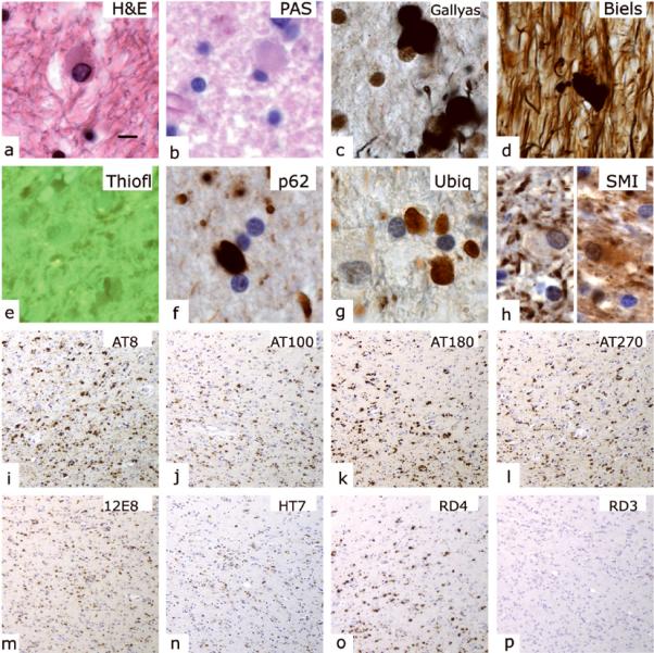
Frontotemporal lobar degenerations are a group of disorders characterized by circumscribed degeneration of the frontal and temporal lobes and diverse histopathologic features. We report clinical, neuropathologic, ultrastructural, biochemical, and genetic data on 7 individuals with a 4-repeat tauopathy characterized by the presence of globular glial inclusions (GGIs) in brain white matter. Clinical manifestations were compatible with the behavioral variant of frontotemporal dementia and included motor neuron symptoms; there was prominent neuronal loss in the frontal and temporal cortex, subiculum, and amygdala. The surrounding white matter showed abundant GGIs composed of abnormal filaments present mostly in oligodendrocytes. The severity of white matter tau abnormalities correlated with a reduction in myelin and axons and with microglial activation. Western blotting of sarkosyl-insoluble tau demonstrated the presence of 2 major tau bands of 64 and 68 kd. No mutations in the microtubule-associated protein tau gene were detected in 2 affected individuals. We propose that 4-repeat tau-immunoreactive GGIs are the neuropathologic hallmark of a distinct sporadic tauopathy with variable clinical presentations that include frontotemporal dementia and occasionally upper motor neuron disease. This type of tauopathy with GGIs expands the group of neurodegenerativedisorders in which oligodendroglial pathology predominates, beyond the synucleinopathy multiple system atrophy disorders.


Figures

Figure 1
Figure 1
Macroscopic overview of representative brains. Note the prominent frontotemporal atrophy in case 1 (a, b) and the asymmetric atrophy (left > right) in case 3 (c, d). Scanned original photographs from 1966 (case 1) and 1975 (case 3).
Figure 2
Figure 2
Common histopathological features in individuals with frontotemporal lobar degeneration associated with abundant white matter tau pathology. Spongiosis in the superficial layers of the frontal cortex (a, H&E). Neuronal loss and gliosis in the frontal cortex (b, H&E). Comparison of areas with few (c, e, f, i, k, l) and abundant (d, g, h, j, m, n) tau-immunoreactive globules in white matter (c, d, upper inset). The areas are evaluated using Luxol-PAS staining (c, d), anti-myelin basic protein (e, g, MBP), anti-proteolipid protein (f, h, PLP), anti-neurofilament (i, j, SMI-31) and anti-microglia/macrophage (k, m, HLA-DR and l, n, CD68) immunostaining. Note the reduction in myelin density, along with the activation of microglia and the appearance of macrophages, along with increasing tau pathology. Using anti-APP immunohistochemistry (i, j upper inset), we observed axonal swellings in the white matter of affected areas (j, upper inset). Scale bar in a indicates 200 μm for a, 100 μm for b, 50 μm for c–n, and 75 μm for insets.
Figure 3
Figure 3
Hemispheric section (middle part of image) of the frontobasal cortex (gyrus rectus and orbital gyrus) at the level of the frontal portion of the lateral ventricles stained with Luxol-fast blue. Note that the region indicated by the arrowhead shows loss of myelin (left upper panel) and numerous tau immunoreactive globular glial inclusions (left lower panel) compared to the region indicated by the double arrowheads, which shows relatively preserved myelin staining (right upper panel) and only a few tau-immunoreactive globular glial inclusions (right lower panel).
Figure 4
Figure 4
Tau-immunoreactive structures and anatomical distribution of neuropathological changes in individuals with frontotemporal lobar degeneration associated with tau-immunopositive globular glial inclusions. The most prominent alteration is the presence of globular glial tau immunoreactivity, together with grain-like structures, predominantly in white matter (a) and, to a lesser extent, also in grey matter (b). Abundant ramified astrocytes (c) showing immunoreactivity with antibody RD4 (right upper inset) are observed in grey matter. Neuronal tau immunoreactivity consists mainly of diffuse cytoplasmic pretangle-type staining (d) and occasional neuronal spherical cytoplasmic inclusions (e) that are immunoreactive with RD4 (left upper inset), but not with RD3 (right upper inset). In cases with short disease duration (f) and cases with long disease duration (i), neurons are relatively preserved in the CA1 subregion (indicated with *, enlarged in g and j), while neuronal loss and gliosis are prominent in the subiculum (indicated with **, enlarged in h and k). The substantia nigra shows a mild degree of neuronal loss and extracellular neuromelanin (l) and mainly diffuse neuronal cytoplasmic tau immunoreactivity (m). Prominent tau-immunoreactive grains, threads, neuronal pretangles and globular glial inclusions are present in the amygdala (n). There are occasional pretangles in locus coeruleus (enlarged in lower inset) (o). The amount of globular glial tau immunoreactivity increases when comparing a case in the early stages (p and r, case 7) with a case of longer disease duration (q and s, case 4), as exemplified in the hippocampus (p, q) and parahippocampal region (r, s). Globular glial tau inclusions are prominent at the border between cortex and white matter (t, frontal). Descending tracts in the pontine basis (u), corpus callosum (v), anterior commissure (w), frontobasal-subaccumbens white matter and internal capsule (x) also consistently show globular glial inclusions. Scale bar in a indicates 60μm for a–d, n; 10 μm for e and all insets; 500μm for f, i, o–s, v–x; 25 μm for g, h, j, k; and 250 μm for t, u.
Figure 5
Figure 5
Characterization of globular glial inclusions. H&E (a), PAS (b), Gallyas (c), Bielschowsky (d, Biels) and Thioflavin (e, Thiofl) stains and immunohistochemistry for p62 (f), ubiquitin (g, Ubiq), neurofilament-SMI-31 (h), Tau-AT8 (i), Tau-AT100 (j), Tau-AT180 (k), Tau-AT270 (l), Tau-12E8 (m), Tau-HT7 (n), Tau-RD4 (o), and Tau-RD3 (p). Scale bar in a indicates 10 μm for a–h; and 100 μm for i–p.
Figure 6
Figure 6
Ultrastructure of a representative white matter oligodendroglia-associated globular inclusion. Note the haphazardly oriented filaments. Scale bars: a: 0.4 μm b: 0.15 μm.
Figure 7
Figure 7
Immunoelectron microscopy and immunoblotting of sarkosyl-insoluble tau from amygdala (A) and hippocampal formation (H) of case number 7. (a) Paired helical filaments (counting from the left, panels 1, 3 and 5) and straight filaments (panels 2, 4 and 6) decorated with anti-tau antibodies 134, AT100 and PS422. Scale bar, 100 nm. (b) Immunoblots using anti-tau antibodies 133, 134, AT8 and PS422 show the presence of strong tau bands of 64 and 68 kDa molecular mass.

References

    1. Cairns NJ, Bigio EH, Mackenzie IR, et al. Neuropathologic diagnostic and nosologic criteria for frontotemporal lobar degeneration: Consensus of the Consortium for Frontotemporal Lobar Degeneration. Acta Neuropathol. 2007;114:5–22. - PMC - PubMed
    1. Boxer AL, Miller BL. Clinical features of frontotemporal dementia. Alzheimer Dis Assoc Disord. 2005;19(Suppl 1):S3–6. - PubMed
    1. Neary D, Snowden JS, Gustafson L, et al. Frontotemporal lobar degeneration: A consensus on clinical diagnostic criteria. Neurology. 1998;51:1546–54. - PubMed
    1. Knibb JA, Kipps CM, Hodges JR. Frontotemporal dementia. Curr Opin Neurol. 2006;19:565–71. - PubMed
    1. Goedert M, Spillantini MG. A century of Alzheimer's disease. Science. 2006;314:777–81. - PubMed

Publication types