. 2009 Mar;34(4):972-86.
doi: 10.1038/npp.2008.144. Epub 2008 Sep 17.

Dopamine D2 and adenosine A2A receptors regulate NMDA-mediated excitation in accumbens neurons through A2A-D2 receptor heteromerization

Affiliations

Dopamine D2 and adenosine A2A receptors regulate NMDA-mediated excitation in accumbens neurons through A2A-D2 receptor heteromerization

Karima Azdad et al. Neuropsychopharmacology. 2009 Mar.

Abstract

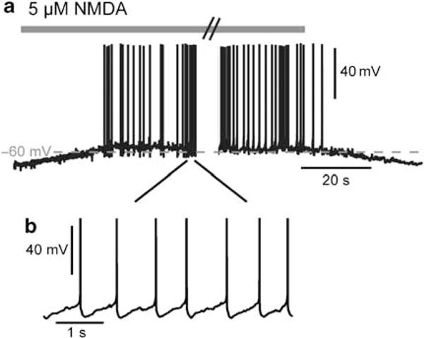
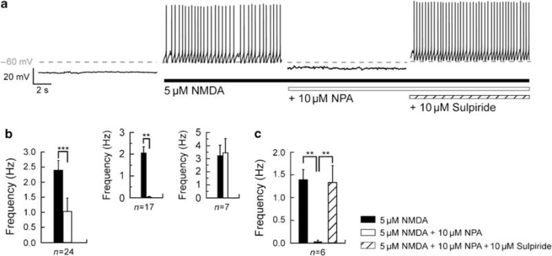
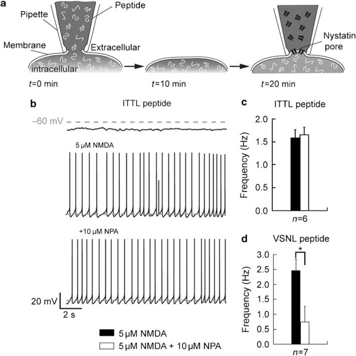
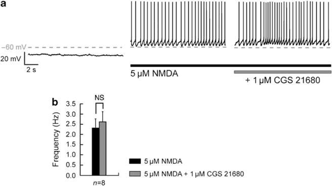
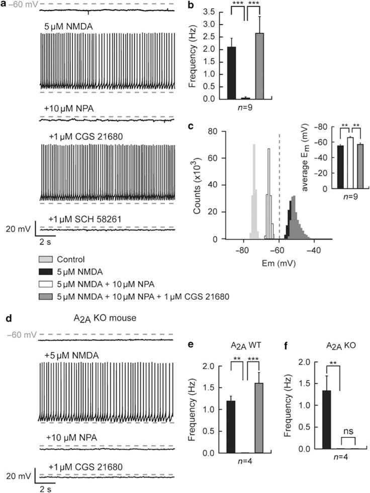
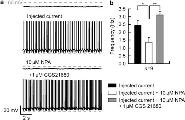
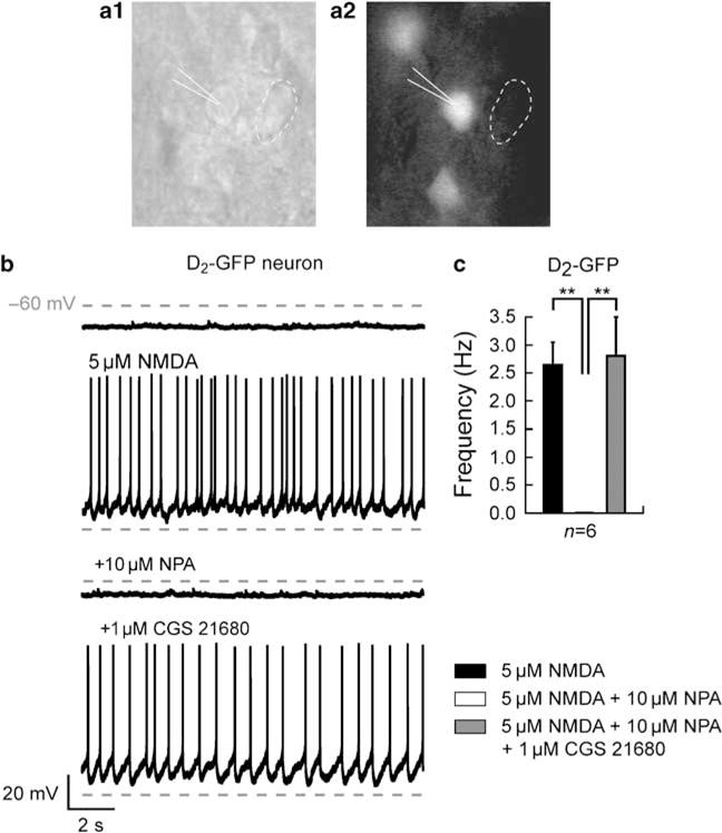
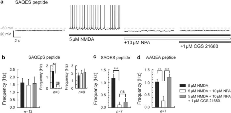
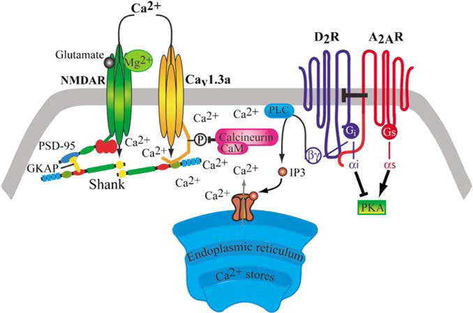
Bursting activity of striatal medium spiny neurons results from membrane potential oscillations between a down- and an upstate that could be regulated by G-protein-coupled receptors. Among these, dopamine D(2) and adenosine A(2A) receptors are highly enriched in striatal neurons and exhibit strong interactions whose physiological significance and molecular mechanisms remain partially unclear. More particularly, respective involvements of common intracellular signaling cascades and A(2A)-D(2) receptor heteromerization remain unknown. Here we show, by performing perforated-patch-clamp recordings on brain slices and loading competitive peptides, that D(2) and A(2A) receptors regulate the induction by N-methyl-D-aspartate of a depolarized membrane potential plateau through mechanisms relying upon specific protein-protein interactions. Indeed, D(2) receptor activation abolished transitions between a hyperpolarized resting potential and a depolarized plateau potential by regulating the Ca(V)1.3a calcium channel activity through interactions with scaffold proteins Shank1/3. Noticeably, A(2A) receptor activation had no effect per se but fully reversed the effects of D(2) receptor activation through a mechanism in which A(2A)-D(2) receptors heteromerization is strictly mandatory, demonstrating therefore a first direct physiological relevance of these heteromers. Our results show that membrane potential transitions and firing patterns in striatal neurons are tightly controlled by D(2) and A(2A) receptors through specific protein-protein interactions including A(2A)-D(2) receptors heteromerization.


Conflict of interest statement

DISCLOSURE/CONFLICT OF INTEREST

The authors declare that except for income received from the primary employer no financial support or compensation has been received from any individual or corporate entity over the past 3 years for research or professional service and there are no personal financial holdings that could be perceived as constituting a potential conflict of interest.

Figures

Figure 1
Figure 1
Membrane potential transition from a hyperpolarized resting potential to a depolarized plateau potential of a medium spiny neuron (MSN) in response to glutamatergic receptor stimulation. (a) Transition in a representative MSN recorded in an acute slice in perforated-patch clamp. Application of 5 μM N-methyl-D-aspartate (NMDA), which mimics cortical synaptic inputs, evoked a reversible membrane potential transition between a hyperpolarized state and a depolarized plateau potential inducing a continuous action potential firing. (b) Periodic spike firing of MSN during the 5 μM NMDA-induced upstate.
Figure 2
Figure 2
Dopamine D2 receptor suppresses the N-methyl-D-aspartate (NMDA)-mediated depolarized plateau potential. (a) Consecutive traces, recorded in a single neuron, showing typical transitions where the action of NMDA (5 μM) was recorded before and in the presence of D2 receptor agonist R(−)-propylnorapomorphine hydrochloride (NPA, 10 μM) and D2 receptor antagonist sulpiride (10 μM). Notice that before NMDA application the recorded medium spiny neuron is in a hyperpolarized resting potential (−78 mV) and in response to NMDA depolarized to plateau potential (−60 mV). In the presence of NMDA, application of NPA suppresses the plateau potential and inhibits the action potential firing. Subsequent application of sulpiride blocks the D2 effect and reestablishes the depolarized plateau potential. (b) Summary histogram obtained from 24 different neurons illustrating the effect of the D2 receptor agonist on the firing frequency. Application of NPA (10 μM) significantly reduced the frequency of action potential firing. In 17 out of 24 recorded neurons, application of NPA totally reverses the depolarized firing plateau, whereas 7 out of 24 recorded neurons do not respond to the activation of D2 receptors. These data show D2-responsive and -unresponsive populations. (c) Summary histogram illustrates the significant reversed effect of D2 receptor antagonist on the firing frequency of D2-responsive neurons (n = 6) (data represent mean ± SEM; **p <0.01, ***p <0.001).
Figure 3
Figure 3
Peptide targeting the Shank-CaVl.3a interaction blocks D2 receptor modulation of the N-methyl-D-aspartate (NMDA)-mediated depolarized plateau potential. (a) Method used to dialyze specific peptide in the recorded medium spiny neuron (MSN) using the whole-cell configuration of the patch-clamp technique. The selective peptide is dialyzed in the neuron through the patch pipette during 5–10 min and then the pipette is gently removed. After 10 min, to allow the neuron to recover, the same neuron is subsequently recorded using the perforated-patch configuration. (b) Consecutive traces of an MSN dialyzed with the ITTL peptide targeting the Shank PDZ-binding domain interacting with CaVl.3a L-type Ca2+ channels. In this dialyzed neuron, D2 receptor activation by R(−)-propylnorapomorphine hydrochloride (NPA, 10 μM) does not affect NMDA receptor-induced depolarized plateau potential. (c) Histogram showing the effect of D2 receptor activation on the firing frequency on neurons dialyzed with the ITTL peptide. Application of NPA (10 μM) did not affect the frequency of action potential firing (n = 6). (d) Effect of NPA on the firing frequency of neurons dialyzed with the VSNL peptide targeting the shank PDZ-binding domain interacting with Cavl.2 L-type Ca2+ channels. On seven recorded neurons, D2 receptor activation strongly decreases the action potential firing frequency (data represent mean ±SEM; *p<0.05).
Figure 4
Figure 4
Adenosine A2A receptor does not affect the N-methyl-D-aspartate (NMDA)-mediated depolarized plateau potential. (a) Typical recording on a single neuron, showing the NMDA-induced depolarized plateau potential before and after application of A2A receptor agonist, 2-[4-[(2-carboxyethyl)-phenyl]ethyl-amino]-5′-N-(ethylcarbamoyl)adenosine (CGS 21680, 1 μM). (b) Statistics illustrated in the histogram show the effect of A2A receptor activation on firing frequency. Application of CGS 21680 (1 μM) does not affect the frequency of action potential firing (n = 8) (data represent mean ± SEM; NS, not significant).
Figure 5
Figure 5
Interaction of dopamine D2 and adenosine A2A receptors modulates the N-methyl-D-aspartate (NMDA)-mediated depolarized plateau potential on D2-responsive neurons. (a) Consecutive traces showing typical transitions where the action of NMDA (5 μM) was recorded before and in the presence of D2 receptor agonist R(−)-propylnorapomorphine hydrochloride (NPA, 10 μM), A2A receptor agonist 2-[4-[(2-carboxyethyl)-phenyl]ethyl-amino]-5′-N-(ethylcarbamoyl)adenosine (CGS 21680, 1 μM), and A2A receptor antagonist 7-(2-phenylethyl)-5-amino-2-(2-furyl)-pyrazolo-[4,3-e]-l,2,4-triazolol[l,5-c]pyrimidine (SCH 58261, 1 μM). On a D2-responsive neuron, subsequent application of CGS 21680 totally counteracts the effect of D2 receptor activation, ie the inhibition of the depolarized plateau potential and firing frequency. The A2A receptor modulation on this D2-responsive neuron was reversed by the selective A2A receptor antagonist SCH 58261 (1 μM). (b) Summary histogram obtained from nine D2-responsive neurons illustrates the antagonistic effect of A2A receptors activation on the action potential firing frequency (n = 9). (c) Typical all-point histogram from a single neuron shows the membrane potential distributions before and after additional application of NMDA, NPA, and CGS 21680. In these conditions, activation of NMDA receptors set and A2A receptors reset the neurons in a depolarized state, whereas activation of D2 receptors holds the neuron in a hyperpolarized state. In inset, a summary histogram illustrates the significant modulation of the average membrane potential after the subsequent application of NMDA, NPA, and CGS 21680. (d) Consecutive traces of a D2-responsive medium spiny neuron (MSN) from an A2A receptor knockout mouse. As expected, subsequent application of CGS 21680 (1 μM) fails to reverse the D2 receptor-induced hyperpolarized potential. (e, f) Summary histograms of the effects of A2A receptor activation on D2-responsive cells of wild-type (e, n = 4 out of 6 recorded neurons) and A2A receptor null mice (f, n = 4 out of 6 recorded neurons). Application of CGS 21680 reverses the D2 receptor-induced hyperpolarized potential in wild-type, whereas it does not affect the firing frequency of D2-responsive MSNs from A2A receptor null mice (data represent mean ± SEM; **p<0.0l, ***p<0.00l).
Figure 6
Figure 6
Modulatory interaction of dopamine D2 and adenosine A2A receptors on the N-methyl-D-aspartate (NMDA)-mediated depolarized plateau potential is not dependent on a presynaptic mechanism. (a) Consecutive traces, recorded in a single neuron submitted to the injection of current (to mimic the action of NMDA), showing typical transitions recorded before and in the presence of D2 receptor agonist R(−)-propylnorapomorphine hydrochloride (NPA, 10μM) and A2A receptor agonist 2-[4-[(2-carboxyethyl)-phenyl]ethyl-amino]-5′-N-(ethylcarbamoyl)adenosine (CGS 21680, 1 μM). The application of NPA suppresses the depolarized the plateau potential and inhibits the action potential firing induced by the injection of current. Subsequent application of CGS 21680 blocks the D2 effect and reestablishes the depolarized plateau potential. (b) Summary histogram obtained from nine different neurons illustrating the effect of the D2 receptor agonist on the firing frequency. Application of NPA (10 μM) significantly reduces the frequency of action potential firing. Activation of A2A receptors by CGS 21680 counteracts the effects of D2 receptor activation on the action potential firing frequency (data represent mean±SEM; *p<0.05, **p<0.0l).
Figure 7
Figure 7
Modulatory interaction of dopamine D2 and adenosine A2A receptors on the N-methyl-D-aspartate (NMDA)-mediated depolarized plateau potential occurs in striatopallidal medium spiny neurons (MSNs). Striatal acute slice from D2-enhanced green fluorescent protein (EGFP) mice in phase contrast (al) and during epifluorescence (a2). The drawing pipette identifies a D2-EGFP-positive neuron and the dashed circle a non-D2-EGFP-positive neuron. (b) Consecutive traces recorded in a single D2-EGFP-positive neuron, where the action of NMDA (5 μM) was recorded before and in the presence of D2 receptor agonist R(−)-propylnorapomorphine hydrochloride (NPA, 10 μM) and A2A receptor agonist 2-[4-[(2-carboxyethyl)-phenyl]ethyl-amino]-5′-N-(ethylcarbamoyl)adenosine (CGS 21680, 1 μM). In the presence of NMDA, application of NPA suppresses the plateau potential and inhibits the action potential firing on the D2-EGFP-positive recorded neuron. Subsequent application of CGS 21680 blocks the D2 effect and reestablishes the depolarized plateau potential. (c) Summary histogram obtained from six D2-EGFP-positive neurons illustrating the effect of the D2 receptor agonist on the firing frequency. Application of NPA (10 μM) totally abolishes the frequency of action potential firing in all recorded neurons, which is fully reversed by A2A receptors activation.
Figure 8
Figure 8
SAQES peptides disrupt functional D2–A2A receptor heteromerization. (a) Consecutive traces in a neuron dialyzed with the SAQES peptide corresponding to the C-terminal epitope of the A2A receptor that interacts with the N-terminal portion of the third intracellular loop of the D2 receptor. In this dialyzed neuron, in presence of N-methyl-D-aspartate (NMDA), application of R(−)-propylnorapomorphine hydrochloride (NPA) suppresses the transition to the depolarized plateau potential and inhibits the firing frequency. The additional application of 2-[4-[(2-carboxyethyl)-phenyl]ethyl-amino]-5′-N-(ethylcarbamoyl)adenosine (CGS 21680) does not have any effect. (b) Summary histogram of NPA effect on the firing frequency of neurons dialyzed by the phosphorylated SAQEpS peptide. NPA (10 μM) and subsequent CGS 21680 (1 μM) do not modify the frequency of action potential firing (n = 12). Histograms in insets show an NPA-induced abolition of the transition to the depolarized plateau potential in three neurons, which is not reversed by subsequent CGS 21680. (c) Data obtained from neurons dialyzed with the SAQES peptide illustrating the effect of the D2 receptor agonist and subsequent A2A receptor agonist on the firing frequency are represented in the graph bar. Application of NPA (10μM) significantly reduces the frequency of action potential firing, whereas A2A receptor activation by CGS 21680 (1 μM) does not counteract D2 receptor activation. (d) Summary histogram of D2 and D2–A2A receptor activation on neurons dialyzed with the AAQEA peptide. D2 receptor activation decreases the action potential firing frequency and this effect is totally reversed by the subsequent A2A receptor activation (data represent mean ±SEM; **p<0.0l, ***p< 0.001).
Figure 9
Figure 9
Schematic presentation of our proposed model for the involvement of protein–protein interactions, at the postsynaptic dendritic spine, in the modulation of the N-methyl-D-aspartate (NMDA)-mediated depolarized plateau potential in the striatopallidal medium spiny neuron. According to our hypothesis the D2R-mediated suppression of NMDA-induced depolarized plateau is mediated by the suppression of Cav1.3a L-type calcium channel current through the D2R-PLC signaling cascade involving the activation of calcineurin and dephosphorylation of these channels. This modulation requires the physical interaction between scaffolding Shank proteins and Cav1.3a calcium channels through a specific PDZ-binding domain. The A2AR counteracts the D2R-mediated suppression of NMDA-induced depolarized plateau via a direct A2AR–D2R interaction at the membrane level through heteromerization. NMDAR, NMDA receptor; Cav1.3a, Cav1.3a L-type calcium channel; A2AR adenosine A2A receptor; D2R, dopamine D2 receptor; Golf, G-protein activating adenylyl cyclase; Gi, G-protein inhibiting adenylyl cyclase; PLC, phospholipase C; PKA, protein kinase A; CaM, calmodulin; IP3, inositol 1,4,5-triphosphate; Shank, multiple ankyrin repeats-SH3 domain-PDZ domain-proline-rich region-sterile-α motif containing protein; PSD-95, postsynaptic density 95; GKAP, guanylate kinase-associated protein.

References

    1. Adermark L, Lovinger DM. Combined activation of L-type Ca2+ channels and synaptic transmission is sufficient to induce striatal long-term depression. J Neurosci. 2007;25:6781–6787. - PMC - PubMed
    1. Agnati LF, Ferré S, Lluis C, Franco R, Fuxe K. Molecular mechanisms and therapeutic implications of intramembrane receptor/receptor interactions among heptahelical receptors with examples from the striatopallidal GABA neurons. Pharmacol Rev. 2003;55:509–550. - PubMed
    1. Alexander GE, Crutcher MD. Functional architecture of basal ganglia circuits: neural substrates of parallel processing. Trends Neurosci. 1990;13:266–271. - PubMed
    1. Bustos VH, Marin O, Meggio F, Cesaro L, Allende CC, Allende JE, et al. Generation of protein kinase Ck1alpha mutants which discriminate between canonical and non-canonical substrates. Biochem J. 2005;391:417–424. - PMC - PubMed
    1. Canals M, Marcellino D, Fanelli F, Ciruela F, de Benedetti P, Goldberg SR, et al. Adenosine A2A-dopamine D2 receptor-receptor heteromerization: qualitative and quantitative assessment by fluorescence and bioluminescence energy transfer. J Biol Chem. 2003;278:46741–46749. - PubMed

Publication types

MeSH terms

LinkOut - more resources