. 2008 Sep 18;3(9):e3232.
doi: 10.1371/journal.pone.0003232.

Imaging mass spectrometry technology and application on ganglioside study; visualization of age-dependent accumulation of C20-ganglioside molecular species in the mouse hippocampus

Affiliations

Imaging mass spectrometry technology and application on ganglioside study; visualization of age-dependent accumulation of C20-ganglioside molecular species in the mouse hippocampus

Yuki Sugiura et al. PLoS One. .

Abstract

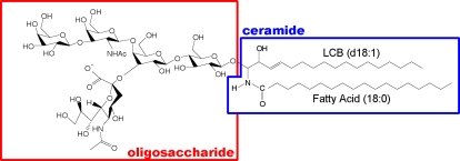
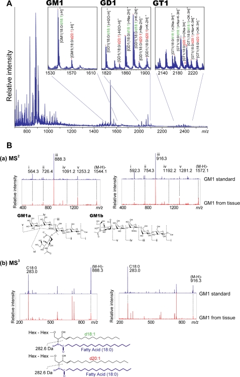
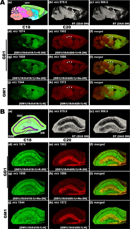
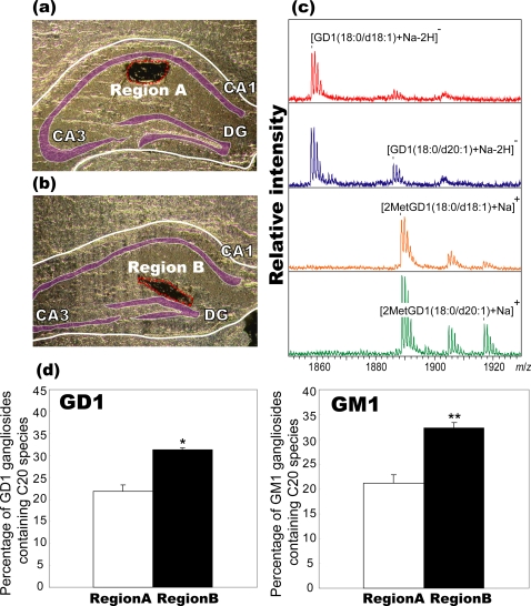
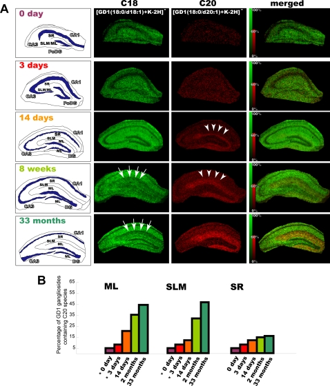
Gangliosides are particularly abundant in the central nervous system (CNS) and thought to play important roles in memory formation, neuritogenesis, synaptic transmission, and other neural functions. Although several molecular species of gangliosides have been characterized and their individual functions elucidated, their differential distribution in the CNS are not well understood. In particular, whether the different molecular species show different distribution patterns in the brain remains unclear. We report the distinct and characteristic distributions of ganglioside molecular species, as revealed by imaging mass spectrometry (IMS). This technique can discriminate the molecular species, raised from both oligosaccharide and ceramide structure by determining the difference of the mass-to-charge ratio, and structural analysis by tandem mass spectrometry. Gangliosides in the CNS are characterized by the structure of the long-chain base (LCB) in the ceramide moiety. The LCB of the main ganglioside species has either 18 or 20 carbons (i.e., C18- or C20-sphingosine); we found that these 2 types of gangliosides are differentially distributed in the mouse brain. While the C18-species was widely distributed throughout the frontal brain, the C20-species selectively localized along the entorhinal-hippocampus projections, especially in the molecular layer (ML) of the dentate gyrus (DG). We revealed development- and aging-related accumulation of the C-20 species in the ML-DG. Thus it is possible to consider that this brain-region specific regulation of LCB chain length is particularly important for the distinct function in cells of CNS.


Conflict of interest statement

Competing Interests: The authors have declared that no competing interests exist.

Figures

Figure 1
Figure 1. Structure of C18-LCB containing GM1a.
Gangliosides comprise a large family; their oligosaccharides structures differ in the glycosidic linkage position, sugar configuration, and the contents of neutral sugars and sialic- acid content. The ceramide moiety of gangliosides, it also has some variation varies with respect to the type of long -chain base (LCB) (sphingosine- base) and fatty acid moiety.
Figure 2
Figure 2. Direct MALDI-MS and MSn allows specific detection of ganglioside molecular species.
A. Averaged mass spectra obtained from the entire hippocampal formation. In the spectra, the mass peaks corresponding to GM1, GD1, and GT1 are detected, and IMS provides distinct signals for molecular species containing C18- and C20-sphingosines. B. MSn structural analysis of ions corresponding to GM1. (a) MS2 product ion spectra show that the ions at m/z 1544 and 1572 had the same oligosaccharide structure, i.e., they contained a sialic acid moiety, but the ceramide mass peaks were observed at different m/z values. (b) MS3 product ion mass spectra of m/z 888.3 and 916.3 were obtained to determine the different structural constituents in the ceramide moieties. Because of the detection of m/z 283.0 (fatty acid-related ion) in both the spectra, the 28-u difference between m/z 1544 and m/z 1572 was attributed to the difference in the sphingosine constituent; m/z 1544 had C18-sphingosine and m/z 1572 had C20-sphingosine.
Figure 3
Figure 3. Localization of C20-sphingosine-containing gangliosides in the hippocampal formation.
IMS at 50 µm raster step size was used to gain an overview of ganglioside distribution in different brain regions (A), and IMS at 15 µm raster size was used to study in detail the distribution pattern of gangliosides in the hippocampus (B). In both panels, schematic diagram of the brain section (a) and ion images of STs (b–c) are presented. For ions corresponding to the GD1 molecular species, we observed the ion distributions of both sodium and potassium complexes, i.e., the ions at m/z 1858 (f) and m/z 1886 (g), which correspond to the [M+Na-H] form of C18- and C20-GD1, and those at m/z 1874 (h) and m/z 1902 (i), which correspond to the [M+K-H] form of C18- and C20-GD1, respectively. The ion distribution patterns corresponding to the GD1-Na salts and GD1-K salts are fairly uniform for both C18- and C20- species. For GM1, m/z 1544 (d) and m/z 1572 (e), which correspond to C18- and C20-sphingosines containing GM1 respectively are shown.
Figure 4
Figure 4. Localization of C20-sphingosine-containing gangliosides was confirmed by MS of extracted and methyl-esterified gangliosides.
To determine the percentage of GM1/GD1 gangliosides containing the C20-species in different regions without allowing sialic acid dissociation during MS measurement, we extracted gangliosides from the dendritic region of the SR (region A, (a)) and the ML/SLM (region B, (b)). They were derivatized to methyl-esterified gangliosides. From the result of MS of underivatized gangliosides and methyl-esterified gangliosides (c), the percentage of GM1/GD1 gangliosides containing the C20-species were calculated (d). Three different mouse brain sections were used, and the data were expressed as mean±S.D. * and ** indicate P<0.05 and P<0.005, respectively, Student's t-test.
Figure 5
Figure 5. Development- and aging-related accumulation of C20-GD1 in the ML and SLM of the hippocampal formation.
We visualized the ion corresponding to GD1 (m/z 1874 and 1902) in the mouse hippocampus at the indicated time points (P0, P3, P14, 1 month, and 33 months). For each time point, intensity scale of C20-GD1 is normalized in order that the brightest pixels of C20-GD1 have 60% of the maximal C18-GD1 intensity value. In the P14 mouse hippocampus, C20-GD1 was concentrated in the narrow area of DG-SMm and began to spread over the medial edge of the region (arrow heads). In contrast, the concentration of the C-18 species decreased in the ML/SLM with aging (arrows). Quantification result of C20-GD1 on the total GD1 signal in the ML, SLM and SR region has also been shown (B). *; At P0 and P3, we could not distinguish between the ML and SLM area; therefore, values obtained from the region corresponding to ML/SLM have been used for both the regions in the graph.

Similar articles

Cited by

References

    1. van Echten G, Sandhoff K. Ganglioside metabolism. Enzymology, Topology, and regulation. J Biol Chem. 1993;268:5341–5344. - PubMed
    1. Rahmann H. Brain gangliosides and memory formation. Behav Brain Res. 1995;66:105–116. - PubMed
    1. Prinetti A, Iwabuchi K, Hakomori S. Glycosphingolipid-enriched signaling domain in mouse neuroblastoma Neuro2a cells. Mechanism of ganglioside-dependent neuritogenesis. J Biol Chem. 1999;274:20916–20924. - PubMed
    1. Ramirez OA, Gomez RA, Carrer HF. Gangliosides improve synaptic transmission in dentate gyrus of hippocampal rat slices. Brain Res. 1990;506:291–293. - PubMed
    1. Kotani M, Terashima T, Tai T. Developmental changes of ganglioside expressions in postnatal rat cerebellar cortex. Brain Res. 1995;700:40–58. - PubMed

Publication types