Limb lengthening and then insertion of an intramedullary nail: a case-matched comparison
- PMID: 18800209
- PMCID: PMC2628243
- DOI: 10.1007/s11999-008-0509-8
Limb lengthening and then insertion of an intramedullary nail: a case-matched comparison
Abstract
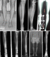
Distraction osteogenesis is an effective method for lengthening, deformity correction, and treatment of nonunions and bone defects. The classic method uses an external fixator for both distraction and consolidation leading to lengthy times in frames and there is a risk of refracture after frame removal. We suggest a new technique: lengthening and then nailing (LATN) technique in which the frame is used for gradual distraction and then a reamed intramedullary nail inserted to support the bone during the consolidation phase, allowing early removal of the external fixator. We performed a retrospective case-matched comparison of patients lengthened with LATN (39 limbs in 27 patients) technique versus the classic (34 limbs in 27 patients). The LATN group wore the external fixator for less time than the classic group (12 versus 29 weeks). The LATN group had a lower external fixation index (0.5 versus 1.9) and a lower bone healing index (0.8 versus 1.9) than the classic group. LATN confers advantages over the classic method including shorter times needed in external fixation, quicker bone healing, and protection against refracture. There are also advantages over the lengthening over a nail and internal lengthening nail techniques.
Level of evidence: Level III, therapeutic study. See the Guidelines for Authors for a complete description of levels of evidence.
Figures


References
-
- {'text': '', 'ref_index': 1, 'ids': [{'type': 'DOI', 'value': '10.1097/00005131-200211000-00006', 'is_inner': False, 'url': 'https://doi.org/10.1097/00005131-200211000-00006'}, {'type': 'PubMed', 'value': '12439195', 'is_inner': True, 'url': 'https://pubmed.ncbi.nlm.nih.gov/12439195/'}]}
- Bhandari M, Schemitsch EH. Bone formation following intramedullary femoral reaming is decreased by indomethacin and antibodies to insulin-like growth factors. J Orthop Trauma. 2002;16:717–722. - PubMed
-
- {'text': '', 'ref_index': 1, 'ids': [{'type': 'PubMed', 'value': '2355035', 'is_inner': True, 'url': 'https://pubmed.ncbi.nlm.nih.gov/2355035/'}]}
- Blachut PA, Meek RN, O’Brien PJ. External fixation and delayed intramedullary nailing of open fractures of the tibial shaft. A sequential protocol. J Bone Joint Surg Am. 1990;72:729–735. - PubMed
-
- {'text': '', 'ref_index': 1, 'ids': [{'type': 'DOI', 'value': '10.2106/JBJS.F.00742', 'is_inner': False, 'url': 'https://doi.org/10.2106/jbjs.f.00742'}, {'type': 'PubMed', 'value': '17200326', 'is_inner': True, 'url': 'https://pubmed.ncbi.nlm.nih.gov/17200326/'}]}
- Brinker MR, O’Connor DP. Exchange nailing of ununited fractures. J Bone Joint Surg Am. 2007;89:177–188. - PubMed
-
- {'text': '', 'ref_index': 1, 'ids': [{'type': 'DOI', 'value': '10.1016/j.bone.2003.12.027', 'is_inner': False, 'url': 'https://doi.org/10.1016/j.bone.2003.12.027'}, {'type': 'PubMed', 'value': '15121017', 'is_inner': True, 'url': 'https://pubmed.ncbi.nlm.nih.gov/15121017/'}]}
- Carvalho RS, Einhorn TA, Lehmann W, Edgar C, Al-Yamani A, Apazidis A, Pacicca D, Clemens TL, Gerstenfeld LC. The role of angiogenesis in a murine tibial model of distraction osteogenesis. Bone. 2004;34:849–861. - PubMed
-
- {'text': '', 'ref_index': 1, 'ids': [{'type': 'PubMed', 'value': '11812486', 'is_inner': True, 'url': 'https://pubmed.ncbi.nlm.nih.gov/11812486/'}]}
- Cole JD, Justin D, Kasparis T, DeVlught D, Knobloch C. The intramedullary skeletal kinetic distractor (ISKD): first clinical results of a new intramedullary nail for lengthening of the femur and tibia. Injury. 2001;32(4):SD129–SD139. - PubMed
Publication types
MeSH terms
LinkOut - more resources
Full Text Sources
Research Materials

