Review
. 2008 Oct 15;415(2):183-96.
doi: 10.1042/BJ20080850.

Conformational changes and flexibility in T-cell receptor recognition of peptide-MHC complexes

Affiliations
Review

Conformational changes and flexibility in T-cell receptor recognition of peptide-MHC complexes

Kathryn M Armstrong et al. Biochem J. .

Abstract

A necessary feature of the immune system, TCR (T-cell receptor) cross-reactivity has been implicated in numerous autoimmune pathologies and is an underlying cause of transplant rejection. Early studies of the interactions of alphabeta TCRs (T-cell receptors) with their peptide-MHC ligands suggested that conformational plasticity in the TCR CDR (complementarity determining region) loops is a dominant contributor to T-cell cross-reactivity. Since these initial studies, the database of TCRs whose structures have been solved both bound and free is now large enough to permit general conclusions to be drawn about the extent of TCR plasticity and the types and locations of motion that occur. In the present paper, we review the conformational differences between free and bound TCRs, quantifying the structural changes that occur and discussing their possible roles in specificity and cross-reactivity. We show that, rather than undergoing major structural alterations or 'folding' upon binding, the majority of TCR CDR loops shift by relatively small amounts. The structural changes that do occur are dominated by hinge-bending motions, with loop remodelling usually occurring near loop apexes. As predicted from previous studies, the largest changes are in the hypervariable CDR3alpha and CDR3beta loops, although in some cases the germline-encoded CDR1alpha and CDR2alpha loops shift in magnitudes that approximate those of the CDR3 loops. Intriguingly, the smallest shifts are in the germline-encoded loops of the beta-chain, consistent with recent suggestions that the TCR beta domain may drive ligand recognition.


Figures
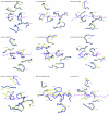
Figure 1
Figure 1. Conformational differences in the CDR loops of bound and free TCRs
The view is of superimposed TCRs from the perspective of the MHC peptide-binding groove, with the peptide from the bound structure shown for reference (purple). Yellow and blue represent the free and bound receptors respectively. For the 2C TCR, only the differences between the free receptor and 2C bound to SIYR/H-2Kb are shown; see Figure 4 for views of loop positions when 2C is bound to other ligands. The discontinuous segment for CDR3α in the unligated ELS4 TCR reflects missing electron density. The image for the unligated D10 TCR, whose structure was determined via NMR, was generated using the best representative conformer of the structural ensemble as identified in the co-ordinate file.
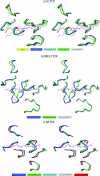
Figure 2
Figure 2. Conformational changes in TCR CDR loops fall into three general classes: hinge-bending movements, loop remodelling or rigid-body movements originating in the TCR framework region
The three classes are illustrated in (a) by CDR3β of the LC13 TCR (hinge bending), (b) by CDR3α of the 1G4 TCR (loop remodelling) and (c) by CDR3α of the E8 TCR (rigid-body shift). Yellow and blue represent the free and bound receptors respectively.
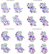
Figure 3
Figure 3. Stereo image of the solvent-accessible surfaces of the 2C, LC13, E8 and 1G4 TCRs from the view of the MHC peptide-binding domains
Colouring is by electrostatic potential, from −25 kT (red) to +25 kT (blue). For each TCR, the top view is of the unligated TCR superimposed on to the bound receptor, whereas the bottom view is of the ligated TCR. Peptides are shown in both the bound and unbound views for orientation. In each case, there are clear differences in structure and electrostatics between the free and bound receptors. Electrostatic potentials were calculated using the program DelPhi [101].
Figure 4
Figure 4. Stereo image showing different CDR loop positions when the same TCR is bound to different pMHC ligands
Orientation and TCR superimposition is through the MHC peptide-binding groove as in Figure 1. (a) View of the CDR loops of the 2C TCR free (yellow) and bound to dEV8/H-2Kb (blue), dEV8/H-2Kbm3 (cyan), SIYR/H-2Kb (green) and QL9/H-2Ld (grey). The two peptides shown reflect the more orthogonal docking angle of the 2C TCR on the QL9 compared with the dEV8/SIYR ligands. (b) View of the CDR loops of the BM3.3 TCR bound to pBM1/H-2Kb (blue), pBM8/H-2Kb (green) and VSV/H-2Kb (grey). (c) View of the CDR loops of the A6 TCR bound to Tax/HLA-A2 (blue), Tax-P6A/HLA-A2 (green), Tax-Y8A/HLA-A2 (cyan), Tax-V7R/HLA-A (grey) and Tax-5K-IBA/HLA-A2 (red). An interactive three-dimensional version of this Figure can be found at http://www.BiochemJ.org/bj/415/0183/bj4150183add.htm.
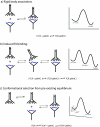
Figure 5
Figure 5. Schematic diagrams indicating three possible TCR–pMHC binding mechanisms
The left-hand side of each panel illustrates protein association, whereas the right-hand side shows traditional free-energy diagrams showing energy as a function of reaction progress. (a) Rigid-body docking, where the structures of the TCR and pMHC are identical in the bound and free states. (b) Induced–fit binding, where a loose intermediate energy complex ([TCR-pMHC]*) is initially formed, followed by rearrangements in the TCR giving rise to the final lowest-energy-bound state. (c) Conformational selection from a pre-existing equilibrium, in which the free TCR samples multiple conformations (two are illustrated here), with only one being binding competent. As shown, binding to the competent TCR conformation follows a rigid-body mechanism, although, as discussed in the text, the association phase could also follow an induced-fit mechanism.

Similar articles

Cited by

References

    1. Mason D. A very high level of crossreactivity is an essential feature of the T-cell receptor. Immunol. Today. 1998;19:395–404. - PubMed
    1. Wilson D. B., Wilson D. H., Schroder K., Pinilla C., Blondelle S., Houghten R. A., Garcia K. C. Specificity and degeneracy of T cells. Mol. Immunol. 2004;40:1047–1055. - PubMed
    1. Maverakis E., van den Elzen P., Sercarz E. E. Self-reactive T cells and degeneracy of T cell recognition: evolving concepts–from sequence homology to shape mimicry and TCR flexibility. J. Autoimmun. 2001;16:201–209. - PubMed
    1. Garcia K. C., Adams E. J. How the T cell receptor sees antigen – a structural view. Cell. 2005;122:333–336. - PubMed
    1. Rudolph M. G., Stanfield R. L., Wilson I. A. How TCRs bind MHCs, peptides, and coreceptors. Annu. Rev. Immunol. 2006;24:419–466. - PubMed

Publication types

MeSH terms

Substances