. 2009 Jun;127(2):196-205.
doi: 10.1111/j.1365-2567.2008.02937.x. Epub 2008 Sep 16.

Altered homeostasis of CD4(+) FoxP3(+) regulatory T-cell subpopulations in systemic lupus erythematosus

Affiliations

Altered homeostasis of CD4(+) FoxP3(+) regulatory T-cell subpopulations in systemic lupus erythematosus

Jau-Ling Suen et al. Immunology. 2009 Jun.

Abstract

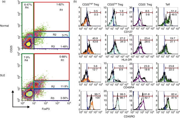
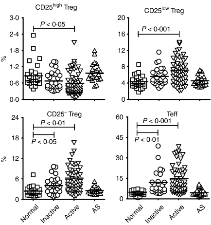
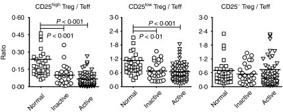
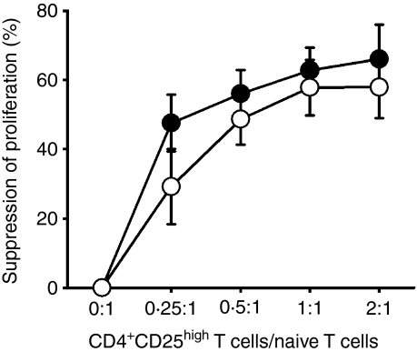
The role of naturally occurring regulatory T cells (Treg), known to be phenotypically heterogeneous, in controlling the expression of systemic lupus erythematosus (SLE) is incompletely defined. Therefore, different subpopulations of CD4(+) FoxP3(+) Tregs in patients with active or inactive SLE were investigated and compared with those of healthy subjects and patients with ankylosing spondylitis (AS). Characterization of different subsets of circulating CD4(+) FoxP3(+) Tregs was examined using flow cytometry. CD4(+) CD25(high) T cells were sorted and examined for suppressive activity in vitro. The results showed first that a significant decrease in the frequency of CD4(+) CD25(high) FoxP3(+) T cells was present in patients with active SLE (n = 58), compared with healthy controls (n = 36) and AS patients (n = 23). In contrast, the frequencies of CD25(low) FoxP3(+) and CD25(-) FoxP3(+) CD4(+) T cells were significantly increased in patients with active SLE by comparison with the control subjects. The elevation of these two putative Treg subpopulations was associated with lower plasma levels of complement C3 and C4 in patients with SLE. In addition, the ratios of the three subsets of CD4(+) FoxP3(+) Tregs versus effector T cells (CD4(+) CD25(+) FoxP3(-)) were inversely correlated with the titer of anti-double-stranded DNA IgG in patients with inactive, but not active, SLE. These results suggest that the pathogenesis of SLE may be associated with a defect in the homeostatic control of different Treg subsets.


Figures

Figure 1
Figure 1
Characterization of different subsets of regulatory T cells (Tregs) and effector T cells (Teffs) in patients with systemic lupus erythematosus (SLE) and in controls. (a) Purified T cells from peripheral blood mononuclear cells (PBMCs) were stained with monoclonal antibodies specific for CD4, CD25 and intracellular forkhead box P3 (FoxP3) and analyzed using a FACScan. Representative dot-plots show the expression of CD25 and FoxP3 on gated CD4+ T cells from one healthy donor (upper panel) and from one patient with SLE (lower panel). Quadrants were established using appropriate isotype controls. CD4+ CD25high FoxP3+ (R1), CD4+ CD25low FoxP3+ (R2) and CD4+ CD25 FoxP3+ (R3) cells were termed as CD25high Tregs, CD25low Tregs and CD25 Tregs, respectively. CD4+ CD25+ FoxP3 (R4) cells were termed as Teffs. (b) PBMCs were stained with monoclonal antibodies specific for CD4, CD25, CD127, HLA-DR, CD45RO, CD45RA and intracellular FoxP3 and analyzed using LSRII. Histograms show surface marker expression on different Treg subsets and Teffs of one healthy donor (black line) and one patient with SLE (color line). The blue lines represent the isotype control. Data are representative of five independent controls and four independent patients.
Figure 2
Figure 2
The percentages of different regulatory T-cell (Treg) subsets and effector T cells (Teffs) in patients with systemic lupus erythematosus (SLE) and in healthy controls. The results are shown as percentages of CD25high Tregs, CD25low Tregs, CD25 Tregs and Teffs in normal controls (n = 36), in patients with inactive SLE (n = 29), in patients with active SLE (n = 58) and in patients with ankylosing spondylitis (AS) (n = 23). Symbols represent the frequencies of Treg subsets and Teffs in individual samples. The line within the vertical points marks the mean for each group. A P-value of<0·05 was considered as significant following analysis of the results using a general linear model adjusted for gender and age. See Fig. 1 for other definitions.
Figure 3
Figure 3
The comparisons of ratios of regulatory T-cell (Treg) subsets versus effector T cells (Teffs) in patients with SLE and in healthy controls. The ratio was defined as the percentage of the indicated Treg subset divided by the percentage of Teffs from the same individual. The ratio was calculated from individual data shown in Fig. 2. A P-value of<0·05 was considered significant following analysis of the results using the Mann–Whitney U-test. See Fig. 1 for other definitions.
Figure 4
Figure 4
The relationships between disease activity and the frequencies or relative ratios of regulatory T-cell (Treg) subsets and effector T cells (Teffs) in patients with systemic lupus erythematosus (SLE). (a) The ratios of different Treg subsets versus Teffs were negatively correlated with the titre of anti-double-stranded DNA immunoglobulin G (anti-dsDNA IgG) in patients with inactive SLE (n = 18). (b) The results are shown as the percentage of CD25high Tregs, CD25low Tregs, CD25 Tregs and Teffs in healthy controls (n = 36); SLE patients exhibiting normal complement levels (Non-low C’) (n = 18); and SLE patients exhibiting low complement levels (Low C’) (n = 69). Lines within the vertical points mark the mean for each group. A P-value of<0·05 was considered as significant following analysis of the results using the Mann–Whitney U-test. *P <0·05 compared with normal subjects; δP <0·05 compared to SLE subjects with normal complement levels. See Fig. 1 for other definitions.
Figure 5
Figure 5
The CD25high regulatory T-cell (Treg) suppressive function in patients with systemic lupus erythematosus (SLE) appears normal in vitro. Naïve T cells (CD4+ CD45RAhigh) were stimulated alone or cocultured with autologous CD4+ CD25high T cells in the presence of mitomycin C-treated autologous antigen-presenting cells (APCs) at the indicated ratios. SLE patients (○, n = 7, three inactive and four active) and age-and gender-matched healthy individuals (•, n = 6) were tested for CD25high Treg activity. The per cent suppression of proliferation was calculated as described in the ‘Materials and methods’. The average proliferative response of APCs was 583 counts per minute (c.p.m.) in the control group and 1224 c.p.m. in the patient group. The average stimulation index of responder T cells was 8·3 in the control group and 9·9 in the patient group. Sorted CD4+ CD25high T cells proliferated poorly after activation with monoclonal anti-CD3, while the average stimulation index of CD25high Tregs in both groups was < 2·0. See Fig. 1 for other definitions.

References

    1. Tan EM. Autoantibodies and autoimmunity: a three-decade perspective. A tribute to Henry G. Kunkel. Ann N Y Acad Sci. 1997;815:1–14. - PubMed
    1. Viglietta V, Baecher-Allan C, Weiner HL, Hafler DA. Loss of functional suppression by CD4+CD25+ regulatory T cells in patients with multiple sclerosis. J Exp Med. 2004;199:971–9. - PMC - PubMed
    1. Kriegel MA, Lohmann T, Gabler C, Blank N, Kalden JR, Lorenz HM. Defective suppressor function of human CD4+ CD25+ regulatory T cells in autoimmune polyglandular syndrome type II. J Exp Med. 2004;199:1285–91. - PMC - PubMed
    1. Crispin JC, Martinez A, Alcocer-Varela J. Quantification of regulatory T cells in patients with systemic lupus erythematosus. J Autoimmun. 2003;21:273–6. - PubMed
    1. Liu MF, Wang CR, Fung LL, Wu CR. Decreased CD4+CD25+ T cells in peripheral blood of patients with systemic lupus erythematosus. Scand J Immunol. 2004;59:198–202. - PubMed

Publication types

MeSH terms