

Effect of sleep stage on interictal high-frequency oscillations recorded from depth macroelectrodes in patients with focal epilepsy
- PMID: 18801037
- PMCID: PMC3792080
- DOI: 10.1111/j.1528-1167.2008.01784.x
Effect of sleep stage on interictal high-frequency oscillations recorded from depth macroelectrodes in patients with focal epilepsy
Abstract
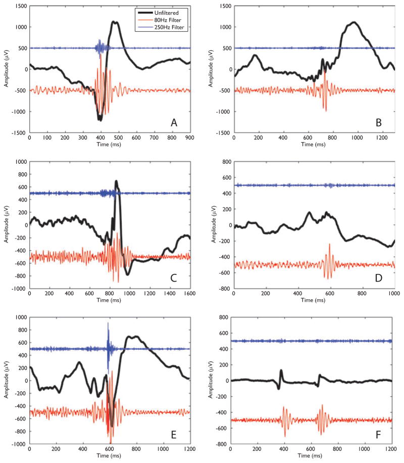
Purpose: To investigate the effect of sleep stage on the properties of high-frequency oscillations (HFOs) recorded from depth macroelectrodes in patients with focal epilepsy.
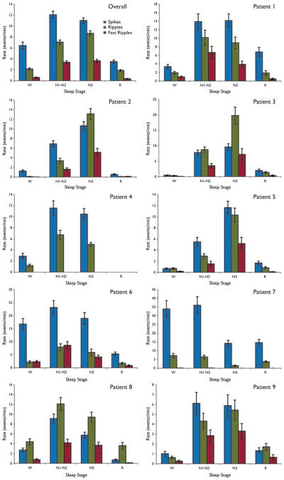
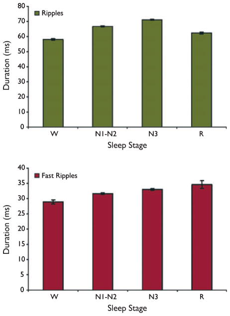
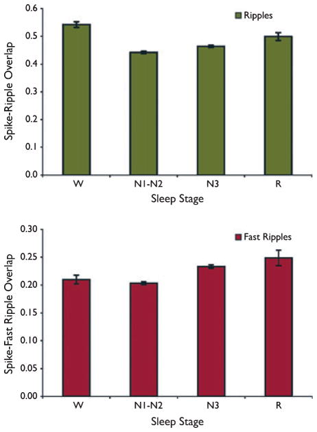
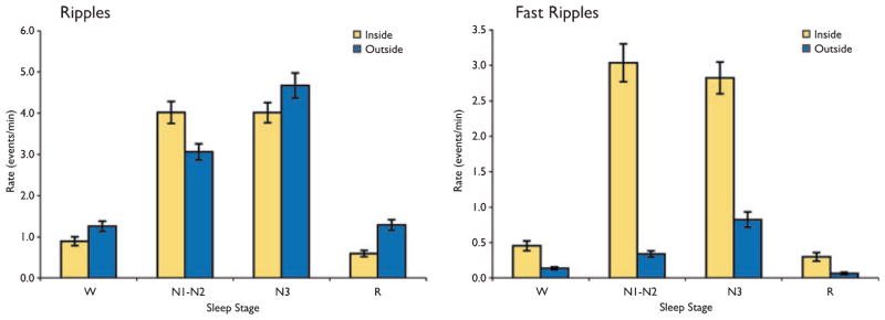
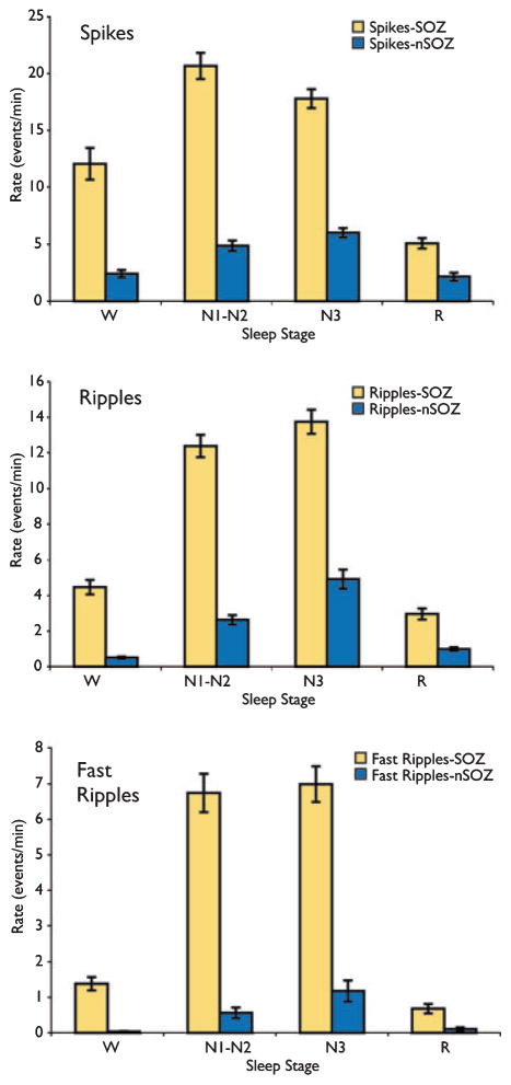
Methods: Ten-minute epochs of wakefulness (W), stage 1-2 non-REM (N1-N2), stage 3 non-REM (N3) and REM sleep (R) were identified from stereo-electroencephalography (SEEG) data recorded at 2 kHz in nine patients. Rates of spikes, ripples (>80 Hz), and fast ripples (>250 Hz) were calculated, as were HFO durations, degree of spike-HFO overlap, HFO rates inside and outside of spikes, and inside and outside of the seizure-onset zone (SOZ).
Results: Ripples were observed in nine patients and fast ripples in eight. Spike rate was highest in N1-N2 in 5 of 9 patients, and in N3 in 4 of 9 patients, whereas ripple rate was highest in N1-N2 in 4 of 9 patients, in N3 in 4 of 9 patients, and in W in 1 of 9 patients. Fast ripple rate was highest in N1-N2 in 4 of 8 patients, and in N3 in 4 of 8 patients. HFO properties changed significantly with sleep stage, although the absolute effects were small. The difference in HFO rates inside and outside of the SOZ was highly significant (p < 0.000001) in all stages except for R and, for fast ripples, only marginally significant (p = 0.018) in W.
Conclusions: Rates of HFOs recorded from depth macroelectrodes are highest in non-REM sleep. HFO properties were similar in stages N1-N2 and N3, suggesting that accurate sleep staging is not necessary. The spatial specificity of HFO, particularly fast ripples, was affected by sleep stage, suggesting that recordings excluding REM sleep and wakefulness provide a more reliable indicator of the SOZ.
Conflict of interest statement
Disclosure: None of the authors has any conflict of interest to disclose.
Figures
References
-
- Billiard M, Besset A, Zachariev Z, Touchon J, Baldy-Moulinier M, Cadilhac J. Relation of seizures and seizure discharges to sleep stages. In: Wolf P, Dam M, Janz D, Dreifuss FE, editors. Advances in epileptology. Vol. 16. Raven Press; New York: 1987. pp. 665–670.
-
- Bragin A, Engel J, Jr, Wilson CL, Fried I, Buzsáki G. High-frequency oscillations in human brain. Hippocampus. 1999a;9:137–142. - PubMed
-
- Bragin A, Engel J, Jr, Wilson CL, Fried I, Mathern GW. Hippocampal and entorhinal cortex high-frequency oscillations (100–500 Hz) in human epileptic brain and in kainic acid-treated rats with chronic seizures. Epilepsia. 1999b;40:127–137. - PubMed
-
- Bragin A, Wilson CL, Staba RJ, Reddick M, Fried I, Engel J., Jr Interictal high-frequency oscillations (80–500 Hz) in the human epileptic brain: entorhinal cortex. Ann Neurol. 2002;52:407–415. - PubMed
-
- Clemens Z, Mçlle M, Erçss L, Barsi P, Halász P, Born J. Temporal coupling of parahippocampal ripples, sleep spindles and slow oscillations in humans. Brain. 2007;130:2868–2878. - PubMed
Publication types
MeSH terms
Grants and funding
LinkOut - more resources
Full Text Sources
Other Literature Sources
