

FDG-PET/CT imaging for staging and radiotherapy treatment planning of head and neck carcinoma
- PMID: 18801181
- PMCID: PMC2559840
- DOI: 10.1186/1748-717X-3-29
FDG-PET/CT imaging for staging and radiotherapy treatment planning of head and neck carcinoma
Abstract
Background: Positron emission tomography (PET) has a potential improvement for staging and radiation treatment planning of various tumor sites. We analyzed the use of 18F-fluorodeoxyglucose (FDG)-PET/computed tomography (CT) images for staging and target volume delineation of patients with head and neck carcinoma candidates for radiotherapy.
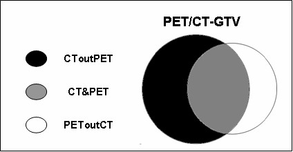
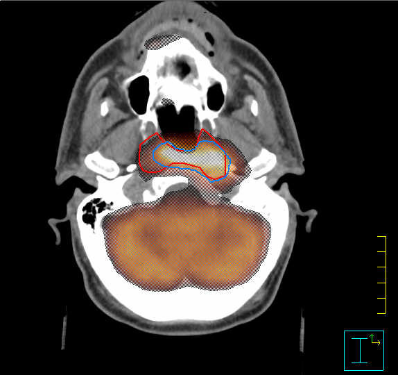
Methods: Twenty-two patients candidates for primary radiotherapy, who did not receive any curative surgery, underwent both CT and PET/CT simulation. Gross Tumor Volume (GTV) was contoured on CT (CT-GTV), PET (PET-GTV), and PET/CT images (PET/CT-GTV). The resulting volumes were analyzed and compared.
Results: Based on PET/CT, changes in TNM categories and clinical stage occurred in 5/22 cases (22%). The difference between CT-GTV and PET-GTV was not statistically significant (p = 0.2) whereas the difference between the composite volume (PET/CT-GTV) and CT-GTV was statistically significant (p < 0.0001).
Conclusion: PET/CT fusion images could have a potential impact on both tumor staging and treatment planning.
Figures
References
-
- Schwartz DL, Ford E, Rejendran J, Yueh B, Coltrera MD, Virgin J, Anzai Y, Haynor D, Lewellyn B, Mattes D, Meyer J, Phillips M, Leblanc M, Kinahan P, Krohn K, Eary J, Laramore GE. FDG-PET/CT imaging for preradiotherapy staging of head and neck cell carcinoma. Int J Radiat Oncol Biol Phys. 2005;61:129–136. - PubMed
-
- Maisey MN. Overview of clinical PET. Br J Radiol. 2002;75:S1–S5. - PubMed
-
- Dizendorf EV, Baumert BG, von Schulthess GK, Lutolf UM, Steinert HC. Impact of whole-body 18F-FDG PET on staging and managing patients for radiation therapy. J Nucl Med. 2003;44:24–29. - PubMed
-
- Schechter NR, Gillenwater AM, Byers MR, Garden AS, Morrison WH, Nquyen LN, Podoloff DA, Ang KK. Can positron emission tomography improve the quality of care for head and neck cancer patients? Int J Radiat Oncol Biol Phys. 2001;51:4–9. - PubMed
Publication types
MeSH terms
Substances
LinkOut - more resources
Full Text Sources
Medical
