. 2008 Nov 21;283(47):32900-12.
doi: 10.1074/jbc.M805749200. Epub 2008 Sep 17.

Molecular recognition of corticotropin-releasing factor by its G-protein-coupled receptor CRFR1

Affiliations

Molecular recognition of corticotropin-releasing factor by its G-protein-coupled receptor CRFR1

Augen A Pioszak et al. J Biol Chem. .

Abstract

The bimolecular interaction between corticotropin-releasing factor (CRF), a neuropeptide, and its type 1 receptor (CRFR1), a class B G-protein-coupled receptor (GPCR), is crucial for activation of the hypothalamic-pituitary-adrenal axis in response to stress, and has been a target of intense drug design for the treatment of anxiety, depression, and related disorders. As a class B GPCR, CRFR1 contains an N-terminal extracellular domain (ECD) that provides the primary ligand binding determinants. Here we present three crystal structures of the human CRFR1 ECD, one in a ligand-free form and two in distinct CRF-bound states. The CRFR1 ECD adopts the alpha-beta-betaalpha fold observed for other class B GPCR ECDs, but the N-terminal alpha-helix is significantly shorter and does not contact CRF. CRF adopts a continuous alpha-helix that docks in a hydrophobic surface of the ECD that is distinct from the peptide-binding site of other class B GPCRs, thereby providing a basis for the specificity of ligand recognition between CRFR1 and other class B GPCRs. The binding of CRF is accompanied by clamp-like conformational changes of two loops of the receptor that anchor the CRF C terminus, including the C-terminal amide group. These structural studies provide a molecular framework for understanding peptide binding and specificity by the CRF receptors as well as a template for designing potent and selective CRFR1 antagonists for therapeutic applications.


Figures

FIGURE 1.
FIGURE 1.
Purification and function of the MBP-CRFR1-ECD-H6 fusion protein. A, analysis of the purified fusion protein by nonreducing SDS-PAGE (lane 1) and nondenaturing (native)-PAGE (lane 2). Molecular mass markers are shown in kDa. The gels were stained with Coomassie Brilliant Blue R-250. B, AlphaScreen assay for association of N-terminally biotinylated CRF-(12-41)-NH2 with MBP-CRFR1-ECD-H6. C, competition AlphaScreen assay assessing the ability of unlabeled, full-length CRF family peptides to inhibit the association of biotin-CRF-(12-41)-NH2 (25 nm) with MBP-CRFR1-ECD-H6 (25 nm). The IC50 values for CRF, UcnI, UcnII, and UcnIII were 1.0, 0.5, 3.6, and 4.1 μm, respectively. D, competition AlphaScreen assay assessing the ability of unlabeled, truncated CRF peptides to inhibit the association of biotin-CRF-(12-41)-NH2 (25 nm) with MBP-CRFR1-ECD-H6 (25 nm). The IC50 values for CRF-(1-41), CRF-(12-41), CRF-(22-41), and CRF-(27-41) were 0.5, 3.5, 6.6, and 6.8 μm, respectively. The AlphaScreen results represent the average of duplicate reactions.
FIGURE 2.
FIGURE 2.
Structure of the ligand-free CRFR1 ECD at 2.75 Å resolution. A, two views of a ribbon diagram showing the CRFR1 ECD (crystal form I) with secondary structure elements labeled. The three disulfide bonds are depicted as sticks. MBP is not shown for clarity. B, residues that stabilize the SCR fold of the CRFR1 ECD. Selected side chains are shown as sticks with their carbon atoms colored according to amino acid sequence conservation among the 15 human class B GPCR ECDs. Cyan indicates invariant residues; magenta indicates conservative substitutions, and slate blue indicates nonconserved residues. Hydrogen bonds are depicted as red dashes. C, alignment of the crystal structure of the ligand-free hCRFR1 ECD with the NMR solution structure of the ligand-free mCRFR2β ECD (PDB code 2JNC). C-α backbone traces are shown with CRFR1 colored slate blue and CRFR2β colored cyan.
FIGURE 3.
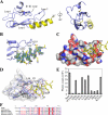
FIGURE 3.
Structural and biochemical analysis of the interaction of CRF with the CRFR1 ECD. A, structure of the CRF-(22-41)-NH2-bound CRFR1 ECD (crystal form II) presented as a ribbon diagram. The ECD is colored slate blue and CRF yellow. MBP is not shown for clarity. B, electron density maps for CRF. The 2Fo - Fc omit map (blue) is contoured at 1 σ and the Fo - Fc omit map (green) is contoured at 3 σ. The maps were prepared as described under “Experimental Procedures” C, molecular surface of the CRFR1 ECD at the peptide-binding site. The surface is colored gray for carbon atoms, blue for nitrogen atoms, and red for oxygen atoms. The CRF peptide is shown as a yellow coil with selected side chains as sticks. D, detail of the interface with the ECD shown as a slate blue ribbon diagram covered by a semi-transparent molecular surface, and CRF shown as a yellow coil. Selected side chains are shown as sticks. Hydrogen bonds are depicted as red dashes, and the red spheres are water molecules. E, AlphaScreen assay assessing the ability of alanine-scan mutants of CRF-(27-41)-NH2 (100 μm) to compete with the interaction of biotin-CRF-(12-41)-NH2 (25 nm) and MBP-CRFR1-ECD-H6 (25 nm). The results shown are the average of triplicate reactions. F, amino acid sequence alignment of selected CRF family peptides. Strictly conserved residues are colored white on a red background and conservative substitutions are colored red on a white background. The antagonist astressin contains the nonstandard amino acids d-phenylalanine (f), and norleucine (B).
FIGURE 4.
FIGURE 4.
Structure of the CRF-(27-41)-NH2-bound CRFR1 ECD at 3.4 Å resolution. A, ribbon diagram of the crystal form III complex with the CRFR1 ECD colored slate blue and CRF yellow. MBP is not shown for clarity. B, electron density maps for CRF. The 2Fo - Fc omit map (blue) is contoured at 1 σ and the Fo - Fc omit map (green) is contoured at 3 σ. The maps were prepared as described under “Experimental Procedures.” C, detail of the interface depicted as in Fig. 3D. D, alignment of the crystal form II and form III structures. C-α backbone traces are shown with the CRF-(22-41)-NH2-bound ECD colored slate blue and CRF-(22-41)-NH2 yellow. The CRF-(27-41)-NH2-bound ECD is colored blue and CRF-(27-41)-NH2 sand.
FIGURE 5.
FIGURE 5.
Conformational changes in the CRFR1 ECD associated with CRF binding. Structural alignment of the ligand-free ECD (crystal form I) and the CRF-(22-41)-NH2-bound ECD (crystal form II). C-α backbone traces of the ECDs are shown with the ligand-free ECD colored cyan and the ligand-bound ECD colored slate blue. CRF is shown as a yellow coil. Selected side chains are shown as sticks, and the red dashes depict hydrogen bonds.
FIGURE 6.
FIGURE 6.
Comparison of the hCRFR1 ECD·CRF complex with the crystal structures of related peptide-class B GPCR ECD complexes. A-C, three views of a structural alignment of the crystal form II CRFR1 ECD·CRF complex with the complexes of PTH (A), GIP (B), and exendin-4 (C) bound to the ECDs of PTH1R (PDB code 3C4M), GIPR (PDB code 2QKH), and GLP1R (PDB code 3C5T), respectively. The ECDs are shown as C-α backbone traces, and the peptides as coils. The CRFR1 ECD is colored slate blue and CRF yellow. The ECDs of PTH1R, GIPR, and GLP1R are colored light gray, and their cognate peptide ligands dark gray. D, amino acid sequence alignment of the human CRFR1 and CRFR2 ECDs with the ECDs of human PTH1R, GIPR, and GLP1R. CRFR1 secondary structure elements are shown at the top and the disulfide bond connectivity at the bottom. The color scheme is the same as in Fig. 3F. PTH1R contains a large insertion in loop 1 that is absent in the other receptors; for clarity, this segment was removed as indicated by four dashes. The N-terminal signal peptides are also not shown.
FIGURE 7.
FIGURE 7.
Comparison of the hCRFR1 ECD·CRF complex and the NMR solution structure of the mCRFR2β ECD·astressin complex. A-C, three views of a structural alignment of the crystal form II complex of CRF-(22-41)-NH2 bound to the hCRFR1 ECD with the NMR solution structure of the mCRFR2β ECD bound to astressin (PDB code 2JND). C-α backbone traces are shown with the CRFR1 ECD·CRF complex colored slate blue and yellow, respectively, and the CRFR2β ECD·astressin complex colored cyan and red, respectively. D, molecular surface of the CRFR1 ECD from crystal form II colored according to sequence conservation between CRFR1 and CRFR2. The surface is colored light blue for residues that are identical, blue for residues that have conservative substitutions, and magenta for residues that differ between the two receptors. CRF-(22-41)-NH2 is shown as a yellow coil. E, amino acid sequence alignment of the human CRFR1 ECD with the human and mouse CRFR2β ECDs. Secondary structure elements are shown at the top and the disulfide bond connectivity at the bottom. The color scheme is the same as in Fig. 3F.

References

    1. Vale, W., Spiess, J., Rivier, C., and Rivier, J. (1981) Science 213 1394-1397 - PubMed
    1. Hsu, S. Y., and Hsueh, A. J. (2001) Nat. Med. 7 605-611 - PubMed
    1. Lewis, K., Li, C., Perrin, M. H., Blount, A., Kunitake, K., Donaldson, C., Vaughan, J., Reyes, T. M., Gulyas, J., Fischer, W., Bilezikjian, L., Rivier, J., Sawchenko, P. E., and Vale, W. W. (2001) Proc. Natl. Acad. Sci. U. S. A. 98 7570-7575 - PMC - PubMed
    1. Reyes, T. M., Lewis, K., Perrin, M. H., Kunitake, K. S., Vaughan, J., Arias, C. A., Hogenesch, J. B., Gulyas, J., Rivier, J., Vale, W. W., and Sawchenko, P. E. (2001) Proc. Natl. Acad. Sci. U. S. A. 98 2843-2848 - PMC - PubMed
    1. Vaughan, J., Donaldson, C., Bittencourt, J., Perrin, M. H., Lewis, K., Sutton, S., Chan, R., Turnbull, A. V., Lovejoy, D., Rivier, C., Rivier, J., Sawchenko, P. E., and Vale, W. (1995) Nature 378 287-292 - PubMed

Publication types