. 2008 Nov 15;586(22):5403-18.
doi: 10.1113/jphysiol.2008.156992. Epub 2008 Sep 18.

TRPML3 mutations cause impaired mechano-electrical transduction and depolarization by an inward-rectifier cation current in auditory hair cells of varitint-waddler mice

Affiliations

TRPML3 mutations cause impaired mechano-electrical transduction and depolarization by an inward-rectifier cation current in auditory hair cells of varitint-waddler mice

Alexander F J van Aken et al. J Physiol. .

Abstract

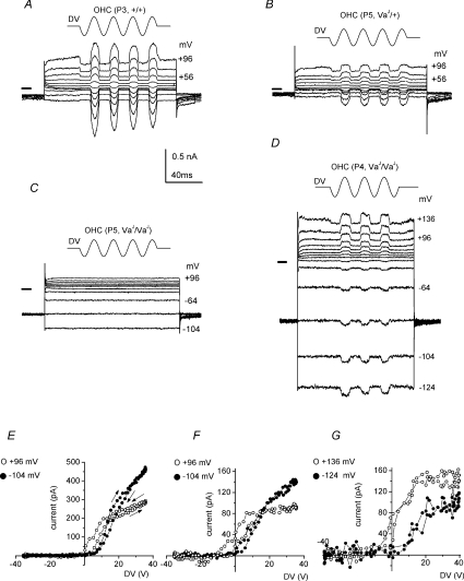
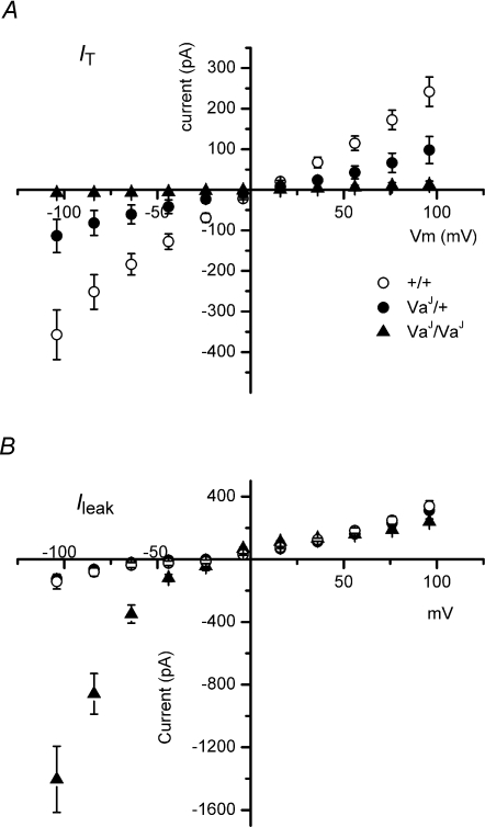
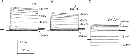
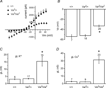
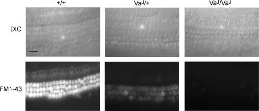
TRPML3 (mucolipin-3) belongs to one of the transient-receptor-potential (TRP) ion channel families. Mutations in the Trpml3 gene cause disorganization of the stereociliary hair bundle, structural aberrations in outer and inner hair cells and stria vascularis defects, leading to deafness in the varitint-waddler (Va) mouse. Here we refined the stereociliary localization of TRPML3 and investigated cochlear hair cell function in varitint-waddler (Va(J)) mice carrying the TRPML3<I362T/A419P> mutations. Using a TRPML3-specific antibody we detected a approximately 68 kDa protein with near-equal expression levels in cochlea and vestibule of wild-type and Va(J) mutants. At postnatal days 3 and 5, we observed abundant localization of TRPML3 at the base of stereocilia near the position of the ankle links. This stereociliary localization domain was absent in Va(J) heterozygotes and homozygotes. Electrophysiological recordings revealed reduced mechano-electrical transducer currents in hair cells from Va(J)/+ and Va(J)/Va(J) mice. Furthermore, FM1-43 uptake and [(3)H]gentamicin accumulation were decreased in hair cells in cultured organs of Corti from Va(J)/+ and Va(J)/Va(J) mice. We propose that TRPML3 plays a critical role at the ankle-link region during hair-bundle growth and that an adverse effect of mutant TRPML3 on bundle development and mechano-electrical transduction is the main cause of hearing loss in Va(J)/+ mutant mice. Outer hair cells of Va(J)/Va(J) mice additionally had depolarized resting potentials due to an inwardly rectifying leak conductance formed by the mutant channels, leading over time to hair-cell degeneration and contributing to their deafness. Our findings argue against TRPML3 being a component of the hair-cell transducer channel.


Figures

Figure 1
Figure 1. TRPML3 expression in vitro and in the inner ear
A, COS-1 cells were transfected with TRPML3-GFP and probed by Western blotting for the presence of fusion protein with HL4460 or anti-GFP. HL4460 detects GFP-tagged TRPML3 (arrowhead) of ∼91 kDa and a putative dimer at ∼215 kDa (arrow). The GFP antibody detects both the 91 kDa (arrowhead) and 215 kDa (arrow) bands. dH2O mock and pcDNA3.1 represent transfection controls. Protein extracts (25 μg) were loaded in each lane. Equal loading and transfer efficiencies were evaluated with β-actin antibody. B, cochlear and vestibular tissue extracts from wild-type (+/+), heterozygous (VaJ/+) and homozygous (VaJ/VaJ) varitint-waddler mice, taken at P21, were probed for the presence of endogenous TRPML3 with HL4460. On Western blots, the 68 kDa TRPML3 species was present in both tissues (arrowhead) for all genotypes. Quantities of 25 μg and 40 μg were loaded in each lane for cochlear and vestibular extracts, respectively. C, transfected COS-1 cells expressing TRPML3-GFP (green) were stained with the HL4460 antibody (red). GFP fluorescence co-localizes with HL4460 labelling (merge). Scale bar in C = 10 μm
Figure 2
Figure 2. TRPML3 localization in cochlear hair cell stereocilia
Confocal images showing stereocilia of IHCs and OHCs of postnatal C3HeB/FeJ mice stained for TRPML3 (HL4460 or PB221; red) and F-actin (phalloidin; green). A, modiolar view of three rows of OHCs (labelled O1 to O3) and one row of IHCs (labelled I) of the apical region of the organ of Corti of a P3 mouse stained for TRPML3 and F-actin shows a discrete band of punctate staining of stereocilia on OHCs and IHCs. B and C, a higher magnification modiolar view of the organ of Corti (apical coil) stained for TRPML3 and F-actin shows discrete labelling at the base of stereocilia on IHCs (B, arrowhead) and OHCs (C, arrowhead). Arrows show tips of the stereocilia. D, top view of three rows of OHCs (basal coil) stained for TRPML3 and F-actin shows two rows of punctate staining (arrowheads) in each hair bundle in between three rows of stereocilia (arrows). E, modiolar view of one row of IHCs from the mid-apical region of P10 mice stained for TRPML3 and F-actin shows no discrete labelling of stereocilia. Scale bar in all images corresponds to 5 μm.
Figure 3
Figure 3. TRMPL3 labelling in the ankle-link region of stereocilia
A, electron micrograph of an OHC hair bundle from a cochlear culture labelled with rabbit antibody PB221 to the ectodomain of TRPML3 followed by 10 nm gold-conjugated goat anti-rabbit. The gold particles are observed in the ankle-link region (arrows) at the base of the hair bundle. B, detail of ankle-link region shown above in A. Scale bars = 200 nm. C, labelling is not seen in the control with non-immune rabbit IgG.
Figure 4
Figure 4. TRPML3 localization in varitint-waddler auditory hair cells
Confocal images showing stereocilia of IHCs and OHCs of P3 mice of +/+ (A), VaJ/+ heterozygotes (B), and VaJ/VaJ homozygotes (C) stained for TRPML3 (HL4460, red) and F-actin (phalloidin, green). Discrete labelling is observed in wild-type +/+ mice at stereocilia of OHCs (arrowhead) and IHCs (arrow), but not in the stereociliary hair bundle of VaJ/+ and VaJ/VaJ mutants. All images taken from middle of apical coil. The star indicates a disoriented hair bundle. Scale bar in all images corresponds to 5 μm.
Figure 5
Figure 5. Transducer currents at different membrane potentials recorded from +/+, VaJ/+ and VaJ/VaJ OHCs
Transducer currents were recorded in response to 45 Hz sinusoidal force stimuli. Cells were held at −84 mV and the membrane potential was stepped to different levels in 20 mV increments. Driver voltage to the fluid jet is shown above the traces, upward being excitatory. Membrane potentials are shown next to some of the traces. A, P3 +/+ OHC; B, P5 VaJ/+ OHC; C, P5 VaJ/VaJ non-transducing OHC; D, P4 VaJ/VaJ transducing OHC. Driver voltage amplitude 40 V in D. Note the large ‘leak’ conductance in C and D. All recordings are from apical coil OHCs. Horizontal bars left of the recordings indicate zero-current level. Some spike artefacts in the currents of B and C have been blanked. E–G, absolute value of transducer currents as a function of driver voltage for the cells of A, B and D, respectively, averaged during two stimulus periods, starting with the negative half-cycle. The resting transducer currents (before stimulus onset) are 11 pA at −104 mV and 87 pA at +96 mV (E); 6 pA at −104 mV and 50 pA at +96 mV (F); 7 pA at −124 mV and 61 pA at +136 mV (G). Filled symbols: currents at most negative potential used; open symbols: currents at most positive potential. Data points are at 200 μs intervals. Arrows in E indicate stimulus direction.
Figure 6
Figure 6. I–V relationships for transducer and leak currents from +/+, VaJ/+ and VaJ/VaJ OHCs
A, averaged mechano-electrical transducer currents for +/+ (n = 3, P3), VaJ/+ (n = 9, P5) and VaJ/VaJ (n = 22, P3–6) OHCs. B, averaged leak currents for +/+ (n = 7, P3–5), VaJ/+ (n = 9, P5) and VaJ/VaJ (n = 23, P0–6) OHCs. Holding potential was −84 mV. From A it is clear that VaJ/VaJ OHCs display negligible transducer currents. Transducer currents in the heterozygous cells are of a lower amplitude when compared to the wild-type control cells. B shows that +/+ and VaJ/+ cells have identical I–V relationships and do not display the inwardly rectifying leak current. The VaJ/VaJ cells display a strong rectifying leak current. Cs+-based intracellular solution. Recordings are from apical (n = 36) and basal (n = 3) OHCs.
Figure 7
Figure 7. Basolateral currents at different membrane potentials recorded from +/+, VaJ/+ and VaJ/VaJ OHCs
Basolateral currents were elicited by voltage steps in 10 mV increments from a holding potential of −84 mV, nominally between −104 mV and +46 mV. Following the steps the potential was returned to −44 mV. Membrane potentials (corrected for series resistance) are shown next to some of the traces. A, P3 +/+ OHC; B, P3 VaJ/+ OHC; C, P4 VaJ/VaJ OHC. Note the large inwardly rectifying leak conductance in C. Recordings are from apical and basal coil OHCs. Horizontal bar left of the recording in A indicates zero-current level for all panels.
Figure 8
Figure 8. Effects of the inwardly rectifying leak conductance on VaJ/VaJ OHCs
A, I–V relationships using K+-based intracellular solution. Averaged currents ±s.e.m. of +/+ (n = 5, P3–5), VaJ/+ (n = 10, P2–5) and VaJ/VaJ (n = 11, P1–6) OHCs. From a holding potential of −84 mV the voltage was stepped in 10 mV increments from −104 mV to +46 mV nominally (error bars for voltage are hidden by the symbols). B, average zero-current potentials with K+-based intracellular solution of +/+ (n = 7, P3–5), VaJ/+ (n = 18, P2–5) and VaJ/VaJ (n = 20, P1–6) OHCs (*P < 0.001). C, average leak conductance with K+-based intracellular solution of +/+ (n = 6, P3–5), VaJ/+ (n = 17, P2–5) and VaJ/VaJ (n = 15, P1–6) OHCs (*P < 0.001). D, average leak conductance using Cs+-based intracellular solution of +/+ (n = 7, P3–5), VaJ/+ (n = 9, P5) and VaJ/VaJ (n = 23, P0–6) OHCs (*P < 0.001).
Figure 9
Figure 9. Failure of homozygous varitint-waddler hair cells to load with FM1-43
Top panels show DIC images from basal coil cochlear cultures (P1, 1 day in vitro) from control (+/+) and mutant (VaJ/+ and VaJ/VaJ) mice. Bottom panels are images taken between 10 and 20 min after a 10 s bath application of 3 μm FM1-43 from the same cultures shown in the top panels. Control hair cells load avidly with FM1-43 whereas heterozygous and homozygous mutant hair cells show reduced dye labelling and fail to load, respectively. Scale bar = 20 μm.
Figure 10
Figure 10. Impaired tritiated gentamicin uptake in VaJ mutants
A progressive decrease in tritiated gentamicin uptake is observed from wild-type to homozygous hair cells. Hair cells in the basal coil of P3 wild-type cochleae take up aminoglycosides. Cells that are labelled (A) correspond with the inner and outer hair cells shown using toluidine staining in B. Heterozygote P3 cochlear basal coil hair cells exhibit less labelling following incubation with gentamicin (C) than those in wild-types. Sections of heterozygote cochleae were also stained with toluidine to show the localization of IHCs and OHCs (D). IHCs in both wild-type (A) and heterozygote (C) sections take up less gentamicin than OHCs. Basal coil hair cells in homozygote cochleae do not take up aminoglycosides (E). Mutant hair cells can be seen using toluidine staining (F). Arrowhead, IHC; arrows, OHCs. Scale bar = 100 μm.

Similar articles

Cited by

References

    1. Adato A, Lefevre G, Delprat B, Michel V, Michalski N, Chardenoux S, Weil D, El-Amraoui A, Petit C. Usherin, the defective protein in Usher syndrome type IIA, is likely to be a component of interstereocilia ankle links in the inner ear sensory cells. Hum Mol Genet. 2005;14:3921–3932. - PubMed
    1. Anniko M, Wroblewski R. Ionic environment of cochlear hair cells. Hear Res. 1986;22:279–293. - PubMed
    1. Assad JA, Hacohen N, Corey DP. Voltage dependence of adaptation and active bundle movement in bullfrog saccular hair cells. Proc Natl Acad Sci U S A. 1989;86:2918–2922. - PMC - PubMed
    1. Atiba-Davies M, Noben-Trauth K. TRPML3 and hearing loss in the varitint-waddler mouse. Biochim Biophys Acta. 2007;1772:1028–1031. - PubMed
    1. Beurg M, Evans MG, Hackney CM, Fettiplace R. A large-conductance calcium-selective mechanotransducer channel in mammalian cochlear hair cells. J Neurosci. 2006;26:10992–11000. - PMC - PubMed

Publication types

MeSH terms