. 2008 Sep 23;105(38):14650-5.
doi: 10.1073/pnas.0801581105. Epub 2008 Sep 18.

Rapid enhancement of two-step wiring plasticity by estrogen and NMDA receptor activity

Affiliations

Rapid enhancement of two-step wiring plasticity by estrogen and NMDA receptor activity

Deepak P Srivastava et al. Proc Natl Acad Sci U S A. .

Erratum in

  • Proc Natl Acad Sci U S A. 2008 Dec 16;105(50):20045.. Woolfrey, Kevin [corrected to Woolfrey, Kevin M]

Abstract

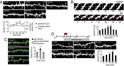
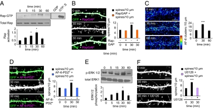
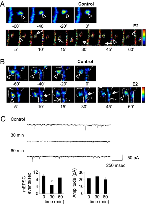
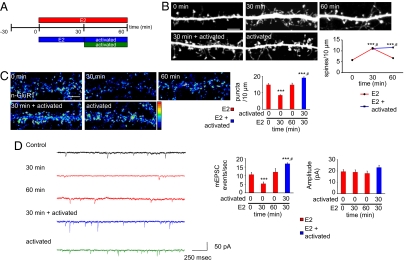
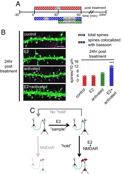
Cortical information storage requires combined changes in connectivity and synaptic strength between neurons, but the signaling mechanisms underlying this two-step wiring plasticity are unknown. Because acute 17beta-estradiol (E2) modulates cortical memory, we examined its effects on spine morphogenesis, AMPA receptor trafficking, and GTPase signaling in cortical neurons. Acute E2 application resulted in a rapid, transient increase in spine density, accompanied by temporary formation of silent synapses through reduced surface GluR1. These rapid effects of E2 were dependent on a Rap/AF-6/ERK1/2 pathway. Intriguingly, NMDA receptor (NMDAR) activation after E2 treatment potentiated silent synapses and elevated spine density for as long as 24 h. Hence, we show that E2 transiently increases neuronal connectivity by inducing dynamic nascent spines that "sample" the surrounding neuropil and that subsequent NMDAR activity is sufficient to stabilize or "hold" E2-mediated effects. This work describes a form of two-step wiring plasticity relevant for cortical memory and identifies targets that may facilitate recovery from brain injuries.


Conflict of interest statement

The authors declare no conflict of interest.

Figures

Fig. 1.
Fig. 1.
E2 rapidly and transiently enhances connectivity by increasing spine turnover. (A) EGFP-expressing cortical neurons (28 days in vitro) were treated with 10 nM E2 for 0, 5, 15, 30, and 60 min. Spine density increased transiently (blue line); average area decreased transiently (red line). (B) Time-lapse imaging of cortical neuron expressing EGFP. Cells were imaged for 60 min before treatment and then at 0, 5, 10, 15, 30, 45, and 60 min after treatment with E2. Red arrows indicate novel spines; red triangles represent persistent spines. (C) E2-treated cells were stained for bassoon. The total number of spines (black bar) and the number of spines colocalizing with bassoon (gray bar) were measured. (D) Schematic of brief, 5-min exposure of E2 experiment. Neurons were treated with E2 for 5 min (E2 pulse); cells were fixed at 0, 5, 15, 30, and 60 min from initial addition of E2. ***, P < 0.001. [Scale bars, 5 μm (A, C, and D); 1 μm (B).]
Fig. 2.
Fig. 2.
Rap activity underlies E2-mediated synaptogenesis. (A) Rap activity after treatment with E2; Rap activity increases in a time-dependent manner. (B) Effect of overexpression of RapGAP on E2-induced increase in spine density; RapGAP (orange bars) blocks rapid E2 actions. (C) Effect of E2 on AF-6 clustering; AF-6 clusters in a transient manner. (D) In the presence of a mutant construct, AF-6-PDZ* (blue bars), E2 is unable to induce a spine-density increase. (E) Time course of ERK1/2 phosphorylation after E2 treatment. (F) Inhibition of ERK1/2 by U0126 (purple bar) inhibits E2-mediated increase in spine density. *, P < 0.05; ***, P < 0.001. (Scale bars, 5 μm.)
Fig. 3.
Fig. 3.
E2 rapidly and transiently induces the formation of silent synapses through trafficking of GluR1 and NR1. (A and B) Time-lapse imaging of neurons expressing GFP-GluR1. Cells were imaged for 60 min before and after administration of E2. Arrowheads indicate GFP-GluR1 in spine heads; arrows indicate GFP-GluR1 in dendritic shaft. Dotted lines indicate neuron outline, as determined by Discosoma red fluorescent protein coexpression; asterisks show transient emergence of novel spines upon E2 treatment. (C) AMPAR mEPSCs after E2 treatment. Frequency and average amplitude of mEPSCs were measured; frequency, but not amplitude, of mEPSCs was significantly reduced at 30 min. *, P < 0.05; ***, P < 0.001. [Scale bars, 1 μm (A and B).]
Fig. 4.
Fig. 4.
Synaptic activity sustains and potentiates E2 effects. (A) Schematic of combined E2 and NMDAR activation experiments. Neurons were treated with E2 for 0, 30, or 60 min; for 30 min with E2 followed by activation of NMDAR for 30 min; or activation of NMDAR alone for 30 min. (B) Effect of E2 priming on dendritic morphology; cells were treated as described in A. (C) Surface staining of GluR1 (n-GluR1) after treatments as in A. The total number of n-GluR1 puncta was measured; n-GluR1 levels are significantly greater than control after combined E2 and NMDAR activity (blue bar). (D) Electrophysiological recording of AMPAR mEPSCs after treatments. Frequency of AMPAR mEPSC events are significantly greater than controls with combined E2 and NMDAR activation (blue bars). AMPAR amplitudes are also increased with treatment with E2 followed by NMDAR activation, but not significantly. **, P < 0.005; ***, P < 0.001; #, different subgroup of significance according to Tukey B post hoc analysis. (Scale bar, 5 μm.)
Fig. 5.
Fig. 5.
Persistent effects of combined E2 and NMDAR activity. (A) Schematic of long-term combined E2 and NMDAR activation experiments. Neurons were treated with E2 for 0 or 30 min; for 30 min with E2 followed by activation of NMDAR for 30 min; or activation of NMDAR alone for 30 min. Cells were fixed 24 h after stimulation. (B) Effect of long-term combined E2 and NMDAR activation on dendritic morphology. Cells were treated as described in A. Treated cells were stained for bassoon. The total number of spines (color stripe bars) and the number of spines colocalizing with bassoon (gray and color stripe bars) were measured. (C) Model of two-step wiring plasticity. E2 treatment increases synaptogenesis, representing the sample step; yellow circles represent nascent connections. If a second stimulus is not applied (i.e., no hold), neuronal connectivity returns to control levels. NMDAR activation alone increases the strength of existing contacts. NMDAR activation after E2 treatment, representing the hold step, sustains and increases the strength of existing and novel connections. ***, P < 0.001. (Scale bar, 5 μm.)

References

    1. Chklovskii DB, Mel BW, Svoboda K. Cortical rewiring and information storage. Nature. 2004;431:782–788. - PubMed
    1. Holtmaat AJ, et al. Transient and persistent dendritic spines in the neocortex in vivo. Neuron. 2005;45:279–291. - PubMed
    1. Le Be JV, Markram H. Spontaneous and evoked synaptic rewiring in the neonatal neocortex. Proc Natl Acad Sci USA. 2006;103:13214–13219. - PMC - PubMed
    1. Kasai H, Matsuzaki M, Noguchi J, Yasumatsu N, Nakahara H. Structure-stability-function relationships of dendritic spines. Trends Neurosci. 2003;26:360–368. - PubMed
    1. Rumpel S, LeDoux J, Zador A, Malinow R. Postsynaptic receptor trafficking underlying a form of associative learning. Science. 2005;308:83–88. - PubMed

Publication types

MeSH terms

LinkOut - more resources