. 2008 Dec;26(12):3194-204.
doi: 10.1634/stemcells.2007-1017. Epub 2008 Sep 18.

Prospective isolation of skeletal muscle stem cells with a Pax7 reporter

Affiliations

Prospective isolation of skeletal muscle stem cells with a Pax7 reporter

Darko Bosnakovski et al. Stem Cells. 2008 Dec.

Abstract

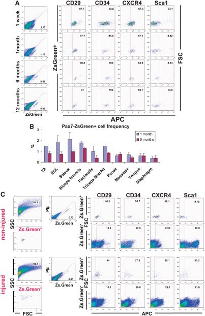
Muscle regeneration occurs through activation of quiescent satellite cells whose progeny proliferate, differentiate, and fuse to make new myofibers. We used a transgenic Pax7-ZsGreen reporter mouse to prospectively isolate stem cells of skeletal muscle by flow cytometry. We show that Pax7-expressing cells (satellite cells) in the limb, head, and diaphragm muscles are homogeneous in size and granularity and uniformly labeled by certain cell surface markers, including CD34 and CD29. The frequency of the satellite cells varies between muscle types and with age. Clonal analysis demonstrated that all colonies arising from single cells within the Pax7-sorted fraction have myogenic potential. In response to injury, Pax7(+) cells reduce CD34, CD29, and CXCR4 expression, increase in size, and acquire Sca-1. When directly isolated and cultured in vitro, Pax7(+) cells display the hallmarks of activation and proliferate, initially as suspension aggregates and later distributed between suspension and adherence. During in vitro expansion, Pax7 (ZsGreen) and CD34 expression decline, whereas expression of PSA-NCAM is acquired. The nonmyogenic, Pax7(neg) cells expand as Sca1(+) PDGRalpha(+) PSA-NCAM(neg) cells. Satellite cells expanded exclusively in suspension can engraft and produce dystrophin(+) fibers in mdx(-/-) mice. These results establish a novel animal model for the study of muscle stem cell physiology and a culture system for expansion of engraftable muscle progenitors.


Figures

Figure 1
Figure 1
ZsGreen fluorescence recapitulates Pax7 expression. (A): ZsGreen expression in embryos from E9.5 to E12.5. Note that green florescence is exclusively localized in the somite, frontonasal processes, and the neural tube. (B): Fluorescence-activated cell sorting profile of muscle digests from Pax7-ZsGreen mice. The upper left panel shows the FSC/SSC profile of total cells. The upper right panel shows fluorescence of live cells (gated using propidium iodide, not shown) in the FSC/SSC gate indicated at the left. Green fluorescence is shown on the x-axis, and red fluorescence is shown on the y-axis. This uncompensated two-dimensional representation allows maximum separation of autofluorescent cells, which track along the diagonal, from the weak true green fluorescent population, which is shifted to the right of the diagonal. The lower panel shows the FSC/SSC profile of total live cells in the ZsGreen gate indicated. Note that green cells were homogeneous in size (FSC) and granularity (SSC). (C): Real-time reverse transcription-polymerase chain reaction on ZsGreen+ or -neg cells directly sorted from hind limb muscle of 3-month-old male mice. Results are presented as fold difference comparing ZsGreen-positive with -neg cells; data represent mean ± SE (n = 3). Abbreviations: E, embryonic day; FSC, forward scatter; neg, negative; SSC, side scatter.
Figure 2
Figure 2
Direct isolation of satellite cells from Pax7-ZsGreen mice. (A): Morphology of ZsGreen+ and ZsGreenneg sorted cells from limb and diaphragm of 3-month-old male and female mice, 3 (top row) and 9 (middle row) days after sorting and expansion in proliferation medium. Bottom row: Morphology of the expanded cells after 2 days in differentiation medium. Note that only ZsGreen+ cells differentiated into myotubes. (B): Real-time reverse transcription-polymerase chain reaction for myogenic genes in Pax7-Zsgreen+ (+) or Pax7-Zsgreen-neg sorted cells expanded in vitro for 7 days in proliferation medium. (C): Immunofluorescence for Pax7, MyoD, and MyHC (in red) in clones obtained from single ZsGreen+ cells. DAPI (blue) was used for counterstaining of the nuclei. Clones were expanded on gelatin-coated surfaces in myogenic proliferation medium for 8 days. (D): Quantification of the plating efficiency of ZsGreen+ and ZsGreenneg sorted cells (top panel). Note that the ZsGreenneg cells could not expand from single cells. All of the colonies obtained from the ZsGreen+ cells were myogenic based on the presence of Pax7-, MyoD- or MyHC-expressing cells within the clone (middle panel). The bottom panel represents the percentage of Pax7-, MyoD-, and MyHC-pos cells within each clone derived from ZsGreen+ cells. Abbreviations: DAPI, 4',6-diamidino-2-phenylindole; GAPDH, glyceraldehyde-3-phosphate dehydrogenase; MyHC, myosin heavy chain; neg, negative; pos, positive.
Figure 3
Figure 3
Surface marker profile of Pax7-ZsGreen+/− cells in limb and diaphragm. Fluorescence-activated cell sorting analyses for different surface markers on Pax7-ZsGreen+ and -negative cells in hind limb and diaphragm in 3-month-old muscle. The x-axis shows the APC channel, and the y-axis shows FSC. Abbreviations: FITC, fluorescein isothiocyanate; FSC, forward scatter; M-CAD, M-cadherin; PDGFRα, platelet-derived growth factor receptor α.
Figure 4
Figure 4
Age-, injury-, and muscle-type-dependent variation in Pax7-ZsGreen+ cells. (A): Surface marker profile of ZsGreen+ cells in 1-week-old and 1-, 6-, and 12-month-old mice. The left column shows green (x-axis) versus red (y-axis) fluorescence. Other columns show APC-conjugated antibody staining (x-axis) versus FSC (y-axis) of gated ZsGreen+ cells. The frequency of ZsGreen+ cells decreased with age, and surface marker profile remained constant, although Pax7 expression increased with age. (B): Frequency of labeled cells in different muscles of 1- and 6-month-old mice. (C): Surface marker profile of ZsGreen+ cells 3 days after injury. One hind limb of each Pax7-ZsGreen mouse was injured with multiple injections of cardiotoxin, and the other (noninjured) was used as a control. Three days after the injury, Pax7-ZsGreen+ cells from injured and noninjured legs were analyzed for expression of various surface markers. Abbreviations: FSC, forward scatter; SSC, side scatter; TA, tibialis anterior.
Figure 5
Figure 5
Characterization of Pax7-ZsGreen+/− cells expanded in vitro. (A): Surface marker profile of ZsGreen+ and -negative cells from limb and diaphragm (diaph) expanded in vitro for 2 weeks. Staining of APC-conjugated antibodies is shown on the x-axis, and FSC is shown on the y-axis. The right column shows green (x-axis) versus red (y-axis) fluorescence. Note that some cells of the initial sorted ZsGreen+ cells remained green in culture. (B): Real-time reverse transcription-polymerase chain reaction for expression of myogenic genes in expanded and differentiated cells from limb and diaphragm. Murine myoblast cell line-C2C12 in proliferation medium (prolif) or differentiation medium (diffe) was used as a control. (C): Immunofluorescence for MyoD, M-CAD, and MyHC in proliferating cells. 4',6-Diamidino-2-phenylindole (blue) labeled the nucleus in the right-hand panel of each pair. Abbreviations: FSC, forward scatter; M-CAD, M-cadherin; MyHC, myosin heavy chain.
Figure 6
Figure 6
Muscle progenitors expand as adherent (adh) and floating cells in vitro and restore dystrophin expression in MDX mice. (A): Cell morphology of ZsGreen+ cells from limb and diaphragm (diaph). expanded in vitro. adh cells had a round to elongated shape. Cells that grew in suspension had a round shape and a tendency to grow in clumps. (B): Fluorescence-activated cell sorting analysis of cultures (susp and adh) initiated with ZsGreen+ cells. The blue trace indicates staining of adh cells; the red trace indicates staining of the susp. (C): Real-time reverse transcription-polymerase chain reaction analyses for myogenic regulators in susp and adh cultures of ZsGreen+ cells. (D): Immunofluorescence for dystrophin expression. ZsGreen+ cells were expanded in susp for 8 days, and 2,000 or 10,000 cells were transplanted into cardiotoxin-injured tibialis anterior muscle of mdx−/− mice (n = 4). The cont-lateral leg was used as a control and was injected with phosphate-buffered saline. Three weeks post-transplantation, muscles were analyzed for dystrophin expression. Red represents dystrophin staining, and blue indicates DAPI staining of the nuclei. Note the presence of clusters of fibers expressing dystrophin in the transplanted leg, but only rare single revertant fibers in the contra-lateral leg. (E): Quantification of the dystrophin-positive fibers in cell-injected and contra-lateral legs. The results represent mean ± SE (p < .01). Abbreviations: DAPI, 4',6-diamidino-2-phenylindole; GAPDH, glyceraldehyde-3-phosphate dehydrogenase; PDGFRα, platelet-derived growth factor receptor α.

Similar articles

Cited by

References

    1. Schultz E. Satellite cells in normal, regenerating and dystrophic muscle. Adv Exp Med Biol. 1985;182:73–84. - PubMed
    1. Bischoff R. A satellite cell mitogen from crushed adult muscle. Dev Biol. 1986;115:140–147. - PubMed
    1. Mauro A. Satellite cell of skeletal muscle fibers. J Biophys Biochem Cytol. 1961;9:493–495. - PMC - PubMed
    1. Schultz E, Gibson MC, Champion T. Satellite cells are mitotically quiescent in mature mouse muscle: An EM and radioautographic study. J Exp Zool. 1978;206:451–456. - PubMed
    1. Snow MH. A quantitative ultrastructural analysis of satellite cells in denervated fast and slow muscles of the mouse. Anat Rec. 1983;207:593–604. - PubMed

Publication types