. 2008 Oct 1;181(7):4733-41.
doi: 10.4049/jimmunol.181.7.4733.

Induction of IL-17+ T cell trafficking and development by IFN-gamma: mechanism and pathological relevance in psoriasis

Affiliations

Induction of IL-17+ T cell trafficking and development by IFN-gamma: mechanism and pathological relevance in psoriasis

Ilona Kryczek et al. J Immunol. .

Abstract

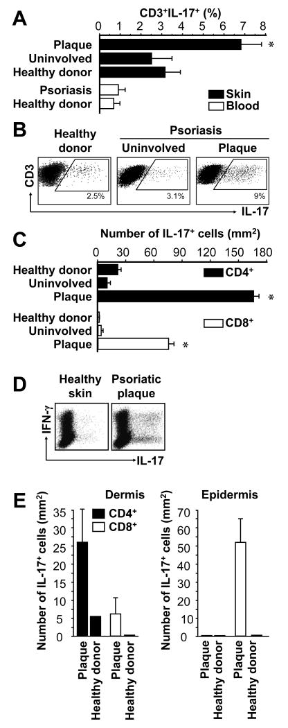
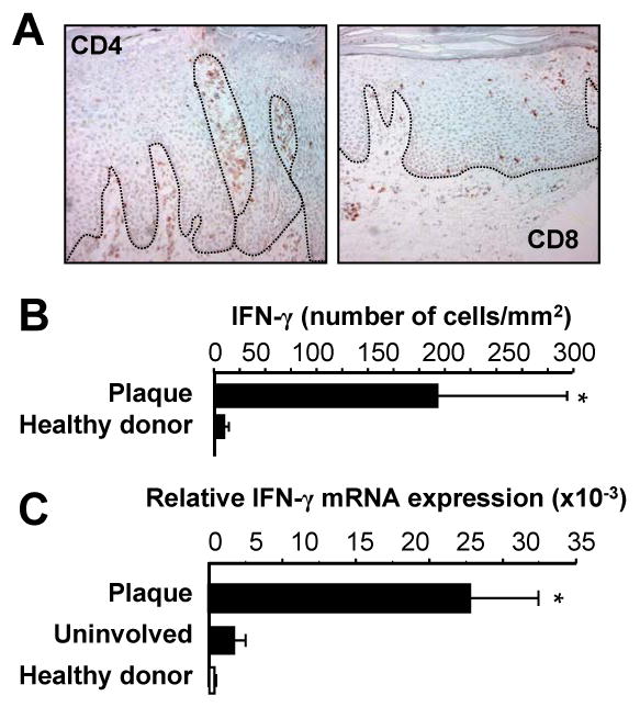
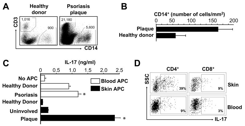
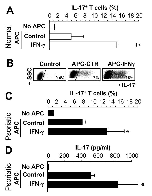
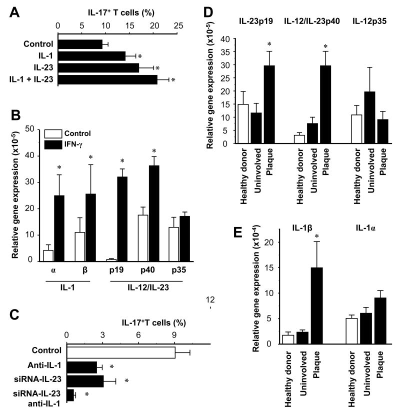
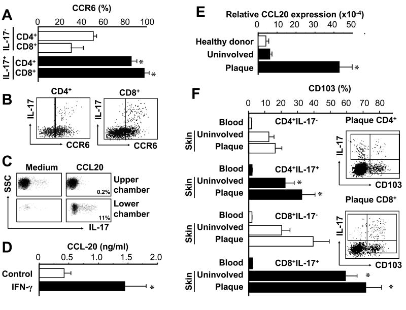
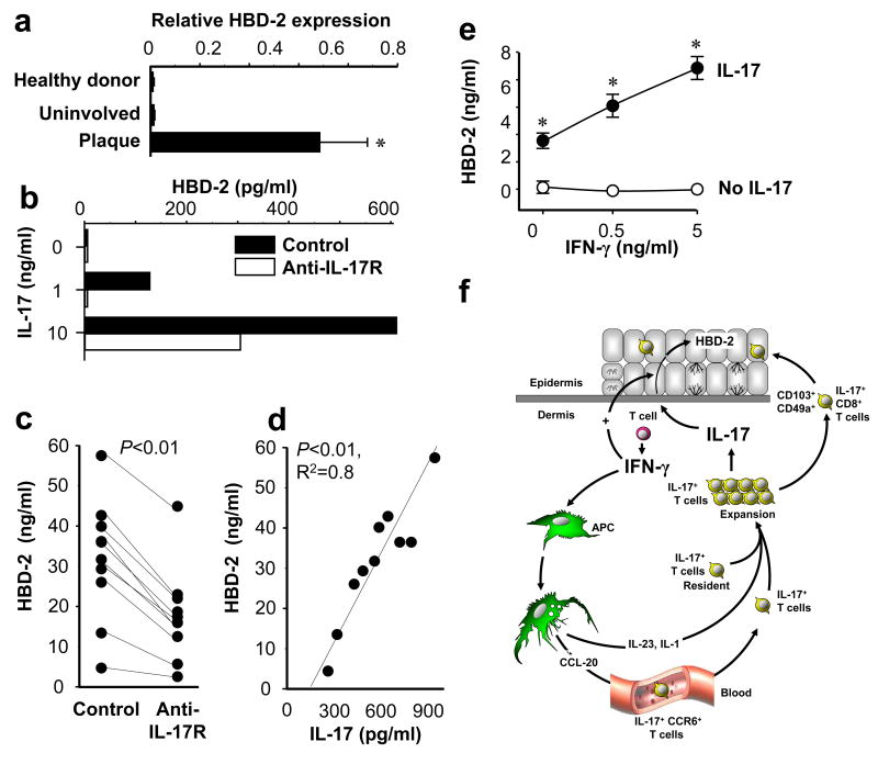
Th1 and Th17 T cells are often colocalized in pathological environments, yet Th1-derived IFN-gamma inhibits Th17 cell development in vitro. We explored the physiologic basis of this paradox in humans. In this study, we demonstrate increased the number of CD4(+) and CD8(+) IL-17(+) T cells in skin lesions of psoriasis. Furthermore, we show that myeloid APCs potently support induction of IL-17(+) T cells, and that this activity is greatly increased in psoriasis. We tested stimuli that might account for this activity. Th1 cells and IFN-gamma are increased in psoriatic blood and lesional skin. We show that IFN-gamma programs myeloid APCs to induce human IL-17(+) T cells via IL-1 and IL-23. IFN-gamma also stimulates APC production of CCL20, supporting migration of IL-17(+) T cells, and synergizes with IL-17 in the production of human beta-defensin 2, an antimicrobial and chemotactic protein highly overexpressed by psoriatic keratinocytes. This study reveals a novel mechanistic interaction between Th1 and IL-17(+) T cells, challenges the view that Th1 cells suppress Th17 development through IFN-gamma, and suggests that Th1 and IL-17(+) T cells may collaboratively contribute to human autoimmune diseases.


Figures

FIGURE 1
FIGURE 1
IL-17+ T cells in psoriasis lesions. Skin biopsies and peripheral blood from healthy donors and patients with psoriasis were stained with specific antibodies as described in Materials and Methods. A-E, IL-17+ T cells were analyzed with FACS. A, IL-17+ T cells. Results were expressed as the mean percent of IL-17+ T cells in T cells, error bars indicate SEM, n = 8-10. B, Representative dot-plots are shown. C, Total number of IL-17+ T cells in the skin. Total number of IL-17+ T cells was calculated by multiplying the percentage of IL-17+ T cells by the absolute number of T cells per mm2 of skin, as determined previously (26). Results are expressed as mean number ± SEM, n = 10. D, Coexpression of IL-17 and IFNγ on IL-17+ T cells. Single cell suspensions were made from skin tissues in healthy donors and patients with psoriasis. The expression of IFNγ and IL-17 was analyzed by intracellular cytokine staining gated on CD3+ T cells. E, Total number of IL-17+ T cell subsets in dermis and epidermis. Total number of IL-17+CD4+ and IL-17+CD8+ T cell subsets was calculated by multiplying the percentage of each IL-17+ T cell subset by the absolute number of T cell subsets per mm2 skin (26) in the separated dermis and epidermis. Results are expressed as mean number ± SEM, n = 5. *P < 0.05 (A, C, E) compared to uninvolved and healthy donor skin.
FIGURE 2
FIGURE 2
IFNγ+ T cells in psoriasis lesions. A, T cells in psoriasis lesions. Psoriasis plaques were stained with anti-CD4 and anti-CD8. One of 5 representative individuals is shown. B, C, IFNγ+ T cells in psoriasis lesions. IFN-γ+ T cells in psoriatic lesions: T cells (CD45+CD3+) were analyzed by FACS in cell suspensions obtained from healthy skin and psoriatic plaque. B, IFN-γ+ T cells were analyzed by FACS and quantified in the skin. Results were expressed as the mean number per mm2 skin ± SEM. (n = 3). C, Expression of IFN-γ in skin from healthy donors and patients with psoriasis. IFN-γ was quantified by real-time PCR. Results are expressed as the mean values of relative expression ± SEM. (n = 5, *P < 0.03 compared to uninvolved and healthy skin).
FIGURE 3
FIGURE 3
Psoriatic myeloid APCs stimulate memory IL-17+ T cell expansion. A, B, Myeloid APCs (CD45+CD14+) were analyzed by FACS in cell suspensions obtained from healthy skin and psoriatic plaque. A, One representative dot-plot of CD45+ leukocytes from the skin of a healthy donor and a patient with psoriasis is shown. Total number of CD3+ or CD14+ cells is indicated. B, Myeloid APCs were analyzed by FACS and quantified in the skin. Results were expressed as the mean number per mm2 skin ± SEM (n = 3). C, D, Psoriatic myeloid APCs induce IL-17+ T cells. Normal peripheral blood T cells were stimulated for 5 days with myeloid APCs derived from skin and blood of healthy donors or patients with psoriasis in the presence of anti-CD3 and anti-CD28. C, IL-17 was detected in the culture supernatants. Results are expressed as mean ± SEM, n = 5, *P<0.05 compared to healthy donor. D, One representative dot-plot of IL-17+ T cells induced by myeloid APCs isolated from psoriatic skin or blood of one patient is shown.
FIGURE 4
FIGURE 4
IFN-γ programs myeloid APCs to stimulate IL-17+ T cells. Normal (A, B) or psoriatic (C, D) blood-derived ex vivo CD11c+ cells were conditioned for 72 hours with or without IFN-γ, and then cultured with normal T cells for 5 days in the presence of anti-CD3 and anti-CD28. A-C, IL-17+ T cells were detected by FACS. Results are expressed as the mean percentage of IL-17+ T cells in T cells ± SEM. D, IL-17 was measured in the supernatants by ELISA. Results are expressed as the mean value of IL-17 ± SEM. *P < 0.05 compared to control. (A, n = 11; B, one of 11; c, d, n = 2).
FIGURE 5
FIGURE 5
Myeloid APCs stimulate IL-17+ T cell expansion through IL-1 and IL-23. A, IL-1 and IL-23 enhance the induction of IL-17+ T cells by CD11c+ cells. Normal peripheral blood T cells were stimulated for 5 days with normal CD11c+ myeloid APCs with or without IL-1 and IL-23 in the presence of anti-CD3 and anti-CD28. IL-17+ T cells were determined by FACS. Results are expressed as mean ± SEM, n = 7, * P < 0.03 compared to control. B, IFN-γ stimulates IL-1 and IL-23 expression by CD11c+ myeloid APCs. CD11c+ myeloid APCs were conditioned with or without IFN-γ. IL-1, IL-12 and IL-23 transcripts were quantified by QRT-PCR. Results are expressed as the mean of relative expression ± SEM, n = 7, *P < 0.01 compared to control. C, Blockade of IL-1 and IL-23 abrogates CD11c+ cell-mediated IL-17+ T cell induction. Blood-derived CD11c+ cells were transfected with IL-23-specific siRNA or control siRNA, then conditioned with IFN-γ. T cells were stimulated for 5 days with these myeloid APCs with or without neutralizing IL-1R antibodies. IL-17+ T cells were detected by FACS. Results are expressed as the mean of IL-17+ T cells in T cells ± SEM, n = 5, *P < 0.05 compared to control. D, E, Expression of IL-23, IL-12 and IL-1 were quantified by real-time PCR in skin from healthy donors and patients with psoriasis. Results are expressed as the mean of relative expression ± SEM, n = 5, *P < 0.03 compared to uninvolved and healthy skin.
FIGURE 6
FIGURE 6
CCR6+IL-17+ T cells and CCL20 in the psoriatic environment. A, B, Psoriatic IL-17+ T cells highly express CCR6. Expression of CCR6 was determined by FACS on IL-17+ and IL-17- T cells in psoriatic skin. A, Results are expressed as the mean of CCR6+ T cells in IL-17+ T cells or IL-17- T cells ± SEM. (n = 6, *P < 0.05 compared to IL-17- T cells). B, One representative dot-plot of 6 is shown. Gated on CD4+ and CD8+ T cells. C, IL-17+ T cells migrate in response to CCL20. Migration assay was performed as described in Methods. The migrated T cells were subjected to intracellular staining for IL-17. The percent of IL-17+ T cells in the upper and lower chambers are shown. D, IFN-γ induces CCL-20 production. Blood CD11c+ cells were stimulated for 3 days with or without IFN-γ. CCL20 was detected by ELISA in the supernatants. Results are expressed as the mean value ± SEM (n = 7, *P < 0.01 compared to control). E, High expression of CCL20 in psoriatic skin. CCL20 transcript was quantified by real-time PCR. Results are expressed as the mean values of relative expression ± SEM. (n = 5, *P < 0.03 compared to uninvolved and healthy skin). F, High expression of CD103 on psoriatic IL-17+ T cells. CD103 expression was determined by FACS on IL-17+ and IL-17- T cell subsets. Results are expressed as the mean of CD103+ T cells in each T cell subset ± SEM. (n = 5, *P < 0.05 compared to IL-17- T cells). Right panel: One representative dot-plot of psoriatic CD103+IL-17+ T cells. Gated on CD4+ and CD8+ T cells.
FIGURE 7
FIGURE 7
Function of IL-17+ T cells in psoriasis. A, High levels of HBD-2 transcript in psoriatic plaque. Expression of HBD-2 was quantified by real-time PCR in the skin from healthy donors and patients with psoriasis. Results are expressed as the mean values of relative expression ± SEM. (n = 5, *P < 0.03 compared to uninvolved and healthy skin). B, IL-17 induces HBD-2 production by NHK. NHKs were cultured for 48 hours with variable concentrations of IL-17 with or without anti-IL-17 receptor. HBD-2 in the supernatants was detected by ELISA. One representative of 3 is shown. C, D, IL-17+ T cells stimulate NHK HBD-2 production through IL-17. T cells were stimulated with psoriatic myeloid APCs for 5 days to generate IL-17+ T cells. C, NHKs were cultured for 48 hours in the presence of 20% concentration of these T cell supernatants in the absence (control) or presence of anti-IL-17 receptor. HBD-2 in the NHK supernatants was detected by ELISA. (n = 10, *P < 0.01 compared to control). D, Strong correlation was observed between the amount of T cell derived IL-17 in the supernatants and NHK HBD-2 production (n = 10, R2 = 0.8, P < 0.01). E, Synergistic effect of IFN-γ on IL-17 mediated HBD-2 production. NHK cells were cultured with IL-17 (10 ng/ml) in the presence of variable concentrations of IFN-γ. HBD-2 was detected in the NHK cell supernatants by ELISA. Results are expressed as the mean values of ± SEM. (n = 3, P < 0.05). F, Proposed schematic model of interaction between Th1, IL-17+ and myeloid APCs. IFN-γ derived from Th1 cells induces CCL-20 production by local APCs, which in turn promotes IL-17+ T cell trafficking to the psoriatic environment. IFN-γ also programs APCs and expands memory IL-17+ T cells through APC-derived IL-1 and IL-23. Additionally, IFN-γ derived from Th1 acts synergistically with IL-17 on keratinocytes. Thus, Th1 and IL-17+ T cells collaboratively contribute to psoriasis pathogenesis.

Similar articles

Cited by

References

    1. Kolls JK, Linden A. Interleukin-17 family members and inflammation. Immunity. 2004;21:467–476. - PubMed
    1. Weaver CT, Harrington LE, Mangan PR, Gavrieli M, Murphy KM. Th17: an effector CD4 T cell lineage with regulatory T cell ties. Immunity. 2006;24:677–688. - PubMed
    1. Dong C. Diversification of T-helper-cell lineages: finding the family root of IL-17-producing cells. Nat Rev Immunol. 2006;6:329–333. - PubMed
    1. Wynn TA. T(H)-17: a giant step from T(H)1 and T(H)2. Nat Immunol. 2005;6:1069–1070. - PubMed
    1. Harrington LE, Hatton RD, Mangan PR, Turner H, Murphy TL, Murphy KM, Weaver CT. Interleukin 17-producing CD4+ effector T cells develop via a lineage distinct from the T helper type 1 and 2 lineages. Nat Immunol. 2005;6:1123–1132. - PubMed

Publication types

MeSH terms