Comparative Study
. 2008 Oct 1;181(7):4815-24.
doi: 10.4049/jimmunol.181.7.4815.

Protein kinase C theta regulates stability of the peripheral adhesion ring junction and contributes to the sensitivity of target cell lysis by CTL

Affiliations
Comparative Study

Protein kinase C theta regulates stability of the peripheral adhesion ring junction and contributes to the sensitivity of target cell lysis by CTL

Allison M Beal et al. J Immunol. .

Abstract

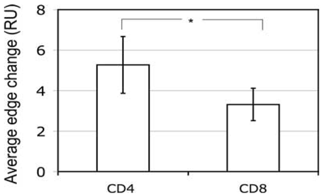
Destruction of virus-infected cells by CTL is an extremely sensitive and efficient process. Our previous data suggest that LFA-1-ICAM-1 interactions in the peripheral supramolecular activation cluster (pSMAC) of the immunological synapse mediate formation of a tight adhesion junction that might contribute to the sensitivity of target cell lysis by CTL. Herein, we compared more (CD8(+)) and less (CD4(+)) effective CTL to understand the molecular events that promote efficient target cell lysis. We found that abrogation of the pSMAC formation significantly impaired the ability of CD8(+) but not CD4(+) CTL to lyse target cells despite having no effect of the amount of released granules by both CD8(+) and CD4(+) CTL. Consistent with this, CD4(+) CTL break their synapses more often than do CD8(+) CTL, which leads to the escape of the cytolytic molecules from the interface. CD4(+) CTL treatment with a protein kinase Ctheta inhibitor increases synapse stability and sensitivity of specific target cell lysis. Thus, formation of a stable pSMAC, which is partially controlled by protein kinase Ctheta, functions to confine the released lytic molecules at the synaptic interface and to enhance the effectiveness of target cell lysis.


Conflict of interest statement

Disclosures

The authors have no financial conflicts of interest.

Figures

FIGURE 1
FIGURE 1
CD4+ CTL are less efficient lytic effectors than are CD8+ CTL. Left, Cytotoxic activity and granule release by human flu-specific CD8+ CTL CER43 on HLA-A2+ target cells (T2-DR1). Middle, Cytotoxic activity and granule release by human HIV Gag-specific CD4+ CTL AC-25 on HLA-DR1+ target cells (T2-DR1). Right, Cytotoxic activity and granule release by CD4+ CTL 161J vs HLA-DR4+ target cells (T2-DR4) sensitized with the PEVIPMFSALSEG (PG13) peptide from the Gag protein. The data shown are representative from three to five experiments.
FIGURE 2
FIGURE 2
Granules isolated from CD4+ and CD8+ CTL exhibit comparable levels of serine esterase and lytic activity. A, Both CD8+ and CD4+ CTL express a similar level of lytic molecules. CD4+ AC-25, CD8+ CER43, and freshly isolated human CD4+ T cells (negative control) were stained intracellularly for either perforin or granzyme A or granzyme B and the level of expression of these lytic molecules was analyzed by flow cytometry. B, CD8+ and CD4+ CTL contain equal amounts of granule contents. Granule extracts from CD8+ and CD4+ CTL were adjusted for cell number so that the number of cells from which the granules were isolated per microliter of the extract was equal. The extracts were then serially diluted and tested for serine protease activity using the BLT (N-benzyloxycarboxyl-l-lysine thiobensyl ester) substrate and results were read at OD 405 nm. C, Granules isolated from CD8+ and CD4+ CTL are equally potent in lysing target cells that do not display peptide Ags. Granule extracts from either CD4+ or CD8+ CTL were tested for the ability to lyse 51Cr-labeled T2-DR1 target cells. The assay was performed in HBSS containing 2 mM CaCl2 for 4 h at 37°C in CO2 incubator. The target cells were incubated with the granules, and after 4 h the culture supernatants were assayed for specific release of 51Cr. The data shown in B and C are representative of three independent assays, in which the granules from separate preparations were tested.
FIGURE 3
FIGURE 3
CD8+ CTL are still effective killers even in response to stimulation by weak agonist ligand. The human HIV-specific CD8+ CTL clone 68A62 recognizes the reverse transcriptase-derived agonist peptide ILKEPVHGV (IV9). A weak agonist peptide that reduces the sensitivity of the cytolytic response was derived by replacement of the IV9 glutamic acid residue at position 4 with alanine. Cytotoxic activity and granule release by 68A62 against target cells loaded with either agonist (IV9) peptide (left) or weak agonist (IV9-A4) peptide (right) are shown. The difference in the sensitivity of the two responses (lysis and granule release) by CD8+ CTL is not affected by stimulation through the TCR by a weak agonist peptide.
FIGURE 4
FIGURE 4
The effect on cytolytic activity by abrogating pSMAC formation with anti-LFA-1 Abs. Cytotoxic activity of CD8+ CTL CER43 (left) and CD4+ CTL AC-25 (right) against T2-DR1 target cells in the presence (+ anti-LFA-1, ■) or absence (○) of Abs against LFA-1. The concentration of cognate peptide (indicated by arrows) required to achieve half-maximal specific lysis of target cells (SD50) by CD8+ CTL is ≈1 order of magnitude lower in the presence of LFA-1 Abs, while SD50 for target cell lysis by CD4+ CTL is largely unaffected (the SD50 value is designated by a single arrow). The data shown are representative of five independent experiments with CD8+ CTL CER43 and three independent experiments with CD4+ CTL AC-25.
FIGURE 5
FIGURE 5
Maintenance and stability of IS formed by CD4+ and CD8+ CTL. A, Representative images of crescent adhesive junctions formed by CD4+ CTL. AC-25 CD4+ CTL were exposed to glass-supported bilayers containing ICAM-1 (300 molecules/µm2) and various concentrations of cognate pMHC (indicated on top). Images are taken at the level of the bilayer. IRM: Interference reflection microscopy images show the CTL contact area. Merge images: ICAM-1 is red and cognate pMHC is green. Scale bar, 5 µm. B, CD4+ and CD8+ CTL (indicated on the left) were exposed to glass-supported bilayers containing ICAM-1 (300 molecules/µm2) and various concentrations of cognate pMHC (indicated on the left). Images are taken at the designated times at the level of the bilayer. The average synapse duration and the percentage of cells forming and reforming synapses were determined and are presented in Table III. ICAM-1 is red and cognate pMHC is green. Scale bar, 5 µm.
FIGURE 6
FIGURE 6
The pSMAC of the synapse formed by AC-25 CD4+ CTL oscillates to a greater extent. CD8+ and CD4+ CTL were added to bilayers containing ICAM-1 (300 molecules/µm2) and pMHC (500 molecules/µm2), and images were acquired from 5 to 10 min at 30-s intervals (see Movie). Modulation of the mature synapse structure was assessed as described in Materials and Methods. Briefly, four lines extending from a central point were drawn for each cell in the first image of the time stack. Using the intensity profile generated for each line, we determined the edge of the pSMAC at each time point. The total change in the position of the pSMAC edge was determined for each of the four lines per cell and then averaged to obtain the data for the graph. Statistical significance was assessed by the Student t test (*, p < 0.01).
FIGURE 7
FIGURE 7
Granules released by CD4+ CTL are less contained within the pSMAC ring. Representative images of the patterns of CD107a accumulation observed by CD8+ (left) and CD4+ (right) CTL over 10 min after CTL exposure to bilayers. At pMHC density of 500 molecules/µm2: 1) 83 ± 4% (CD8+) and 37 ± 1% (CD4+) of CTL show CD107a staining confined to the cSMAC; 2) 6 ± 5% (CD8+) and 28 ± 9% (CD4+) of CTL show CD107a staining spilling out of an unstable synapse; 3) 2 ± 2% (CD8+) and 23 ± 10% (CD4+) CTL show CD107a staining when a multifocal contact is formed. At pMHC density of 25 molecules/µm2: 1) 83 ± 7% (CD8+) and 35 ± 5% (CD4+) of CTL show CD107a staining confined to the cSMAC; 2) 3 ± 3% (CD8+) and 33 ± 9% (CD4+) of CTL show CD107a staining spilling out of an unstable synapse; 3) 9 ± 8% (CD8+) and 20 ± 6% (CD4+) CTL show CD107a staining when a multifocal contact is formed. ICAM-1 is green and CD107a is red. Scale bar, 5 µm. These images are representative of three independent experiments (total number of cells analyzed was >60 cells per experimental group).
FIGURE 8
FIGURE 8
The ability to form a stable peripheral ring junctions affects target cell lysis by CTL that release granules in response to anti-CD3 stimulation. In these experiments, we assessed the ability of the CD4+ and CD8+ CTL to lyse ICAM-1-positive target cells (C1R), not expressing cognate pMHC, in response to anti-CD3 stimulation to trigger granule release as described in Materials and Methods. A, Specific lysis of C1R target cells by AC-25 CD4+ CTL and CER43 CD8+ CTL in response to anti-CD3 Fab stimulation. Anti-CD3 Fab was added at the indicated concentrations. B, Serine esterase release was detected in the supernatant of CTL incubated with target cells plus anti-CD3 Fab at the indicated concentrations, and the OD at 405 nm is shown. The data shown are representative from three experiments.
FIGURE 9
FIGURE 9
Improved sensitivity of target cell lysis by CD4+ CTL in the presence of a PKCθ inhibitor. Target cell lysis and granule release by AC-25 CD4+ CTL were analyzed at various peptide concentrations (as indicated) in the presence (right) or absence (left) of a specific inhibitor of PKCθ, and the results are depicted as percentage specific lysis and percentage granule release in the graph. This inhibitor increased the amount of stable synapses formed by CD4+ CTL, as shown in Table III. Inhibitor-treated CD4+ CTL demonstrated more effective target cell lysis, that is, increased lysis of target cells without releasing more granules. The data shown are representative of four independent experiments.
FIGURE 10
FIGURE 10
Inhibition of PKCθ also enhances lysis triggered by anti-CD3 Fab. We assessed the ability of the CD4+ CTL to lyse ICAM-1-positive target cells (C1R), not expressing cognate pMHC, in response to anti-CD3 stimulation to trigger granule release. A, Specific lysis of C1R target cells by AC-25 CD4+ CTL in response to anti-CD3 Fab stimulation in the presence (PKCθ inhibitor) or absence (untreated) of a PKCθ inhibitor. Anti-CD3 Fab was added at the indicated concentrations. B, Serine esterase release was detected in the supernatant of CTL with target cells plus anti-CD3 Fab at the indicated concentrations, and the OD at 405 nm is shown. CTL were treated as indicated in A. Statistical significance was assessed by the Student t test (*, p ≤ 0.01).

Similar articles

Cited by

References

    1. Mentzer SJ, Smith BR, Barbosa JA, Crimmins MA, Herrmann SH, Burakoff SJ. CTL adhesion and antigen recognition are discrete steps in the human CTL-target cell interaction. J. Immunol. 1987;138:1325–1330. - PubMed
    1. Shaw S, Luce GE, Quinones R, Gress RE, Springer TA, Sanders ME. Two antigen-independent adhesion pathways used by human cytotoxic T-cell clones. Nature. 1986;323:262–264. - PubMed
    1. Spitz H, van Schooten W, Keizer H, van Seventer G, van de Rijn M, Terhorst C, de Vries J-E. Alloantigen recognition is preceded by nonspecific adhesion of cytotoxic T cells and target cells. Science. 1986;232:403–405. - PubMed
    1. Grakoui A, Bromley SK, Sumen C, Davis MM, Shaw AS, Allen PM, Dustin ML. The immunological synapse: a molecular machine controlling T cell activation. Science. 1999;285:221–227. - PubMed
    1. Monks C, Freiberg B, Kupfer H, Sciaky N, Kupfer A. Three-dimensional segregation of supramolecular activation clusters in T cells. Nature. 1998;395:82–86. - PubMed

Publication types

MeSH terms