. 2008 Oct 1;181(7):4840-51.
doi: 10.4049/jimmunol.181.7.4840.

Distinct regulation of integrin-dependent T cell conjugate formation and NF-kappa B activation by the adapter protein ADAP

Affiliations

Distinct regulation of integrin-dependent T cell conjugate formation and NF-kappa B activation by the adapter protein ADAP

Brandon J Burbach et al. J Immunol. .

Abstract

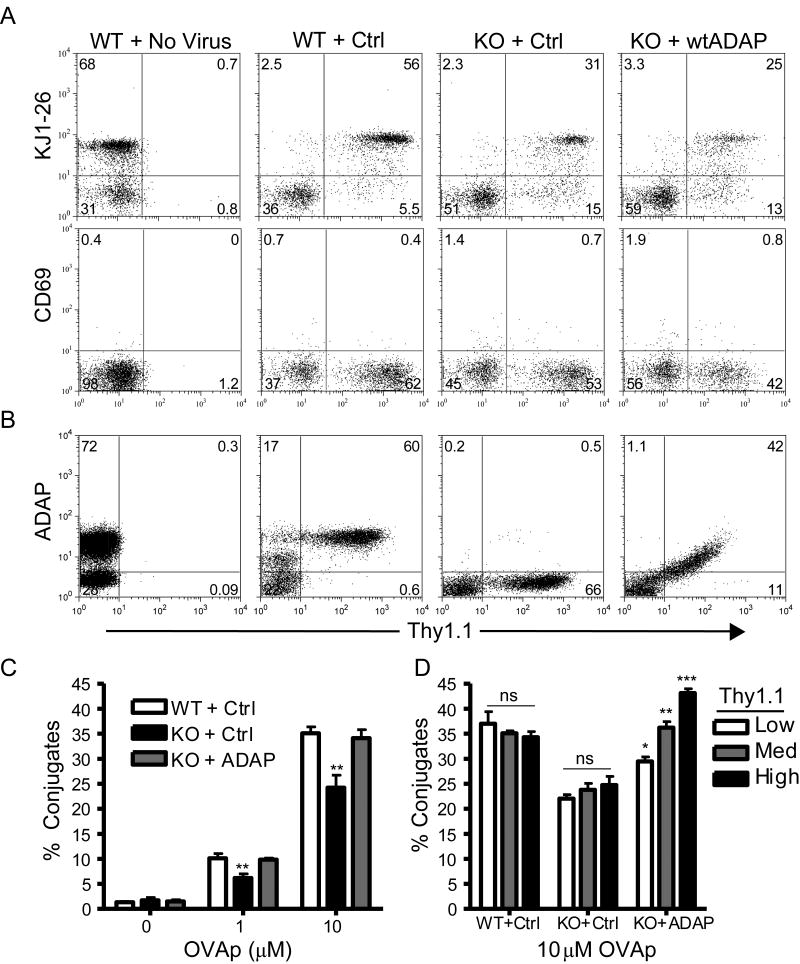
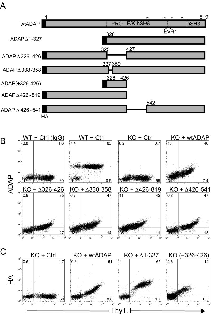
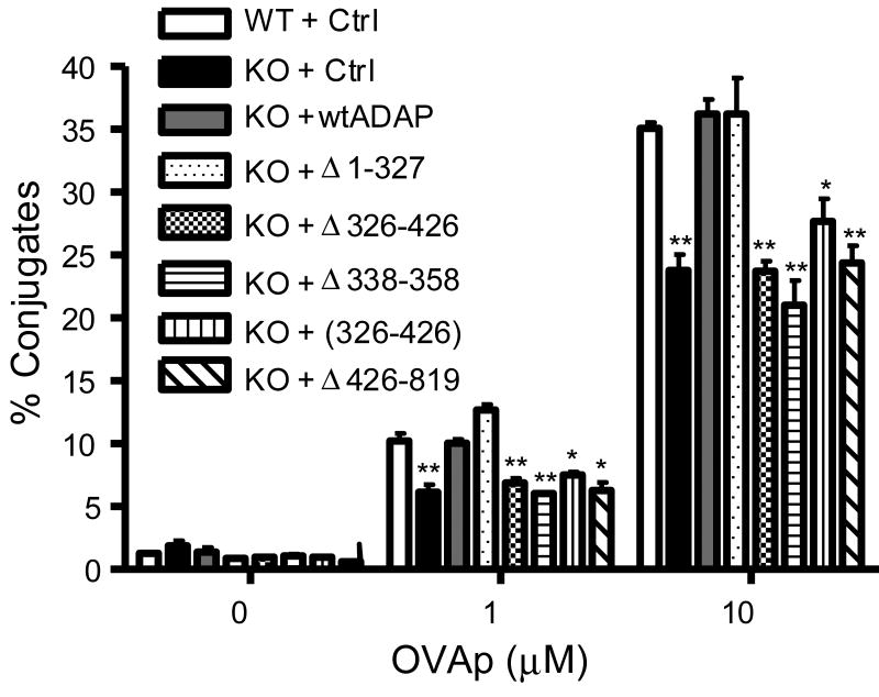
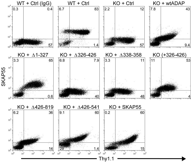
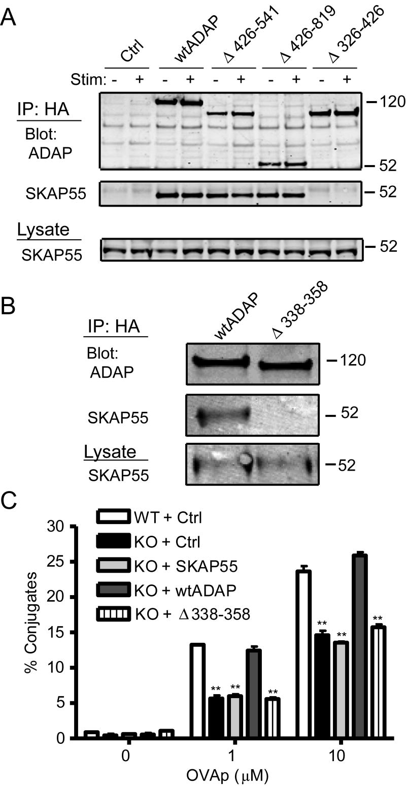
Following TCR stimulation, T cells utilize the hematopoietic specific adhesion and degranulation-promoting adapter protein (ADAP) to control both integrin adhesive function and NF-kappaB transcription factor activation. We have investigated the molecular basis by which ADAP controls these events in primary murine ADAP(-/-) T cells. Naive DO11.10/ADAP(-/-) T cells show impaired adhesion to OVAp (OVA aa 323-339)-bearing APCs that is restored following reconstitution with wild-type ADAP. Mutational analysis demonstrates that the central proline-rich domain and the C-terminal domain of ADAP are required for rescue of T:APC conjugate formation. The ADAP proline-rich domain is sufficient to bind and stabilize the expression of SKAP55 (Src kinase-associated phosphoprotein of 55 kDa), which is otherwise absent from ADAP(-/-) T cells. Interestingly, forced expression of SKAP55 in the absence of ADAP is insufficient to drive T:APC conjugate formation, demonstrating that both ADAP and SKAP55 are required for optimal LFA-1 function. Additionally, the ADAP proline-rich domain is required for optimal Ag-induced activation of CD69, CD25, and Bcl-x(L), but is not required for assembly of the CARMA1/Bcl10/Malt1 (caspase-recruitment domain (CARD) membrane-associated guanylate kinase (MAGUK) protein 1/B-cell CLL-lymphoma 10/mucosa-associated lymphoid tissue lymphoma translocation protein 1) signaling complex and subsequent TCR-dependent NF-kappaB activity. Our results indicate that ADAP is used downstream of TCR engagement to delineate two distinct molecular programs in which the ADAP/SKAP55 module is required for control of T:APC conjugate formation and functions independently of ADAP/CARMA1-mediated NF-kappaB activation.


Figures

Figure 1
Figure 1. Rescue of antigen-dependent T:APC conjugate formation in ADAP-/- T cells following reconstitution of wtADAP
Freshly harvested, naïve lymphocytes from transgenic DO11.10/hCAR/ADAP+/+ (WT) or ADAP-/- (KO) mice were transduced with adenovirus encoding Thy1.1 alone (Ctrl), Thy1.1 plus wild-type murine ADAP (wtADAP), or mock transduced (No Virus) and cultured ex vivo for 3d as described in Materials and Methods. (A) Cells were stained for Thy1.1 and either the DO11.10 TCR (KJ-126) or CD69 and analyzed by flow cytometry. (B) Cells were fixed and intracellular staining was performed with a sheep anti-ADAP antibody. (C) T:APC conjugate formation between KJ-126+/Thy1.1+ T cells and Balb/c splenocytes pre-pulsed with the indicated concentrations of OVAp (left panel) was performed as described in Materials and Methods. The same KJ-126+/Thy1.1+ population of T cells was gated into three subpopulations expressing either low, medium, or high levels of Thy1.1, and T:APC conjugate efficiency with 10μM OVAp was determined (right panel). Results are representative of at least three independent experiments performed.
Figure 2
Figure 2. Schematic and expression of ADAP mutant constructs
(A) Scale diagram of ADAP expression constructs used in this study. Amino acid numbering is given for the murine ADAP p130 isoform. Abbreviations: PRO, proline-rich domain; E/K, glutamic acid and lysine-rich domain; hSH3, N-terminal (N) or C-terminal (C) helical SH3 domain; EVH1, Ena/Vasp homology domain. Asterisks (*) are used to denote the position of tyrosines 547, 549, 584, 615, 687 found in phosphorylation consensus motifs. (B) Freshly harvested naïve DO11.10/hCAR/ADAP+/+ (WT) or ADAP-/- (KO) lymphocytes were transduced with adenoviruses encoding the indicated constructs or Thy1.1 control adenovirus (ctrl) and fixed and stained with an anti-Thy1.1 antibody and either anti-ADAP antibody or non-immune sheep serum (IgG) and analyzed by flow cytometry. (C) Same as in (B) except the indicated constructs were stained with an anti-hemaglutinin (HA) antibody. Expression profiles are representative of at least three independent analyses performed for each construct shown.
Figure 3
Figure 3. The ADAP proline rich domain is required for T:APC conjugate formation
Naïve DO11.10/hCAR/ADAP-/- (KO) or ADAP+/+ (WT) lymphocytes expressing the indicated ADAP constructs were analyzed for T:APC conjugate formation between KJ-126+/Thy1.1+ T cells and Balb/c splenocytes pre-pulsed with the indicated concentrations of OVAp. Results are shown for a single representative experiment and are representative of at least four independent experiments performed for each construct.
Figure 4
Figure 4. The ADAP proline rich domain controls SKAP55 expression in ADAP-/- T cells
Naïve DO11.10/hCAR/ADAP-/- (KO) or ADAP+/+ (WT) lymphocytes expressing the indicated ADAP constructs, wild-type SKAP55, or the control adenovirus (Ctrl) were fixed and intracellular staining was performed with rabbit anti-SKAP55 antibody or control rabbit immunoglobulin (IgG) and analyzed by flow cytometry. Results are shown for a single representative experiment and are representative of at least four independent experiments performed for each construct.
Figure 5
Figure 5. SKAP55 expression in ADAP-/- T cells fails to rescue T:APC conjugate formation
(A) Jurkat T cells were transduced with control adenovirus (Ctrl) or with the indicated HA-tagged ADAP constructs. After 2d, 106 cells were left unstimulated or stimulated for 5 min with anti-TCR mAb OKT3, lysed, and immunoprecipitated with agarose-conjugated anti-HA and western blots for ADAP and SKAP55 performed. The relative molecular mass of size standards is shown on the right. Similar results were observed in four independent experiments. (B) Naïve hCAR/ADAP+/+ lymphocytes expressing endogenous levels of SKAP55 were transduced with either full-length murine ADAP (wtADAP) or ADAP lacking the restricted proline rich domain (Δ338-358). Cells were lysed as described in Materials and Methods, immunprecipitated with agarose-conjugated anti-HA antibodies, and Western blots were performed with anti-HA or anti-SKAP55 antibodies. (C) Naïve DO11.10/hCAR/ADAP-/- (KO) or ADAP+/+ (WT) lymphocytes expressing the indicated constructs were analyzed for T:APC conjugate formation between KJ-126+/Thy1.1+ T cells and Balb/c splenocytes pre-pulsed with the indicated concentrations of OVAp. Results are shown for a single representative experiment and are representative of at least four independent experiments performed for each construct.
Figure 6
Figure 6. ADAP is not required for recruitment of Rap1 or SKAP55 to the contact site between T cells and anti-TCR beads
(A) Naïve DO11.10/hCAR/ ADAP+/+ (WT) or ADAP-/- (KO) lymphocytes were transduced with GFP-Rap1 and conjugates with anti-TCR (2C11) coated beads were formed as described in Materials and Methods. GFP-Rap1 expressing cells were gated and photographed by image-scanning cytometry and a representative image from each sample is shown. An example of GFP-Rap1 expression in a cell absent of bead stimulation is also shown (Unstim). (B) Graphical display of the percentage of total GFP-Rap1 signal in each cell that is concentrated against the anti-TCR coated bead, quantified as described in Materials and Methods. (C) Naïve DO11.10/hCAR/ADAP+/+ (WT) T cells expressing the control virus (Ctrl) or ADAP-/- (KO) lymphocytes expressing wtADAP, ADAPΔ426-819, or SKAP55 were stimulated with anti-TCR coated beads as described in (A), fixed, and stained for SKAP55 as described for Fig. 4. (D) Graphical display of the percentage of total SKAP55 signal in each cell that is concentrated against the anti-TCR coated bead.
Figure 7
Figure 7. The ADAP proline rich domain is required for antigen-dependent T cell activation
Naïve DO11.10/hCAR/ADAP+/+ (WT) or ADAP-/- (KO) lymphocytes expressing the indicated ADAP constructs were stimulated with fresh Balb/C splenocytes pulsed with the indicated doses of OVAp as described in Materials and Methods. (A-B) Cells were stimulated for 18h, stained for KJ-126, Thy1.1, CD69, and CD25 as indicated, fixed, and analyzed by flow cytometry. (C) Cells were stimulated for 48h with OVAp, stained for KJ-126 and Thy1.1, fixed, permeablized with saponin, stained for anti-Bcl-xL, and analyzed by flow cytometry. (A-C) The percentage of CD69+, CD25+, or Bcl-xL+ cells within the KJ1-26+Thy1.1+ gate is shown on each histogram. Results are shown for a single representative experiment and are representative of at least four independent experiments performed for CD69 and CD25 and three experiments for Bcl-xL.
Figure 8
Figure 8. The ADAP/SKAP55 interaction is not required for assembly of the CARMA1/Bcl10 complex or TCR induced NF-κB activation
(A) Naïve DO11.10/hCAR/ADAP-/- (KO) or ADAP+/+ (WT) lymphocytes expressing the indicated ADAP constructs or the control (Ctrl) were left untreated (-) or stimulated with PMA (+) and lysed as described in Materials and Methods. Lysates were subjected to immunoprecipitation with an anti-Bcl10 antibody, and western blots were performed with antibodies to CARMA1, ADAP, or Bcl10. (B) Freshly harvested resting ADAP+/+ (WT) or ADAP-/- (KO) lymphocytes were left unstimulated or stimulated with PMA and lysed as in (A) and immunoprecipiated in parallel with antibodies to either ADAP (left panel) or Bcl-10 (right panel). Western blots were performed with antibodies to ADAP, CARMA1, Bcl10, and SKAP55 as indicated between the panels. Results are representative of three (A) or two (B) independent experiments. (C) Naïve DO11.10/hCAR/ADAP-/- (KO) or ADAP+/+ (WT) lymphocytes expressing the indicated constructs were stimulated with anti-CD3 plus anti-CD28 antibodies as described in Materials and Methods or left untreated. The samples were fixed and stained with antibodies to Thy1.1 and NF-κB (p65), and nuclei were stained with 7-AAD. Cells were analyzed on a multispectral image-scanning flow cytometer as described in Materials and Methods. Nuclear localization of p65 in unstimulated T cells was set to 1 and results show the increase in p65 nuclear translocation in stimulated relative to unstimulated Thy1.1+ cells from three independent experiments.

Similar articles

Cited by

References

    1. Simeoni L, Kliche S, Lindquist J, Schraven B. Adaptors and linkers in T and B cells. Curr Opin Immunol. 2004;16:304–313. - PubMed
    1. Samelson LE. Signal transduction mediated the T cell antigen receptor: the role of adapter proteins. Annu Rev Immunol. 2002;20:371–394. - PubMed
    1. Jordan MS, Singer AL, Koretzky GA. Adaptors as central mediators of signal transduction in immune cells. Nat Immunol. 2003;4:110–116. - PubMed
    1. Sommers CL, Samelson LE, Love PE. LAT: a T lymphocyte adapter protein that couples the antigen receptor to downstream signaling pathways. BioEssays. 2004;26:61–67. - PubMed
    1. Zhang WG, Sommers CL, Burshtyn DN, Stebbins CC, DeJarnette JB, Trible RP, Grinberg A, Tsay HC, Jacobs HM, Kessler CM, Long EO, Love PE, Samelson LE. Essential role of LAT in T cell development. Immunity. 1999;10:323–332. - PubMed

Publication types

MeSH terms