Comparative Study
. 2008 Oct 1;181(7):4955-64.
doi: 10.4049/jimmunol.181.7.4955.

Multifunctional, high-level cytokine-producing Th1 cells in the lung, but not spleen, correlate with protection against Mycobacterium tuberculosis aerosol challenge in mice

Affiliations
Comparative Study

Multifunctional, high-level cytokine-producing Th1 cells in the lung, but not spleen, correlate with protection against Mycobacterium tuberculosis aerosol challenge in mice

Emily K Forbes et al. J Immunol. .

Abstract

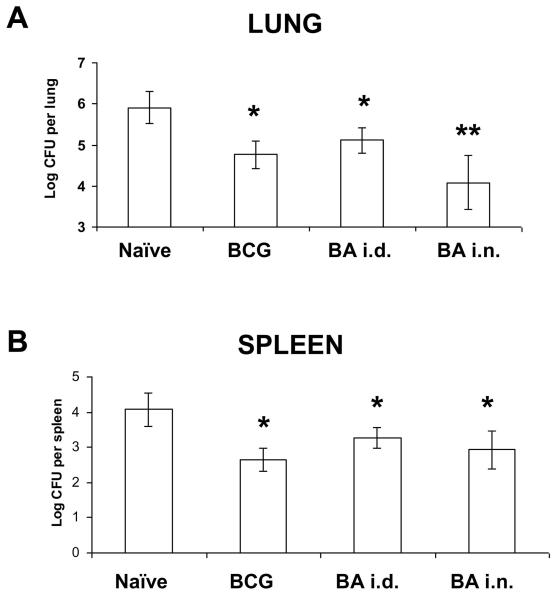
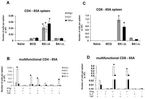
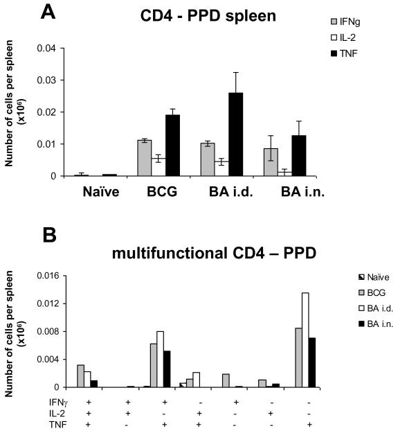
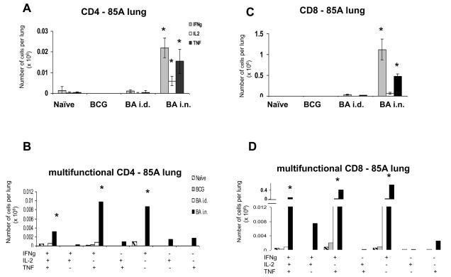
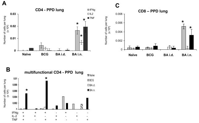
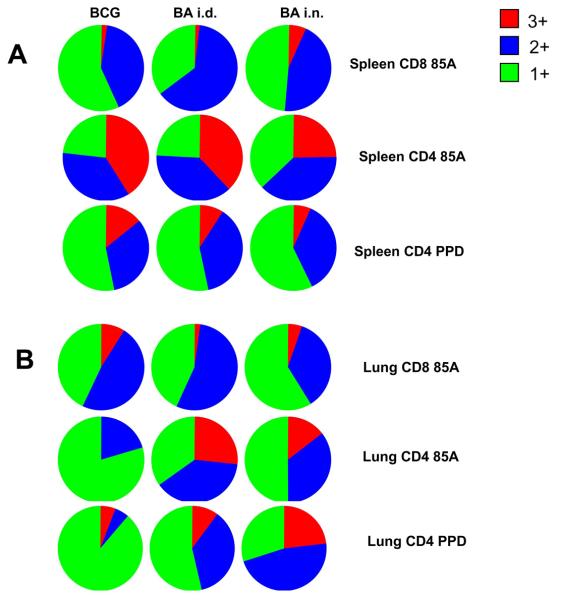
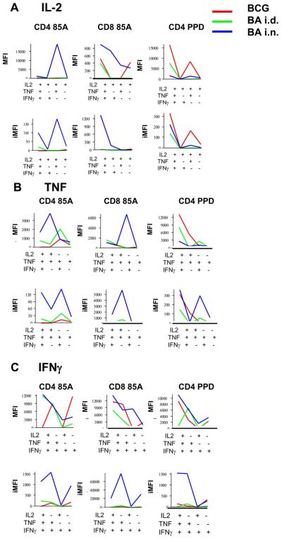
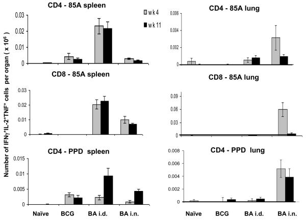
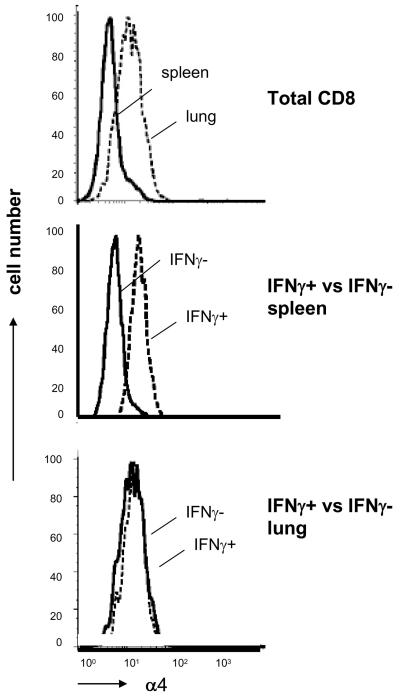
Boosting bacillus Calmette-Guérin (BCG)-primed mice with a recombinant adenovirus expressing Mycobacterium tuberculosis Ag 85A by different administration routes has very different effects on protection against aerosol challenge with M. tuberculosis. Mice boosted intradermally make very strong splenic CD4 and CD8 Th1 cytokine responses to Ag 85A, but show no change in lung mycobacterial burden over BCG primed animals. In contrast, intranasally boosted mice show greatly reduced mycobacterial burden and make a much weaker splenic response but a very strong lung CD4 and CD8 response to Ag 85A and an increased response to purified protein derivative. This effect is associated with the presence in the lung of multifunctional T cells, with high median fluorescence intensity and integrated median fluorescence intensity for IFN-gamma, IL-2, and TNF. In contrast, mice immunized with BCG alone have few Ag-specific cells in the lung and a low proportion of multifunctional cells, although individual cells have high median fluorescence intensity. Successful immunization regimes appear to induce Ag-specific cells with abundant intracellular cytokine staining.


Figures

Figure 1
Figure 1. Mycobacterial CFU after Ad85A boosting of BCG primed mice
BALB/c mice were immunised with BCG and 10 weeks later boosted with Ad85A given intradermally (BA i.d.) or intranasally (BA i.n.). Mice were challenged with M. tuberculosis by aerosol 4 weeks after the boost. Mice were sacrificed 6 weeks later and lungs and spleen CFU enumerated. Data represent the mean +/− SD from 6-8 mice per group. * p<0.05 versus naïve, **p<0.05 versus BCG and BA i.d. Similar results were obtained in two other experiments one harvested at 4 weeks and another at 6 weeks.
Figure 2
Figure 2. Cytokine responses of splenic T cells to antigen 85A
Mice were primed with BCG and boosted with Ad85A either intradermally (BA i.d) or intranasally (BA i.n.). Splenocytes were isolated 4 weeks after the boost and stimulated with pooled 85A peptides for 6 hours. The frequencies of IFNγ, IL-2 and TNF producing cells were determined by flow cytometry on CD4 (A) or CD8 (C) gated cells and total number of cells per organ calculated. The number of cells expressing each of the seven possible combinations of the cytokines is also shown for CD4 (B) and CD8 (D) cells. Results are expressed as the mean +/− SEM of three mice per group, representative of two independent experiments. * p< 0.05 compared to B and BA i.n.
Figure 3
Figure 3. Cytokine responses of splenic T cells to PPD
Mice were primed with BCG and boosted with Ad85A either intradermally (BA i.d) or intranasally (BA i.n.). Splenocytes were isolated 4 weeks after the boost and stimulated with PPD for 6 hours. The frequencies of IFNγ, IL-2 and TNF producing cells were determined by flow cytometry on CD4 (A) gated cells and total numbers of cells per organ calculated. The number of cells expressing each of the seven possible combinations of the cytokines is shown for CD4 (B) cells. CD8 responses are very low and not shown. Results are expressed as the mean +/− SEM of three mice per group, representative of two independent experiments.
Figure 4
Figure 4. Cytokine responses of lung T cells to antigen 85A
Mice were primed with BCG and boosted with Ad85A either intradermally (BA i.d) or intranasally (BA i.n.). Lung cells were isolated 4 weeks after the boost and stimulated with pooled 85A peptides for 6 hours. The frequencies of IFNγ, IL-2 and TNF producing cells were determined by flow cytometry on CD4 (A) or CD8 (C) gated cells and total numbers of cells per organ calculated. The number of cells expressing each of the seven possible combinations of the cytokines is shown for CD4 (B) and CD8 (D) cells. Results are expressed as the mean +/− SEM of three mice per group, representative of two independent experiments. * p< 0.05 compared to B and BA i.d.
Figure 5
Figure 5. Cytokine responses of lung T cells to PPD
Mice were primed with BCG and boosted with Ad85A either intradermally (BA i.d) or intranasally (BA i.n.). Lung cells were isolated 4 weeks after the boost and stimulated with PPD for 6 hours. The frequencies of IFNγ, IL-2 and TNF producing cells were determined by flow cytometry on CD4 (A) or CD8 (C) gated cells and total numbers of cells per organ calculated. The number of cells expressing each of the seven possible combinations of the cytokines is shown only for CD4 (B) cells, but not for CD8 because the numbers are too low for reliable analysis. Results are expressed as the mean +/− SEM of three mice per group, representative of two independent experiments. * p< 0.05 compared to B and BA i.d.
Figure 6
Figure 6. Multifunctional cytokine producing cells in the spleen and lungs
Pie charts indicate the proportions of single (1+ green), dual (2+ blue) and triple (3+ red) producers of IFNγ, TNF and IL-2 in the spleen (A) and lungs (B) of immunized mice.
Figure 7
Figure 7. MFIs and iMFIs for intracellular cytokines of lung CD4 and CD8 T cells of immunized mice
MFIs indicate the intensity of staining for intracellular cytokines in single, dual or triple cytokine producing cells. Integrated MFIs (iMFIs) are the product of the intensity of staining and the number of cytokine producing cells. MFIs and iMFIs are shown for cells producing (A) IL-2, (B) TNF and (C) IFNγ from mice immunized with BCG alone (red line) BA i.d. (green line) or BA i.n. (blue line) and restimulated in vitro with either pooled antigen 85A peptides or PPD.
Figure 8
Figure 8. Multifunctional cells at 4 and 11 weeks after Ad85A boost
The number of multifunctional 3+ cells expressing IFNγ, IL-2 and TNF in spleen (A) and lung (B) in response to pooled 85A peptides or PPD at 4 and 11 weeks post boost are shown.
Figure 9
Figure 9. Expression of the alpha4 integrin chain on spleen and lung CD8 cells
Expression of alpha 4 on CD8 gated T cells from spleen or lung (top panel), on intracellular IFNγ+ /− splenic CD8 cells stimulated in vitro with pooled 85A peptides (middle panel) and on IFNγ +/− lung CD8 cells (bottom panel). All cells are from BA i.n. immunized mice.

References

    1. WHO/HTM/TB/2007.376.

    1. Mittrucker HW, Steinhoff U, Kohler A, Krause M, Lazar D, Mex P, Miekley D, Kaufmann SH. Poor correlation between BCG vaccination-induced T cell responses and protection against tuberculosis. Proc Natl Acad Sci U S A. 2007;104:12434–12439. - PMC - PubMed
    1. Palma C, Iona E, Giannoni F, Pardini M, Brunori L, Orefici G, Fattorini L, Cassone A. The Ag85B protein of Mycobacterium tuberculosis may turn a protective immune response induced by Ag85B-DNA vaccine into a potent but non-protective Th1 immune response in mice. Cell Microbiol. 2007;9:1455–1465. - PubMed
    1. Goonetilleke NP, McShane H, Hannan CM, Anderson RJ, Brookes RH, Hill AV. Enhanced immunogenicity and protective efficacy against Mycobacterium tuberculosis of bacille Calmette-Guerin vaccine using mucosal administration and boosting with a recombinant modified vaccinia virus Ankara. J Immunol. 2003;171:1602–1609. - PubMed
    1. Romano M, D'Souza S, Adnet PY, Laali R, Jurion F, Palfliet K, Huygen K. Priming but not boosting with plasmid DNA encoding mycolyltransferase Ag85A from Mycobacterium tuberculosis increases the survival time of Mycobacterium bovis BCG vaccinated mice against low dose intravenous challenge with M. tuberculosis H37Rv. Vaccine. 2006;24:3353–3364. - PubMed

Publication types

MeSH terms