. 2008 Oct 1;181(7):4989-5000.
doi: 10.4049/jimmunol.181.7.4989.

Cathelicidin administration protects mice from Bacillus anthracis spore challenge

Affiliations

Cathelicidin administration protects mice from Bacillus anthracis spore challenge

Mark W Lisanby et al. J Immunol. .

Abstract

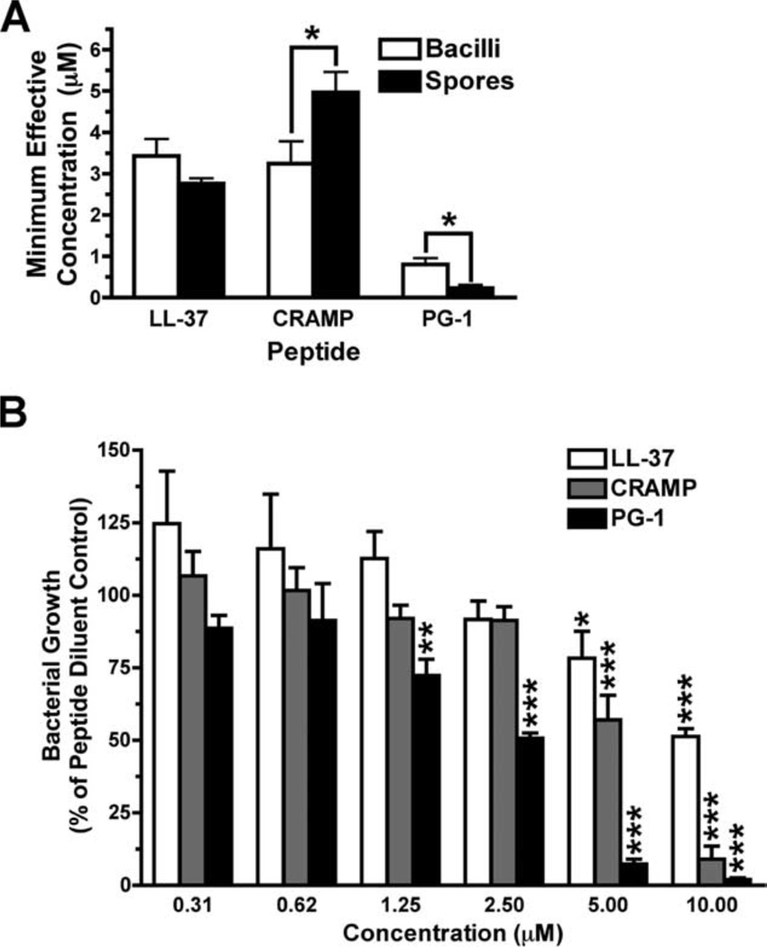
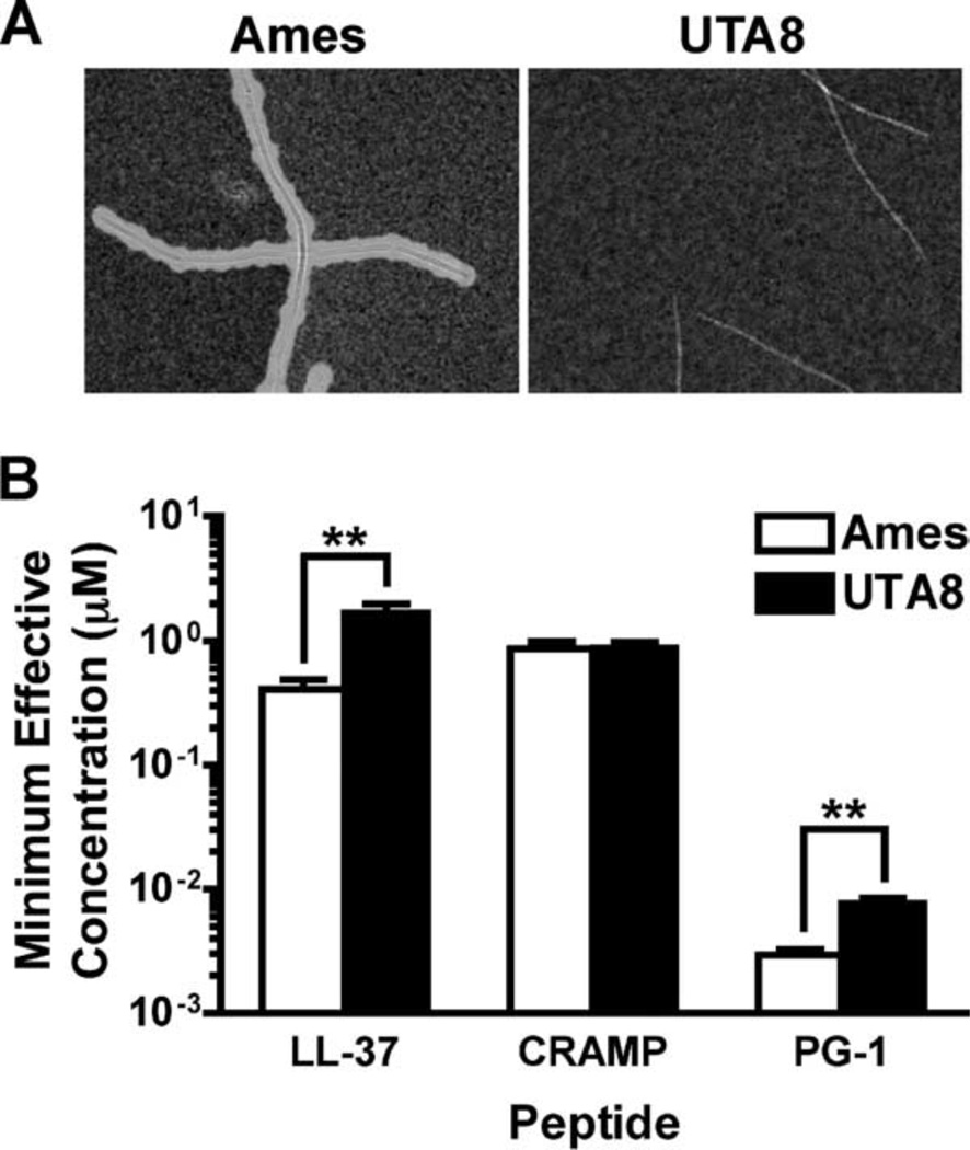
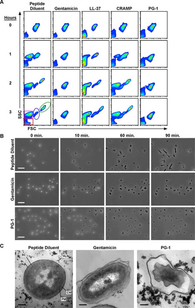
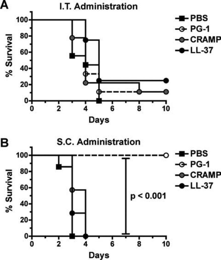
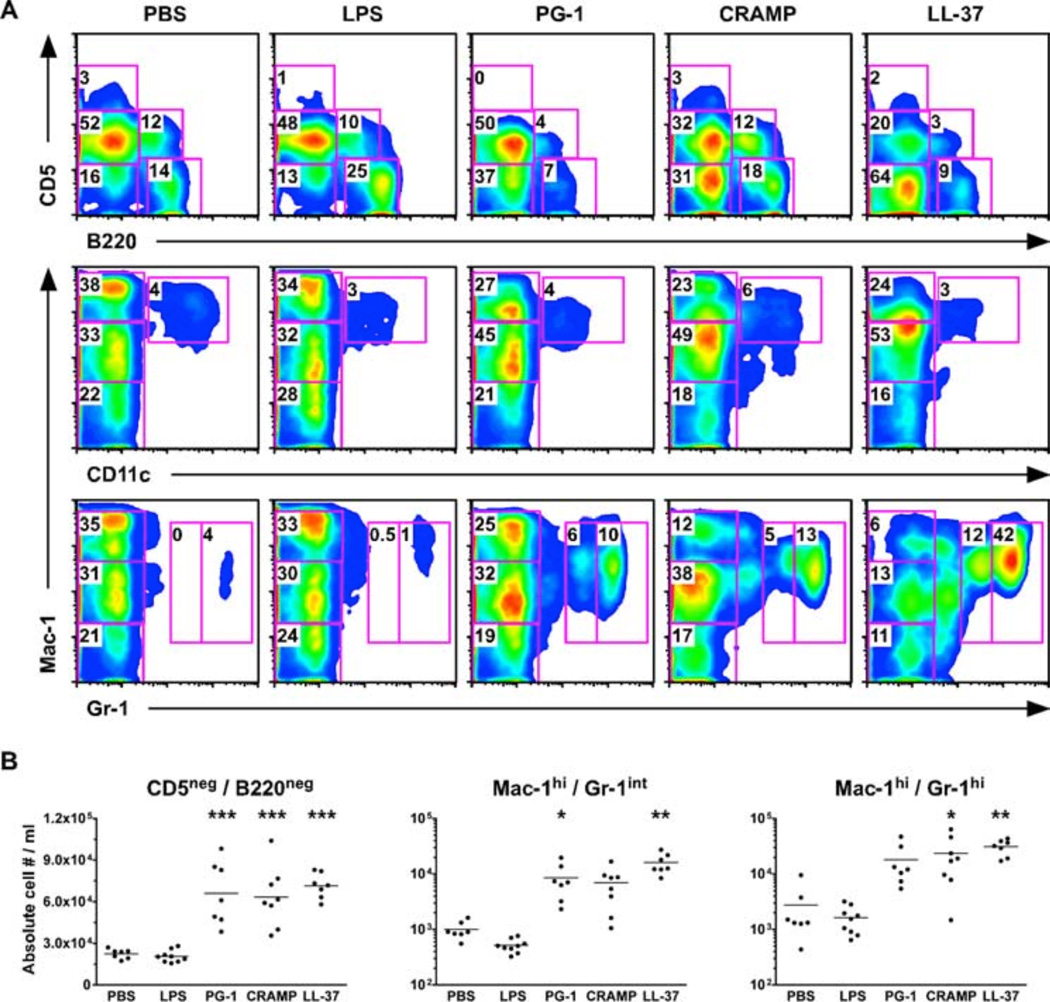
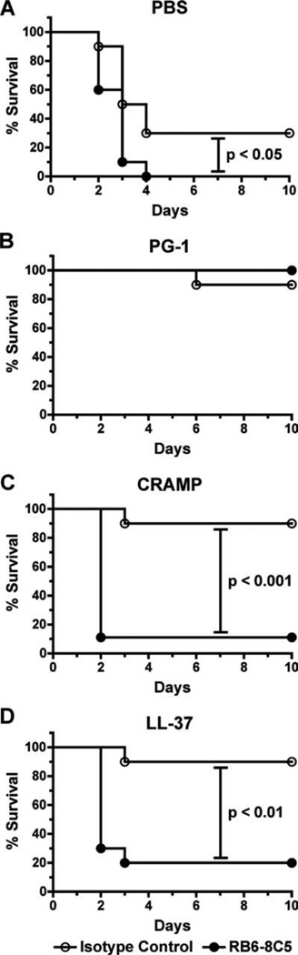
Cathelicidins are a family of cationic peptides expressed in mammals that possess numerous bactericidal and immunomodulatory properties. In vitro analyses showed that human, mouse, and pig cathelicidins inhibited Bacillus anthracis bacterial growth at micromolar concentrations in the presence or absence of capsule. Combined in vitro analyses of the effects of each peptide on spore germination and vegetative outgrowth by time lapse phase contrast microscopy, transmission electron microscopy, and flow cytometric analysis showed that only the pig cathelicidin was capable of directly arresting vegetative outgrowth and killing the developing bacilli within the confines of the exosporium. C57BL/6 mice were protected from spore-induced death by each cathelicidin in a time- and dose-dependent manner. Protection afforded by the porcine cathelicidin was due to its bactericidal effects, whereas the human and mouse cathelicidins appeared to mediate protection through increased recruitment of neutrophils to the site of infection. These findings suggest that cathelicidins might be utilized to augment the initial innate immune response to B. anthracis spore exposure and prevent the development of anthrax.


Conflict of interest statement

Disclosures

The authors have no financial conflict of interest.

Figures

FIGURE 1
FIGURE 1
Cathelicidins inhibit B. anthracis growth. A, RDAs were conducted by incubating 2 × 10⁶ ungerminated Sterne strain spores or mid-log phase bacilli with serially diluted peptides (100-0.1 µM). After an overnight incubation to allow for visible colony growth, zones of growth clearance were measured, and the MEC was calculated using linear regression analysis. Graph values represent the mean calculated MEC from at least eight plates ± SEM. Unpaired, two-tailed Student t tests were used to determine statistical significance. *, p < 0.05; **, p < 0.01; ***, p < 0.001. B, MBDAs were performed in which 1 × 10⁶ bacilli/ml were ali-quoted into wells containing serially diluted peptides. Percent growth was calculated by measuring the OD600 of peptide-treated samples normalized to the peptide diluent control samples, which were set to 100% for each experimental repetition. Values represent the mean percentages pooled from three experiments conducted in quintuplicate 6 SEM. Statistical significance was determined by two-tailed, one-sample t tests in which each sample was compared against the peptide diluent control. *, p < 0.05; **, p < 0.01; ***, p < 0.001.
FIGURE 2
FIGURE 2
Capsule expression does not prevent bactericidal activity of peptides. Spores were grown for 4 h in MHB at 37°C in 5% CO2 and capsule expression was verified by India Ink exclusion (A) before being utilized in RDAs (B) in which the MEC of each peptide was determined against 2 × 10⁶ Ames and UTA8 bacilli. Graph values represent the mean MEC calculated from four plates ± SEM. Unpaired, two-tailed Student’s t tests were used to determine statistical significance. **, p < 0.01.
FIGURE 3
FIGURE 3
PG-1 treatment prevents vegetative outgrowth by disrupting the plasma membrane of the developing bacilli. In all experimental conditions, 1 × 10⁶ spores were incubated in peptide diluent control, genta-micin (5 µg/ml), or peptides (10 µM) diluted in MHB at 37°C for the indicated lengths of time. A, Flow cytom-etry was used to monitor the effects of each peptide on germination and outgrowth. Density plots depict the forward scatter (FSC-H) and side scatter (SSC-H) profiles of germinating spores in which debris (red), exosporium remnants (purple), spores (blue), and bacilli (green) can all be monitored as distinct populations. Scatter profiles are representative of three experiments conducted in duplicate. B, Representative photomicrograph excerpts taken from time lapse phase contrast microscopic analyses in which spores were incubated at 37°C and imaged every 10 s for 90 min. Photomicrographs are representative of three experiments. Bars = 10 µm. C, Representative transmission electron photomicrographs of spores treated with peptide diluent, gentamicin, or PG-1 for 1 h at 37°C. The plasma membrane (PM) and cell wall (CW) of the developing bacilli are shown in relation to the spore coat (SC) and basal layer (BL) and hair-like nap (HLN) of the exosporium. Bars = 0.2 µm.
FIGURE 4
FIGURE 4
Cathelicidin administration protects C57BL/6 mice from s.c. spore challenge. A–C, Survival of mice inoculated s.c. with 5 LD50 (5 × 10⁵) of spores in 200 µl of PBS (n = 12 mice) or 50 µM solutions of PG-1 (A), CRAMP (B), or LL-37 (C) simultaneously (0 h) or 4 or 24 h after infection (n = 10 mice per group). D–F, Survival of mice inoculated s.c. with 5 LD50 (5 × 10⁵) of spores in 200 µl of PBS (n = 20 mice) or 50 µM or 10 µM solutions of PG-1 (D), CRAMP (E), or LL-37 (F; = 5 10 mice per group). In both experiments, mice were monitored for 10 days, and log rank tests were utilized to determine whether differences in survival were statistically significant.
FIGURE 5
FIGURE 5
PG-1 but not CRAMP or LL-37 protects A/J mice against s.c. spore challenge, whereas none protect against i.t. administration. A, Survival of mice challenged i.t. with 5 LD50 (5 × 10⁵) of spores suspended in 30 µl of PBS or 50 µM peptides (diluted in PBS) and monitored for 10 days. n = 8–9 mice per group. B, Survival of mice inoculated with 5 LD50 (2.5 × 10³) of spores suspended in 200µl of PBS or 50µM peptides (diluted in PBS) s.c. and monitored for 10 days. n = 7 mice per group. Log rank tests were utilized in both experiments to determine whether differences in survival were statistically significant.
FIGURE 6
FIGURE 6
Administration of pep-tides i.p. to C57BL/6 mice causes recruitment of neutrophils. Mice were injected i.p. with 200 µl of PBS, LPS (20 µg), or peptides (50 µM), and the peritoneal cavity was lavaged 4 h later. A, Cell populations were determined based on the relative expression levels of the cell surface markers B220, CD5, Mac-1, CD11c, and Gr-1. B, The absolute numbers of myeloid cells (CD5B220) and immature (Mac-1highGr-1intermediate) and mature (Mac-1highGr-1high) neutrophils were significantly larger in mice that received peptides than those injected with PBS as determined by a one-way ANOVA with a Dunnett posttest. *, p < 0.05; **, p < 0.01; ***, p < 0.001.
FIGURE 7
FIGURE 7
Recruitment of neutrophils i.p. following LL-37 administration reduces spore burden in Mac-1+ cells and increases spore clearance. Mice were injected i.p. with PBS or peptides as described in Fig. 6 and after a 4-h incubation, 1 × 10⁷ Alexa Fluor 555-labeled spores were injected i.p. Peritoneal lavages were performed 1 h later, and cells were subsequently stained for cytospin analysis or plated to determine spore survival. Graph values are pooled from three experiments. A, Representative photomicrographs of cytospins prepared from the PECs collected from PBS-treated mice illustrating differential staining that allows intracellular spores (red only) to be distinguished from extracellular spores (green and red) within Mac-1+ cells (white) and the total cell population (phase and Hoechst). Bars, 20 µm. B, Intracellular spore burden was determined from 300 randomly selected cells from cytospins shown in A by counting the number of Alexa Fluor 555-labeled spores (red) that did not costain with the Alexa Fluor 488-labeled anti-BclA mAb (EF12; green). Values represent the mean number of intracellular spores per Mac-1+ peritoneal cell ± SEM. Statistical significance was determined with a one-way ANOVA and Dunnett posttest. n = 5 mice per group. C, Aliquots of 1 × 10⁵ cells from each mouse were lysed, serially diluted, and plated overnight. The percent recovered was calculated by normalizing the number of CFU quantitated from PBS-pretreated mice to 100% for each experimental repetition. Values represent the mean percentages ± SEM. n = 9 mice per group. *, p < 0.05; **, p < 0.01; ***, p < 0.001.
FIGURE 8
FIGURE 8
Cathelicidins do not protect macrophages from anthrax LeTx-induced cytolysis. RAW 264.7 cells were incubated for 5 h with PBS or serially diluted HNP-1 (A), PG-1 (B), CRAMP (C), or LL-37 (D) with low or high levels of LeTx (400 ng/ml lethal factor and 200 ng/ml or 1600 ng/ml protective Ag, respectively). Cell viability was determined by alamarBlue reduction. Points represent the mean values pooled from three experiments conducted in duplicate ± SEM.
FIGURE 9
FIGURE 9
Gr-1 depletion abrogates CRAMP- and LL-37-, but not PG-1-, induced protection of C57BL/6 mice challenged s.c. with spores. Survival curves of mice injected i.p. with 50 µg of an anti-GR-1 mAb to deplete neutrophils or an isotype-matched control Ab. The following day, 5 LD50 (5 × 10⁵) of spores were coadministered s.c. in 200 µl of PBS (A) or 50 µM solutions of PG-1 (B), CRAMP (C), or LL-37 (D), and survival was monitored for 10 days. Log rank tests were conducted to determine whether differences in survival were statistically relevant. n = 10 mice per group.

Similar articles

Cited by

References

    1. Ganz T. Defensins: antimicrobial peptides of innate immunity. Nat. Rev. Immunol. 2003;3:710–720. - PubMed
    1. Yang D, Chertov O, Oppenheim JJ. Participation of mammalian defensins and cathelicidins in anti-microbial immunity: receptors and activities of human defensins and cathelicidin (LL-37) J. Leukocyte Biol. 2001;69:691–697. - PubMed
    1. Jenssen H, Hamill P, Hancock RE. Peptide antimicrobial agents. Clin. Microbiol. Rev. 2006;19:491–511. - PMC - PubMed
    1. Finlay BB, Hancock RE. Can innate immunity be enhanced to treat microbial infections? Nat. Rev. Microbiol. 2004;2:497–504. - PubMed
    1. Hancock RE, Diamond G. The role of cationic antimicrobial peptides in innate host defenses. Trends Microbiol. 2000;8:402–410. - PubMed

Publication types

MeSH terms