. 2008 Oct;118(10):3403-10.
doi: 10.1172/JCI32132.

Ectopic expression of neural autoantigen in mouse liver suppresses experimental autoimmune neuroinflammation by inducing antigen-specific Tregs

Affiliations

Ectopic expression of neural autoantigen in mouse liver suppresses experimental autoimmune neuroinflammation by inducing antigen-specific Tregs

Stefan Lüth et al. J Clin Invest. 2008 Oct.

Abstract

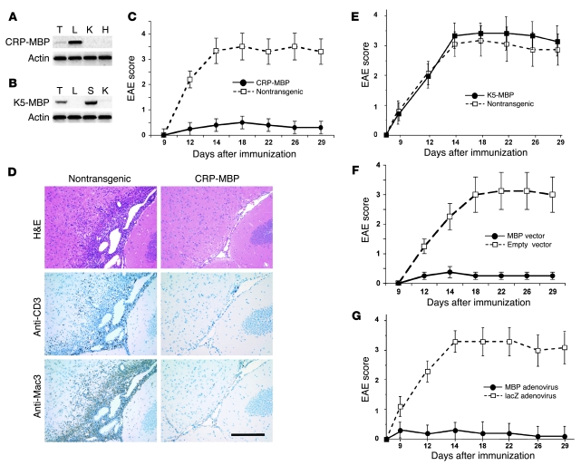
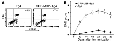
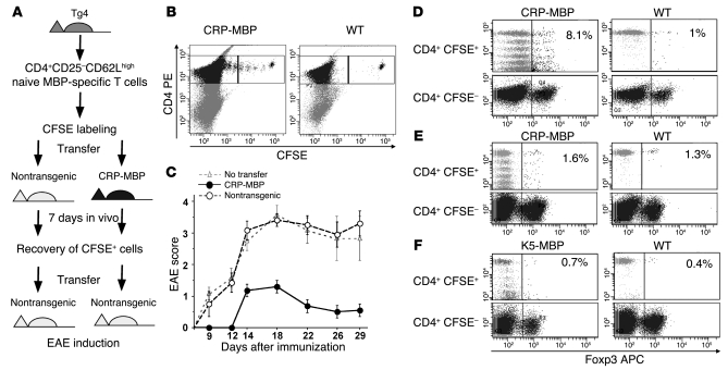
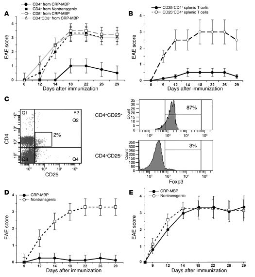
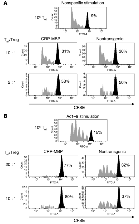
Tregs are important mediators of immune tolerance to self antigens, and it has been suggested that Treg inactivation may cause autoimmune disease. Therefore, immunotherapy approaches that aim to restore or expand autoantigen-specific Treg activity might be beneficial for the treatment of autoimmune disease. Here we report that Treg-mediated suppression of autoimmune disease can be achieved in vivo by taking advantage of the ability of the liver to promote immune tolerance. Expression of the neural autoantigen myelin basic protein (MBP) in the liver was accomplished stably in liver-specific MBP transgenic mice and transiently using gene transfer to liver cells in vivo. Such ectopic MBP expression induced protection from autoimmune neuroinflammation in a mouse model of multiple sclerosis. Protection from autoimmunity was mediated by MBP-specific CD4+CD25+Foxp3+ Tregs, as demonstrated by the ability of these cells to prevent disease when adoptively transferred into nontransgenic mice and to suppress conventional CD4+CD25- T cell proliferation after antigen-specific stimulation with MBP in vitro. The generation of MBP-specific CD4+CD25+Foxp3+ Tregs in vivo depended on expression of MBP in the liver, but not in skin, and occurred by TGF-beta-dependent peripheral conversion from conventional non-Tregs. Our findings indicate that autoantigen expression in the liver may generate autoantigen-specific Tregs. Thus, targeting of autoantigens to hepatocytes may be a novel approach to prevention or treatment of autoimmune diseases.


Figures

Figure 1
Figure 1. Ectopic expression of MBP in liver induces suppression of EAE.
(A) CRP-MBP mice expressed transgenic MBP specifically in the liver (L) and weakly in the thymus (T), but not in other organs, such as kidney (K) and heart (H). (B) K5-MBP mice expressed transgenic MBP specifically in the skin (S) and in the thymus, but not in liver or kidney. (C) CRP-MBP mice (n = 24) and nontransgenic littermates (n = 25) were immunized with Ac1–9 to induce EAE. Data are mean ± SEM from 3 independent experiments. (D) Brain histology at day 18 of EAE showed dense perivascular and subpial inflammatory infiltrates in nontransgenic mice, but not in CRP-MBP mice. Staining with H&E or immunostaining for macrophages (anti-Mac3) or T cells (anti-CD3) is shown. Scale bar: 200 μm. (E) K5-MBP mice (n = 11) and nontransgenic littermates (n = 10) were immunized with Ac1–9 to induce EAE. Data are mean ± SEM from 2 independent experiments. (F) Transient hepatic MBP expression facilitated by hydrodynamics-based gene transfer with MBP-encoding vector (n = 10), but not by empty control vector (n = 8), suppressed EAE. Data are mean ± SEM. (G) Transient hepatic MBP expression facilitated by adenoviral gene transfer of MBP (n = 10), but not by control adenovirus (lacZ; n = 10), suppressed EAE. Data are mean ± SEM.
Figure 2
Figure 2. Protection from EAE by ectopic autoantigen expression in the liver does not depend on deletion of autoreactive lymphocytes.
(A) Expression of encephalitogenic T cell receptor by splenic CD4+ T cells of FVB/N×Tg4 or CRP-MBP×Tg4 mice, detected by cytometry with Vβ8.2-specific antibody. Percentages indicate the relative amount of peripheral CD4+Vβ8.2+ T cells. (B) CRP-MBP×Tg4 mice (n = 8) and Tg4 littermates (n = 10) were immunized with Ac1–9 to induce EAE. Data are mean ± SEM.
Figure 4
Figure 4. Protective Tregs are generated by TGF-β–dependent peripheral conversion from naive CD4+CD25 T cells.
(A) Experimental procedure for assessing in vivo conversion of naive MBP-specific T cells to Tregs. Naive splenic T cells from Tg4 mice were labeled with CFSE and transferred to nontransgenic or CRP-MBP mice; after 7 days, they were recovered again for transfer to nontransgenic mice, in which EAE was induced. (B) Analysis of transferred CFSE+ and residual CFSE cells retrieved from CRP-MBP or nontransgenic mice. The CD4+CFSE+ cells in the respective upper right gates were selected for transfer into wild-type mice. (C) CD4+CFSE+ cells retrieved from nontransgenic (n = 10) or CRP-MBP (n = 11) mice were transferred to nontransgenic recipient mice (2 × 10⁴ cells per mouse), in which EAE was induced. Mice without cell transfer (n = 8) served as a control. Data are mean ± SEM. (DF) Analysis of CFSE-labeled T cells (D and F) and CFSE-labeled TGF-β–insensitive T cells (E). Cells were retrieved from spleen and liver 7 days after transfer to CRP-MBP (D and E), K5-MBP (F), or nontransgenic mice. Percent Foxp3+CFSE+ T cells is indicated.
Figure 3
Figure 3. Liver-induced protection from EAE is mediated by peripheral Tregs.
(A) Suppression of EAE by adoptive transfer into wild-type mice of 105 splenic CD4+ cells — but not CD8+ or CD4CD8 cells — from CRP-MBP mice, or of CD4+ T cells from nontransgenic mice. Data are mean ± SEM. (B) Protection of wild-type mice from EAE by adoptive transfer of 105 splenic CD25+CD4+ T cells from CRP-MBP mice, but not by transfer of 105 splenic CD25CD4+ T cells from CRP-MBP mice. Data are mean ± SEM. (C) Spleen cells from CRP-MBP mice were gated for expression of CD4 and CD25; percentage indicates CD4+CD25+ proportion of the total splenocytes. Expression of Foxp3 in CD4+CD25+ and CD4+CD25 T cells is shown at right; percentages indicate Foxp3+ proportion of the splenic CD4+CD25+ and CD4+CD25 cells. (D) Neonatal thymectomy and 6.5-Gy irradiation of CRP-MBP mice (n = 14) or nontransgenic littermates (n = 12), followed by reconstitution with bone marrow and embryonic thymus from wild-type mice, was performed to bypass possible thymic immune tolerance to MBP before EAE induction. Data are mean ± SEM. (E) Transfer of 105 CD8CD4+CD25+ thymic Tregs from CRP-MBP or nontransgenic mice (n = 10 per group) into wild-type mice failed to protect the recipient mice from subsequent induction of EAE. Data are mean ± SEM.
Figure 5
Figure 5. Selective expansion of autoantigen-specific Tregs in mice expressing the autoantigen ectopically in the liver.
Conventional CD25CD4+ effector T cells (Teff) from Tg4 mice were labeled with CFSE and activated in the absence or presence of CD25+CD4+ Tregs from CRP-MBP or nontransgenic mice at the indicated ratios. (A) Tregs of CRP-MBP and nontransgenic mice showed an equal capacity to suppress after nonspecific stimulation with antibody to CD3. (B) Tregs from CRP-MBP mice showed greatly increased efficacy to suppress after being stimulated with Ac1–9 compared with Tregs from nontransgenic mice, indicating selective expansion of MBP-specific Tregs in CRP-MBP mice. Percentages indicate the proportion of cells that did not proliferate (black peaks).

Comment in

References

    1. Hogquist K.A., Baldwin T.A., Jameson S.C. Central tolerance: learning self-control in the thymus. Nat. Rev. Immunol. 2005;5:772–782. doi: 10.1038/nri1707. - DOI - PubMed
    1. Goodnow C.C., Sprent J., Fazekas de St. Groth B., Vinuesa C.G. Cellular and genetic mechanisms of self tolerance and autoimmunity. Nature. 2005;435:590–597. doi: 10.1038/nature03724. - DOI - PubMed
    1. Jonuleit H., Schmitt E. The regulatory T cell family: distinct subsets and their interrelations. J. Immunol. 2003;171:6323–6327. - PubMed
    1. Cohen, I.R. 2000. Tending Adam’s garden: evolving the cognitive immune self. Academic Press. New York, New York, USA. 266 pp.
    1. Viglietta V., Baecher-Allan C., Weiner H.L., Hafler D.A. Loss of functional suppression by CD4+CD25+ regulatory T cells in patients with multiple sclerosis. J. Exp. Med. 2004;199:971–979. doi: 10.1084/jem.20031579. - DOI - PMC - PubMed

Publication types