. 2008 Oct;118(10):3453-61.
doi: 10.1172/JCI36089.

Neutrophil activation by the tissue factor/Factor VIIa/PAR2 axis mediates fetal death in a mouse model of antiphospholipid syndrome

Affiliations

Neutrophil activation by the tissue factor/Factor VIIa/PAR2 axis mediates fetal death in a mouse model of antiphospholipid syndrome

Patricia Redecha et al. J Clin Invest. 2008 Oct.

Abstract

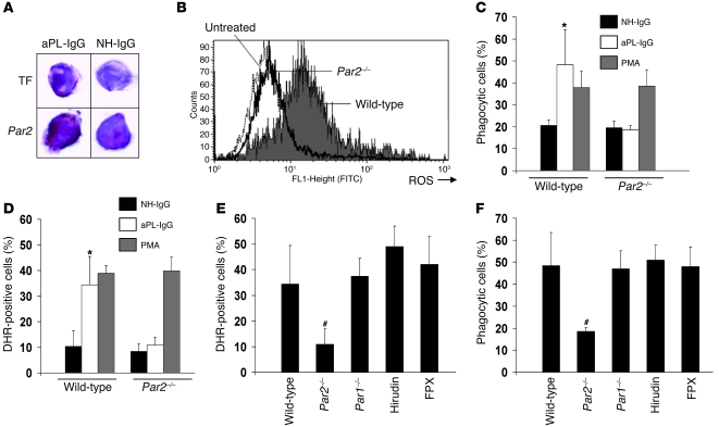
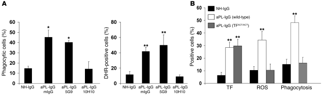
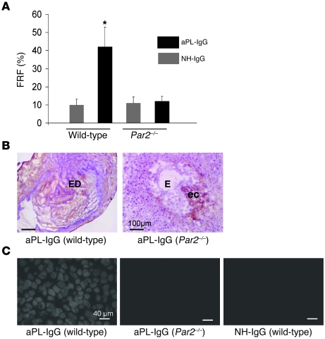
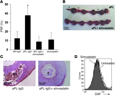
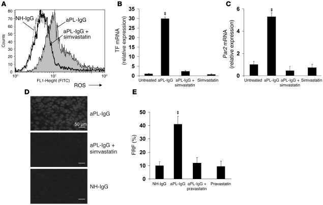
Women with antiphospholipid syndrome (APS), a condition characterized by the presence of antiphospholipid antibodies (aPL), often suffer pregnancy-related complications, including miscarriage. We have previously shown that C5a induction of tissue factor (TF) expression in neutrophils contributes to respiratory burst, trophoblast injury, and pregnancy loss in mice treated with aPL. Here we analyzed how TF contributes to neutrophil activation and trophoblast injury in this model. Neutrophils from aPL-treated mice expressed protease-activated receptor 2 (PAR2), and stimulation of this receptor led to neutrophil activation, trophoblast injury, and fetal death. An antibody specific for human TF that has little impact on coagulation, but potently inhibits TF/Factor VIIa (FVIIa) signaling through PAR2, inhibited aPL-induced neutrophil activation in mice that expressed human TF. Genetic deletion of the TF cytoplasmic domain, which allows interaction between TF and PAR2, reduced aPL-induced neutrophil activation in aPL-treated mice. Par2-/- mice treated with aPL exhibited reduced neutrophil activation and normal pregnancies, which indicates that PAR2 plays an important role in the pathogenesis of aPL-induced fetal injury. We also demonstrated that simvastatin and pravastatin decreased TF and PAR2 expression on neutrophils and prevented pregnancy loss. Our results suggest that TF/FVIIa/PAR2 signaling mediates neutrophil activation and fetal death in APS and that statins may be a good treatment for women with aPL-induced pregnancy complications.


Figures

Figure 1
Figure 1. PAR2 is required for aPL-IgG–induced neutrophil activity.
(A) Immunohistochemical detection of TF and PAR2 on neutrophils from aPL-IgG– and NH-IgG–treated mice. Original magnification, ×400. (B) ROS production, measured as DHR-positive cells, on whole blood neutrophils from aPL-IgG–treated mice. The number of DHR-positive neutrophils increased in aPL-IgG–treated wild-type mice compared with untreated mice. ROS production in neutrophils did not increase in aPL-IgG–treated Par2–/– mice. (C and D) Phagocytic cells and DHR-positive cells in wild-type and Par2–/– mice. The percentage of phagocytic (C) and DHR-positive (D) neutrophils increased in aPL-IgG–treated wild-type mice compared with NH-IgG–treated mice. The absence of PAR2 prevented aPL-IgG–induced ROS production and phagocytosis. Incubation of neutrophils from Par2–/– mice with PMA induced increased ROS generation and phagocytosis, which indicates that the capacity of Par2–/– mice to generate oxidants or in phagocytosis is normal. (E and F) ROS production and phagocytosis in neutrophils from aPL-IgG–treated mice. (E) Neutrophils from Par1–/– mice treated with aPL-IgG showed increased ROS generation (E) as well as phagocytosis similar to aPL-IgG–treated wild-type mice (F). Increased ROS production was also observed in neutrophils from mice treated with hirudin or FPX in addition to aPL-IgG. n = 5–7 per group. *P < 0.05 versus NH-IgG; #P < 0.05 versus wild type. Data in CF are mean ± SD.
Figure 2
Figure 2. TF/FVIIa/PAR2 signaling is required for aPL-IgG–induced increase in neutrophil activity.
(A) Effect of 10H10 and 5G9 on neutrophil phagocytosis and neutrophil ROS generation in aPL-IgG–treated mice, as determined by FACS. aPL-IgG–induced phagocytosis and ROS generation were prevented by 10H10, but not 5G9. Mouse IgG1 (mIgG) was used as isotype control. (B) TF expression, ROS production, and phagocytosis in neutrophils from NH-IgG–treated wild-type mice, aPL-IgG–treated wild-type mice, and aPL-IgG–treated TFΔCT/ΔCT mice. aPL-IgG–induced ROS production and phagocytosis was not observed in TFΔCT/ΔCT mice. *P < 0.05, **P < 0.01 versus NH-IgG. n = 5 mice per group. All data are mean ± SD.
Figure 3
Figure 3. Par2–/– mice are protected from trophoblast oxidative injury and fetal death.
Pregnant Par2–/– or wild-type mice were given 10 mg aPL-IgG or NH-IgG i.p. on days 8 and 12. (A) Mice were killed on day 15 of pregnancy, uteri were dissected, and FRF was calculated as described in Methods. In matings of wild-type mice, approximately 40% of the embryos of mice treated with aPL-IgG were resorbed; in contrast, Par2–/– mice showed a reduction in aPL-IgG–induced FRF. *P < 0.05 versus NH-IgG. n = 5–7 mice per group. Data are mean ± SD. (B and C) Mice were killed on day 8, 2 h after aPL-IgG or NH-IgG injection. Uteri were dissected, and decidua sections were cut and stained with an anti-C3 antibody (B) or DHE (C) to measure superoxide generation. (B) The chromogen was DAB (brown), and the counterstain was hematoxylin. In aPL-IgG–treated wild-type mice, there was extensive C3 staining (brown) in deciduae as well as embryo debris (ED). In contrast, the decidual tissue from aPL-IgG–treated Par2–/– mice showed minimal staining for C3 at the ectoplacental cone (ec) and intact embryo (E). (C) aPL-IgG–induced superoxide production in wild-type mice was attenuated in Par2–/– mice. Oxidative damage in deciduae from aPL-IgG–treated PAR2 mice was minimal and not different from that in NH-IgG–treated wild-type mice. Scale bars: 100 μm (B); 40 μm (C).
Figure 4
Figure 4. Simvastatin prevents pregnancy loss in aPL-IgG–treated mice.
(A and B) Pregnant C57BL/6 mice were given 10 mg aPL-IgG or NH-IgG i.p. on days 8 and 12, and some also received 20 μg simvastatin i.p. 18 h before administration of aPL-IgG (n = 5–11 mice per group). (A) Mice were killed on day 15 of pregnancy, uteri were dissected, and FRF was calculated. Treatment with simvastatin prevented fetal loss. P < 0.01 versus aPL-IgG plus simvastatin. Data are mean ± SD. (B) Uteri from day 15 of pregnancy. There were 5 fetuses and 3 resorptions (asterisks) in the uterus of an aPL-IgG–treated mouse, while the uterus of a mouse that received aPL-IgG plus simvastatin contained 7 fetuses and no resorptions, similar to mice treated with NH-IgG (not shown). Data are representative of observations in 5–8 mice per group. (C) Mice were killed on day 8, 2 h after aPL-IgG or NH-IgG injection. Decidua sections were stained with an anti-C3 antibody as in Figure 3. In aPL-IgG–treated wild-type mice, there was extensive C3 staining (brown) in deciduae as well as embryo debris. In contrast, the decidual tissue from mice treated with aPL-IgG plus simvastatin showed minimal staining for C3 and intact embryo. (D) FACS analysis of DAF expression on trophoblast-like BeWo cells. There was no difference in DAF expression between untreated BeWo cells and BeWo cells incubated with 10 μg/ml simvastatin. Scale bars: 1 cm (B); 200 μm (C, left); 50 mm (C, right).
Figure 5
Figure 5. Statins inhibit TF and PAR2 synthesis and prevent neutrophil oxidative burst and oxidative damage in aPL-IgG–treated mouse placentas.
Pregnant C57BL/6 mice were given aPL-IgG or NH-IgG as in Figure 3; some also received 20 μg simvastatin or 5 μg pravastatin i.p. 18 h before aPL-IgG administration (n = 5–10 mice per group). (A) On day 8, 2 h after aPL-IgG or NH-IgG injection, a blood sample was drawn in order to determine ROS production and phagocytosis in neutrophils by FACS, measured as the number of DHR-positive cells. aPL-IgG increased ROS production, whereas simvastatin prevented neutrophil oxidative burst in aPL-IgG–treated mice. The number of DHR-positive neutrophils was similar between NH-IgG treated and aPL-IgG plus simvastatin–treated mice. (B and C) RT-PCR analysis was performed in isolated neutrophils to quantify TF and PAR2 gene expression. Neutrophils from aPL-IgG–treated mice showed a 28-fold increase in TF mRNA (B) and a 5-fold increase in Par2 mRNA (C). Simvastatin prevented aPL-IgG–induced increase in TF and PAR2 synthesis. (D) Mice were killed on day 8, and deciduae were removed to determine superoxide generation using DHE fluorescence. Increased free radical–mediated lipid peroxidation was observed in deciduae from aPL-IgG–treated mice. No oxidative damage was observed in NH-IgG and aPL-IgG plus simvastatin–treated mice. Scale bars: 50 μm. (E) Mice were killed on day 15 of pregnancy, uteri were dissected, and FRF was calculated. Similar to the effects of simvastatin, pravastatin treatment prevented fetal loss. P < 0.05 versus aPL-IgG plus simvastatin or pravastatin as appropriate.

Comment in

References

    1. Redecha P., et al. Tissue factor: a link between C5a and neutrophil activation in antiphospholipid antibody induced fetal injury. Blood. 2007;110:2423–2431. doi: 10.1182/blood-2007-01-070631. - DOI - PMC - PubMed
    1. Monroe D.M., Key N.S. The tissue factor-factor VIIa complex: procoagulant activity, regulation, and multitasking. J. Thromb. Haemost. 2007;5:1097–1105. doi: 10.1111/j.1538-7836.2007.02435.x. - DOI - PubMed
    1. Strukova S. Blood coagulation-dependent inflammation. Coagulation-dependent inflammation and inflammation-dependent thrombosis. Front. Biosci. 1006;11:59–80. doi: 10.2741/1780. - DOI - PubMed
    1. Camerer E., Huang W., Coughlin S.R.2000. . Tissue factor- and factor X-dependent activation of protease-activated receptor 2 by factor VIIa. Proc. Natl. Acad. Sci. U. S. A. 975255–5260.10.1073/pnas.97.10.5255 - DOI - PMC - PubMed
    1. Ruf W., Dorfleutner A., Riewald M. Specificity of coagulation factor signaling. J. Thromb. Haemost. 2003;1:1495–1503. doi: 10.1046/j.1538-7836.2003.00300.x. - DOI - PubMed

Publication types

MeSH terms