

Inherited pelvic organ prolapse in the mouse: preliminary evaluation of a new murine model
- PMID: 18802654
- PMCID: PMC3796144
- DOI: 10.1007/s00192-008-0723-7
Inherited pelvic organ prolapse in the mouse: preliminary evaluation of a new murine model
Abstract
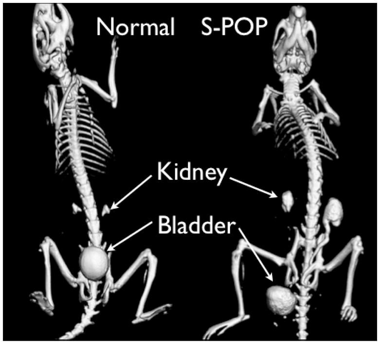
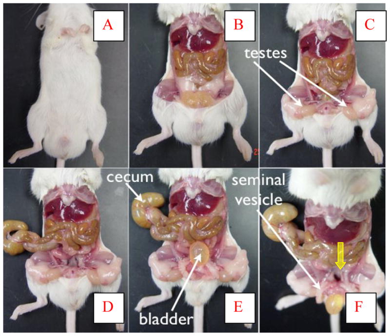
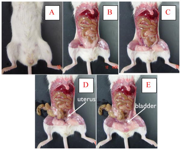
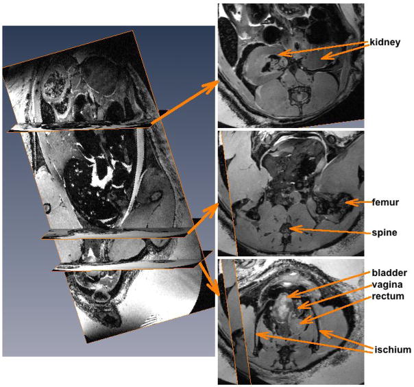
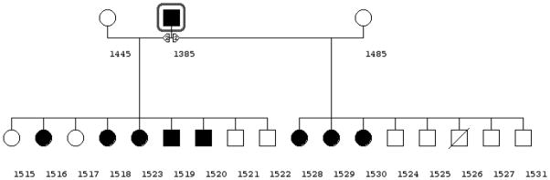
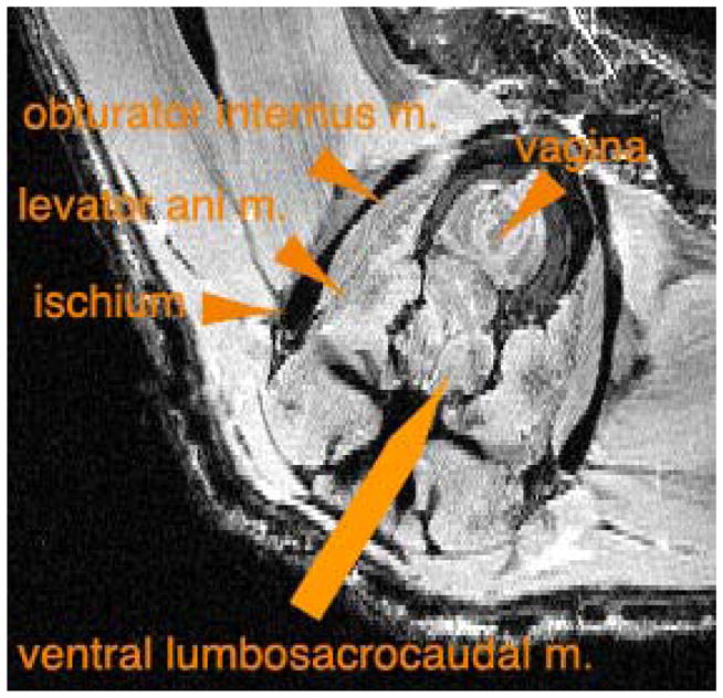
The objective of this study was to report the initial anatomic, radiographic, and genetic evaluations of a novel form of spontaneous pelvic organ prolapse (S-POP) in mice. We observed S-POP in a colony of UPII-SV40T transgenic mice developed for studies on bladder cancer. We utilized magnetic resonance imaging and necropsy to characterize this finding. We have established a breeding colony to identify inheritance patterns and for future studies. Selective breeding isolated the S-POP phenotype from the transgene. In contrast to other animal models, the S-POP mouse does not require an obligatory antecedent event to manifest pelvic organ prolapse. Necropsy and imaging demonstrate significant displacement of the pelvic organs distal to the pelvic floor in both sexes. The appearance of the POP is similar to that seen in the human female phenotype. Preliminary breeding studies indicate an autosomal dominant inheritance pattern. This mouse may be an effective animal model for the study of POP in humans.
Conflict of interest statement
No conflicts of interest.
Figures
Similar articles
-
Functional pelvic floor anatomy in Nepali women attending a general gynaecology clinic.Int Urogynecol J. 2018 Oct;29(10):1435-1440. doi: 10.1007/s00192-017-3534-x. Epub 2017 Dec 21. Int Urogynecol J. 2018. PMID: 29270722
-
Lower urogenital tract anatomical and functional phenotype in lysyl oxidase like-1 knockout mice resembles female pelvic floor dysfunction in humans.Am J Physiol Renal Physiol. 2008 Aug;295(2):F545-55. doi: 10.1152/ajprenal.00063.2008. Epub 2008 May 21. Am J Physiol Renal Physiol. 2008. PMID: 18495804
-
Computed tomography evaluation of pelvic organ prolapse. Techniques and applications.J Comput Assist Tomogr. 2003 Sep-Oct;27(5):779-85. doi: 10.1097/00004728-200309000-00016. J Comput Assist Tomogr. 2003. PMID: 14501370
-
Diagnostic imaging of pelvic floor dysfunction.Curr Opin Urol. 2001 Jul;11(4):423-8. doi: 10.1097/00042307-200107000-00015. Curr Opin Urol. 2001. PMID: 11429505 Review.
-
Dynamic MR imaging of the pelvic floor: a pictorial review.Radiographics. 2009 May-Jun;29(3):e35. doi: 10.1148/rg.e35. Epub 2009 Mar 6. Radiographics. 2009. PMID: 19270071 Review.
Cited by
-
Mouse Knockout Models for Pelvic Organ Prolapse: a Systematic Review.Int Urogynecol J. 2022 Jul;33(7):1765-1788. doi: 10.1007/s00192-021-05066-5. Epub 2022 Jan 28. Int Urogynecol J. 2022. PMID: 35088092 Free PMC article.
-
A Review and Case Study of 3D Imaging Modalities for Female Amniote Reproductive Anatomy.Integr Comp Biol. 2022 May 10;62(3):542-58. doi: 10.1093/icb/icac027. Online ahead of print. Integr Comp Biol. 2022. PMID: 35536568 Free PMC article.
-
Comparative histology of mouse, rat, and human pelvic ligaments.Int Urogynecol J. 2016 Nov;27(11):1697-1704. doi: 10.1007/s00192-016-3008-6. Epub 2016 Apr 18. Int Urogynecol J. 2016. PMID: 27091643
-
Current practice in animal models for pelvic floor dysfunction.Int Urogynecol J. 2023 Apr;34(4):797-808. doi: 10.1007/s00192-022-05387-z. Epub 2022 Oct 26. Int Urogynecol J. 2023. PMID: 36287229 Review.
-
Postpartum stress urinary incontinence: lessons from animal models.Expert Rev Obstet Gynecol. 2010 Sep 1;5(5):567-580. doi: 10.1586/eog.10.48. Expert Rev Obstet Gynecol. 2010. PMID: 21113428 Free PMC article.
References
-
- Bump RC, Norton PA. Epidemiology and natural history of pelvic floor dysfunction. Obstet Gynecol Clin North Am ; 1998;25:723–46. - PubMed
-
- Subak LL, Waetjen LE, van den Eeden S. Cost of pelvic organ prolapse surgery in the United States. Obstet Gynecol ; 2001;98:646–61. - PubMed
-
- Buchsbaum GM, Duecy EE, Kerr LA, et al. Urinary incontinence in nulliparous women and their parous sisters. Obstet Gynecol ; 2005;106:1253–58. - PubMed
-
- Jack GS, Nikolova G, Vilain E. Familial transmission of genitovaginal prolapse. Int Urogynecol J Pelvic Floor Dysfunct ; 2005;20:1–4. - PubMed
-
- Kim S, Harvey MA, Johnston S. A review of the epidemiology and pathophysiology of pelvic floor dysfunction: do racial differences matter? J Obstet Gynecol. 2005;27:251–59. - PubMed
Publication types
MeSH terms
Grants and funding
LinkOut - more resources
Full Text Sources
Medical