. 2008 Oct;46(10):507-14.
doi: 10.1002/dvg.20398.

Pitx2 deletion in pituitary gonadotropes is compatible with gonadal development, puberty, and fertility

Affiliations

Pitx2 deletion in pituitary gonadotropes is compatible with gonadal development, puberty, and fertility

Michael A Charles et al. Genesis. 2008 Oct.

Abstract

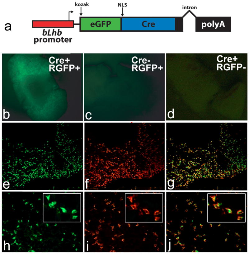
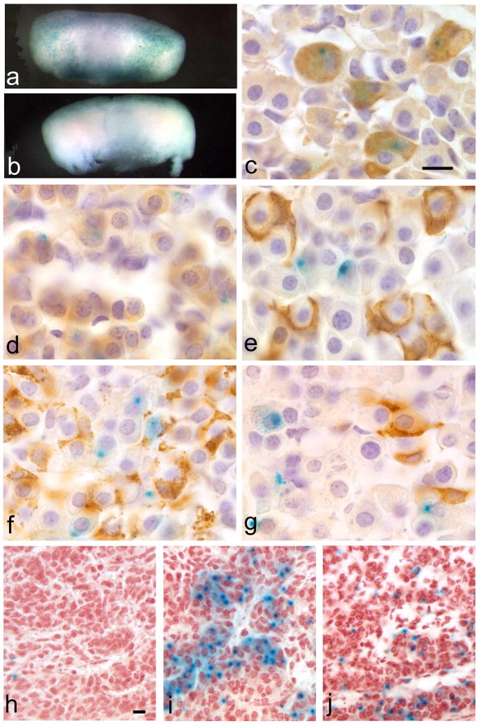
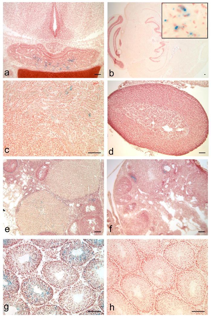
This report introduces a gonadotrope-specific cre transgenic mouse capable of ablating floxed genes in mature pituitary gonadotropes. Initial analysis of this transgenic line, Tg(Lhb-cre)1Sac, reveals that expression is limited to the pituitary cells that produce luteinizing hormone beta, beginning appropriately at e17.5. Cre activity is detectable by a reporter gene in nearly every LHbeta-producing cell, but the remaining hormone-producing cell types and other organs exhibit little to no activity. We used the Tg(Lhb-cre)1Sac strain to assess the role Pitx2 in gonadotrope function. The gonadotrope-specific Pitx2 knockout mice exhibit normal expression of LHbeta, sexual maturation, and fertility, suggesting that Pitx2 is not required for gonadotrope maintenance or for regulated production of gonadotropins.


Figures

Figure 1
Figure 1. Gonadotrope-specific cre transgenic construct and expression
The Tg(Lhb-cre)1Sac transgene (a) is controlled by the bovine Lhb promoter, containing a KOZAK translation initiation site, eGFP and cre recombinase fusion gene with a nuclear localization signal (NLS), and a poly adenylation signal and intron from the rabbit β-globin gene. Pituitaries from mice with the Tg(Lhb-cre)1Sac transgene (Cre+) and the Rosa26-GFP reporter allele (RGFP+) (b, e-j) were compared with pituitaries from Cre-, RGFP+ (c) and Cre+, RGFP- (d). GFP fluorescence is detected in Cre+, RGFP+ mice (b), but not in mice carrying only the reporter (cre-, RGFP+) (c) or only the transgene (Cre+, RGFP-) (d). Frozen pituitary sections of a mouse carrying the Tg(Lhb-cre)1Sac transgene and the Rosa26-GFP reporter allele (e-j) at 10× (e-g) and 20× (h-j) power objective lenses, with high magnification insets. Colabeling with antibody for GFP (e, h) and LHβ (f, i) is detected by yellow color in merged picture (g, j) in over 80% of anterior pituitary cells.
Figure 2
Figure 2. Tg(Lhb-cre)1Sac transgene is active in late gestation and is specific to pituitary gonadotropes
Adult pituitaries of Tg(Lhb-cre)1Sac; Rosa26LacZ reporter progeny (a) and non-transgenic (b) stained with X-gal. Staining is detectable in the adult anterior lobe of the Tg(Lhb-cre)1Sac;Rosa26LacZ reporter progeny (a, c-g) but not in the non-transgenic (b). Immunohistochemical staining of pituitary hormones shows colocalization of the X-gal staining with LHβ immunoreactivity (c), but very little or no colocalization with GH (d), POMC (e), PRL (f) or TSHβ immunoreactivity (g). Tg(Lhb-cre)1Sac; Rosa26LacZ embryos reveal the onset of transgene activity by X-gal staining in only one or two cells at e16.5 (h), and increased penetrance of X-gal staining comparable to normal LHβ expression at e17.5 (i) and e18.5 (j). Scale bars represent 10μm for panels c-g (100× magnification) and panels h-j (40× magnification).
Figure 3
Figure 3. Limited ectopic activity of Tg(Lhb-cre)1Sac transgene
Tissues from Tg(Lhb-cre)1Sac;Rosa26-LacZ reporter animals stained with X-gal reveal that cre activity is high in the e17.5 anterior pituitary, but is absent from the hypothalamus (a). Limited evidence of cre activity is detected by X-gal staining in rare cells in the adult brain (b) and kidney (c), but not in the adrenal gland (d). Low levels of X-gal staining are detected in a few ovarian follicles and corpora lutea of from Tg(Lhb-cre)1Sac;Rosa26LacZ double transgenic mice (e) above background X-gal staining in Rosa26LacZ mice lacking the cre transgene (f). X-gal staining is present in some seminiferous tubules of the double-transgenic mice (g), but not in wild-type testis (h). Scale bars represent 100μm for all panels. Inset (b) width represents 100μm.
Figure 4
Figure 4. Normal LH expression in gonadotrope-specific Pitx2 knockout mice
(a) Most LHβ immunostained cells (red) co-stain with PITX2-specific antibodies (green) in Pitx2flox/+ adult mouse pituitary (white arrows), but about half of the PITX2 immuno-positive cells do not co-stain with LHβ-specific antibodies (yellow arrows). (b) Pitx2flox/-; Tg(Lhb-cre)1Sac adult mouse pituitary lacks cells that co-stain with antibodies for PITX2 and LHβ. Cells immunostained for PITX2 only (yellow arrow), or LHβ only (pink arrows) predominate, while PITX2 and LHβ co-stained cells are quite rare (white arrow).

Similar articles

Cited by

References

    1. Berger P, Kranewitter W, Madersbacher S, Gerth R, Geley S, Dirnhofer S. Eutopic production of human chorionic gonadotropin beta (hCG beta) and luteinizing hormone beta (hLH beta) in the human testis. FEBS Lett. 1994;343:229–233. - PubMed
    1. Bingham NC, Verma-Kurvari S, Parada LF, Parker KL. Development of a steroidogenic factor 1/Cre transgenic mouse line. Genesis. 2006;44:419–424. - PubMed
    1. Brinkmeier ML, Gordon DF, Dowding JM, Saunders TL, Kendall SK, Sarapura VD, Wood WM, Ridgway EC, Camper SA. Cell-specific expression of the mouse glycoprotein hormone alpha-subunit gene requires multiple interacting DNA elements in transgenic mice and cultured cells. Mol Endocrinol. 1998;12:622–633. - PubMed
    1. Burns KH, Matzuk MM. Minireview: genetic models for the study of gonadotropin actions. Endocrinology. 2002;143:2823–2835. - PubMed
    1. Burrows HL, Douglas KR, Seasholtz AF, Camper SA. Genealogy of the Anterior Pituitary Gland: Tracing a Family Tree. Trends Endocrinol Metab. 1999;10:343–352. - PubMed