. 2009 Mar;57(4):444-53.
doi: 10.1002/glia.20772.

Low-density lipoprotein receptors regulate microglial inflammation through c-Jun N-terminal kinase

Affiliations

Low-density lipoprotein receptors regulate microglial inflammation through c-Jun N-terminal kinase

Ana Pocivavsek et al. Glia. 2009 Mar.

Abstract

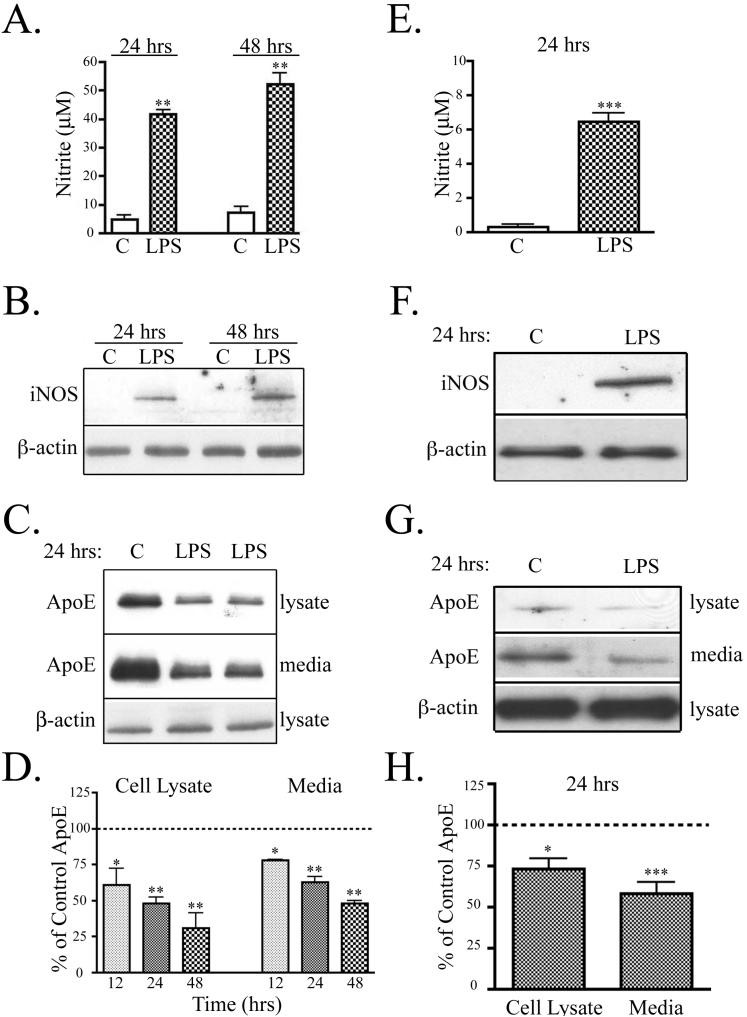
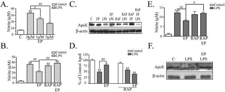
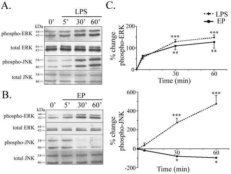
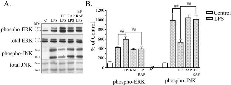
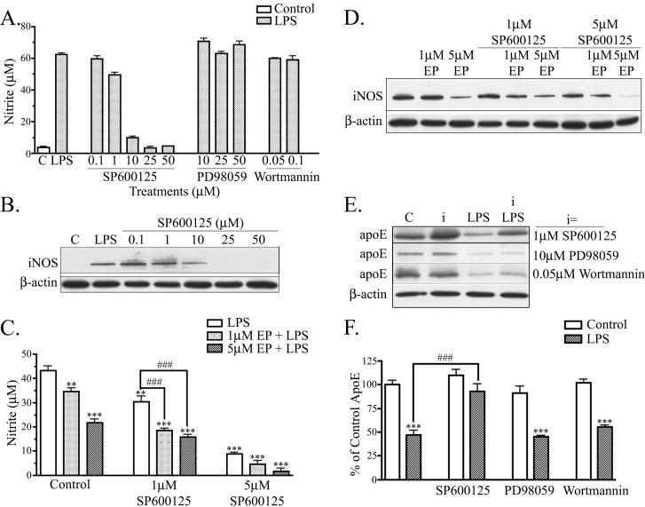
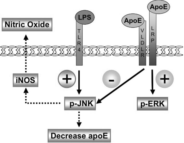
Apolipoprotein E (apoE) has been implicated in modulating the central nervous system (CNS) inflammatory response. However, the molecular mechanisms involved in apoE-dependent immunomodulation are poorly understood. We hypothesize that apoE alters the CNS inflammatory response by signaling via low-density lipoprotein (LDL) receptors in glia. To address this hypothesis, we used a small bioactive peptide formed from the receptor-binding domain of apoE, apoE peptide (EP), to study LDL receptor signaling in microglia. To model glial activation, we treated primary mouse microglia and the microglial cell line BV2 with lipopolysaccharide (LPS) and studied two inflammatory responses: an increase in nitric oxide production (NO) and a decrease in apoE production. We found that treatment of primary microglia and BV2 cells with EP attenuated LPS-induced NO accumulation and apoE reduction in a dose-dependent manner. Using the receptor-associated protein to block ligand binding to members of the LDL receptor family, we found that EP attenuated both of these LPS-induced inflammatory responses via LDL receptors. We studied two intracellular signaling cascades associated with apoE: c-Jun N-terminal kinase (JNK) and extracellular signal-regulated kinase (ERK). LPS induced both ERK and JNK activation, whereas EP induced ERK activation, but drastically reduced JNK activation. Inhibition of JNK with SP600125 reduced LPS-induced NO production and apoE reduction in a dose-dependent manner. Treatment of microglia with suboptimal EP in combination with JNK inhibitor enhanced attenuation of LPS-induced NO production. These data suggest that microglial LDL receptors regulate JNK activation, which is necessary for apoE modulation of the inflammatory response.


Figures

Figure 1
Figure 1. Activation of microglia by LPS increased NO and decreased apoE
BV2 microglia and primary microglia were treated with 100 ng/ml LPS. A. In BV2 cells, LPS promoted nitrite accumulation in the conditioned media at 24 hrs and 48 hrs (mean ± SEM; **P<0.01; n = 6). B. BV2 cell lysates were analyzed by Western blotting with antibodies to iNOS and β-actin (as a control). A representative blot shows that LPS increased iNOS levels (n = 4). C. BV2 cell lysates were analyzed for apoE and β-actin and conditioned media was analyzed for apoE. A representative blot shows that treatment of cells with LPS for 24 hrs reduced apoE in both cell lysates and media. D. Quantification of Western blots showed that apoE in cell lysate and media of BV2 cells was significantly decreased at 12 hrs, 24 hrs, and 48 hrs. The data were quantified as percent of control (mean ± SEM; *P<0.05; **P<0.01 compared with corresponding control cultures; n = 6). E. Primary microglia treated with LPS for 24 hrs accumulated a significant amount of nitrite in the conditioned media (mean ± SEM; ***P<0.001; n=4). F. Cell lysates from primary microglia were analyzed by Western blotting with antibodies to iNOS and β-actin (as a control). A representative blot shows that LPS increased iNOS levels (n=3). G. Representative Western blots of primary microglia cell lysate analyzed for apoE and β-actin and conditioned media analyzed for apoE is shown. Microglia treated with LPS for 24 hrs showed reduced apoE in both cell lystes and media. H. Quantification of Western blot (Panel G) showed that apoE in cell lysate and media of primary microglia was significantly decreased at 24 hrs (mean ± SEM; *P<0.05; ***P<0.001 compared with untreated cultures; n = 4).
Figure 2
Figure 2. ApoE peptide attenuates LPS induced NO increase and apoE decrease
A. BV2 cells were treated with 100 ng/ml LPS and nitrite was measured in the conditioned media at 24 hrs. LPS induced nitrite accumulation and EP showed dose-dependent attenuation of LPS-induced nitrite production (n = 6). B. Cells were treated as in Panel A with 5 μM EP and LPS. EP attenuated LPS-induced nitrite production and 50 nM RAP prevented the effect of EP, while RAP alone had no effect (mean ± SEM; **P<0.01 compared with corresponding control cultures; #P<0.05; ##P<0.01 compared with indicated cultures; n = 6). C. BV2 cell lysates were analyzed by Western blot analysis for apoE and β-actin. A representative blot is shown. LPS decreased apoE levels and 1 μM EP prevented that decrease, while EP alone had no effect on apoE levels. 50 nM RAP alone had no effect, but RAP prevented EP from reversing the effects of LPS. β-actin levels were the same across treatment conditions. D. Western blot data were quantified as percent of control (mean ± SEM; *P<0.05; **P<0.01; ***P<0.001 compared with control cultures; ## P<0.01 compared with indicated cultures; n = 3). E. Primary microglia were treated with 100 ng/ml LPS and nitrite was measured in the conditioned media at 24 hrs. LPS induced nitrite accumulation and 5 μM EP showed attenuation of LPS-induced nitrite production. 50 nM RAP prevented the effect of EP, while RAP alone had no effect (mean ± SEM; ***P<0.001 compared with corresponding control cultures; #P<0.05; ##P<0.01 compared with indicated cultures; n = 3). F. Cells lysates from primary microglia were analyzed by Western blot analysis for apoE and β-actin. LPS decreased apoE levels and 1 μM EP prevented that decrease. β-actin levels were the same across treatment conditions (n = 3).
Figure 3
Figure 3. ApoE peptide increased ERK activation and reduced JNK activation
BV2 microglia were treated with 1 μM EP or 100 ng/ml LPS from 5 to 60 min. Cell lysates were analyzed by Western blotting with antibodies to phospho-ERK, total ERK, phospho-JNK, and total JNK. A. Cells treated with LPS showed a time-dependent increase in both phospho-ERK and phospho-JNK. Total levels of ERK and JNK protein remain unchanged. B. Cells treated with EP showed a time-dependent increase in phospho-ERK and decrease in phospho-JNK. Total levels of ERK and JNK protein remain unchanged. C. Western blot data were quantified as percent change of phospho-ERK (upper graph) and percent change of phospho-JNK (lower graph) comparing EP and LPS treatment. LPS significantly increased phospho-ERK and phospho-JNK by 30 min. EP significantly increased phospho-ERK and decreased phospho-JNK by 30 min (mean ± SEM; *P < 0.05; **P < 0.01; ***P < 0.001; n = 3).
Figure 4
Figure 4. ApoE peptide signaling effects are LDL receptor family mediated
BV2 microglia were treated with various combinations of PBS (control), 1 μM EP, 100 ng/ml LPS, and 50 nM RAP for 1 hr. Cell lysates were analyzed by Western blotting with antibodies to phospho-ERK, total ERK, phospho-JNK, and total JNK. A. Cells treated with EP and LPS showed increased phospho-ERK and reduced phospho-JNK compared to cells treated with LPS alone. Treatment of BV2 cells with RAP prevented EP-induced potentiation of phospho-ERK and EP-induced attenuation of phospho-JNK. Total levels of ERK and JNK protein were unchanged. B. Western blot data were quantified as percent of control for phospho-ERK and phospho-JNK (mean ± SEM; ##P < 0.01 compared with indicated cultures; n = 4).
Figure 5
Figure 5. JNK modulates NO production and apoE levels
A. BV2 cells were treated with indicated doses of a JNK inhibitor (SP600125), ERK inhibitor (PD98059), or PI3K/AKT inhibitor (Wortmannin), and stimulated with 100 ng/ml LPS for 24 hrs. SP600125 treatment reduced nitrite production in a dose-dependent manner. PD98059 and Wortmannin did not effect LPS induced nitrite production (n = 3). B. Cell lysates were analyzed by Western blotting with antibodies to iNOS and β-actin. A representative blot shows that SP600125 reduced iNOS levels in a dose-dependant manner (n = 3). C. BV2 cells were treated with suboptimal doses of EP and SP600125. Each alone showed attenuation of LPS-induced nitrite accumulation; in combination, they showed significant additive effects (***P < 0.001; **P < 0.01 compared with control cultures. ###P<0.001 compared with indicated cultures; n = 6). D. Cell lysates of BV2 cells treated with suboptimal doses of EP and SP600125 were analyzed by Western blotting antibodies to iNOS and β-actin. Combined EP and SP600125 treatment reduced of LPS-induced iNOS accumulation (n = 3). E. BV2 cells were treated with LPS and/or kinase inhibitors (i) and cell lysates were analyzed for apoE and β-actin expression. A representative blot shows that SP600125 increased apoE levels, but neither other inhibitor had any effects. F. Western blot data were quantified as percent of control for apoE in the cell lysate. Cells treated with 1 μM SP600125 and LPS showed an increase in lysate apoE compared to cells treated with LPS alone, whereas cells treated with 10 μM PD98059 or 0.05 μM Wortmannin and LPS did not show an increase in lysate apoE compared to cells treated with LPS alone (mean ± SEM; ***P < 0.001 compared with corresponding control cultures; ###P < 0.001 compared with indicated cultures; n = 3).
Figure 6
Figure 6. LDL receptor immunomodulatory signaling in microglia
LPS binds TLR4 receptors on the cell surface and signals an increase in phospho-JNK, leading to an increase in iNOS synthesis, an accumulation of NO extracellularly, and an independent decrease of intracellular apoE. ApoE binds LDL receptors on the cell surface and signals an increase in ERK activation and decrease in JNK activation. The decrease in JNK activation suppresses iNOS synthesis, reduces NO accumulation extracellularly, and suppresses a reduction of intracellular apoE.

Similar articles

Cited by

References

    1. Aderem A, Ulevitch RJ. Toll-like receptors in the induction of the innate immune response. Nature. 2000;406(6797):782–7. - PubMed
    1. Beffert U, Stolt PC, Herz J. Functions of lipoprotein receptors in neurons. J Lipid Res. 2004;45(3):403–9. - PubMed
    1. Bhat NR, Zhang P, Lee JC, Hogan EL. Extracellular signal-regulated kinase and p38 subgroups of mitogen-activated protein kinases regulate inducible nitric oxide synthase and tumor necrosis factor-alpha gene expression in endotoxin-stimulated primary glial cultures. J Neurosci. 1998;18(5):1633–41. - PMC - PubMed
    1. Blasi E, Barluzzi R, Bocchini V, Mazzolla R, Bistoni F. Immortalization of murine microglial cells by a v-raf/v-myc carrying retrovirus. J Neuroimmunol. 1990;27(2-3):229–37. - PubMed
    1. Boyles JK, Pitas RE, Wilson E, Mahley RW, Taylor JM. Apolipoprotein E associated with astrocytic glia of the central nervous system and with nonmyelinating glia of the peripheral nervous system. J Clin Invest. 1985;76(4):1501–13. - PMC - PubMed

Publication types

MeSH terms