Review
. 2008 Nov 1;56(14):1590-1605.
doi: 10.1002/glia.20776.

How does the Schwann cell lineage form tumors in NF1?

Affiliations
Review

How does the Schwann cell lineage form tumors in NF1?

Steven L Carroll et al. Glia. .

Abstract

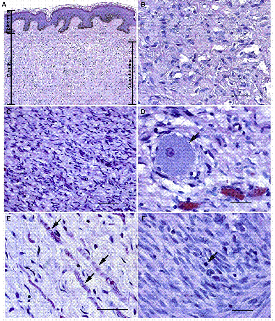
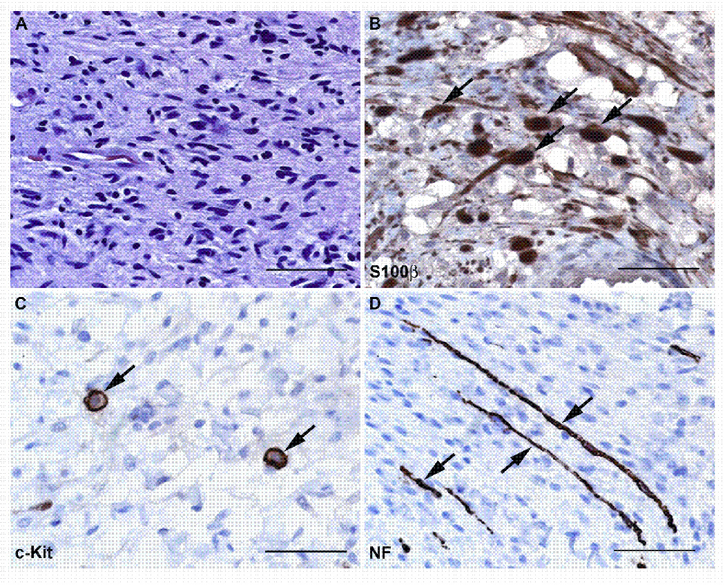
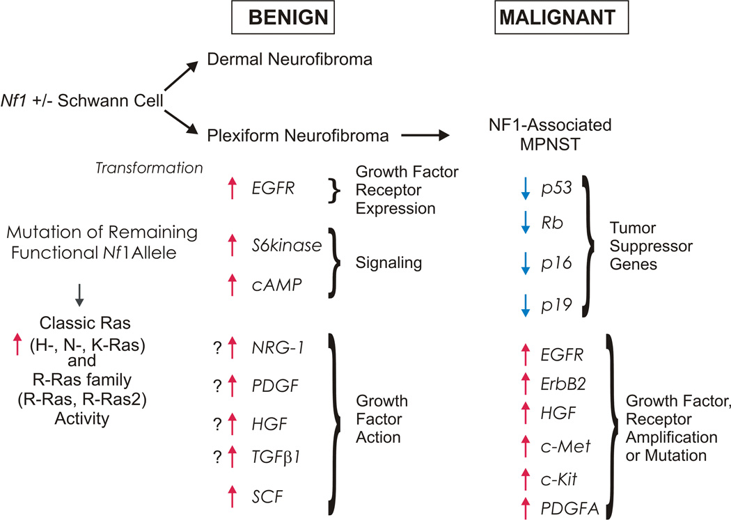
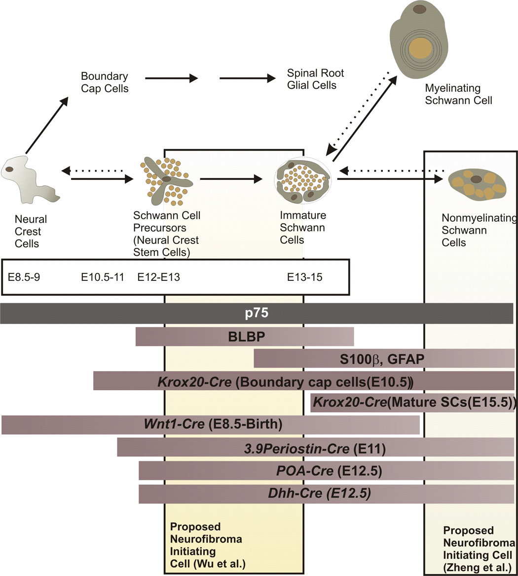
Neurofibromas are benign tumors of peripheral nerve that occur sporadically or in patients with the autosomal dominant tumor predisposition syndrome neurofibromatosis type 1 (NF1). Multiple neurofibroma subtypes exist which differ in their site of occurrence, their association with NF1, and their tendency to undergo transformation to become malignant peripheral nerve sheath tumors (MPNSTs), the most common malignancy associated with NF1. Most NF1 patients carry a constitutional mutation of the NF1 tumor suppressor gene. Neurofibromas develop in these patients when an unknown cell type in the Schwann cell lineage loses its remaining functional NF1 gene and initiates a complex series of interactions with other cell types; these interactions may be influenced by aberrant expression of growth factors and growth factor receptors and the action of modifier genes. Cells within certain neurofibroma subtypes subsequently accumulate additional mutations affecting the p19(ARF)-MDM2-TP53 and p16INK4A-Rb signaling cascades, mutations of other as yet unidentified genes, and amplification of growth factor receptor genes, resulting in their transformation into MPNSTs. These observations have been validated using a variety of transgenic and knockout mouse models that recapitulate neurofibroma and MPNST pathogenesis. A new generation of mouse models is also providing important new insights into the identity of the cell type in the Schwann cell lineage that gives rise to neurofibromas. Our improving understanding of the mechanisms underlying the pathogenesis of neurofibromas and MPNSTs raises intriguing new questions about the origin and pathogenesis of these neoplasms and establishes models for the development of new therapies targeting these neoplasms.


Figures

Fig. 1
Fig. 1
Microscopic anatomy of NF1-associated peripheral nerve sheath tumors. (A, B) Panel A illustrates a low power (10x) view of a localized cutaneous neurofibroma that presented as a pedunculated mass on the left shoulder of a 60 year old woman with NF1. This tumor (indicated by the bar on the right of the panel) spanned almost the entire thickness of the dermis and infiltrated into the underlying subcutis (a layer of loose fibroadipose tissue underlying the dermis; not shown in this image). Note that the tumor does not involve the most superficial portion of the dermis or the overlying epidermis (the maximal extent of the epidermis and the dermis are indicated by bars on the left of the panel). Panel B is a representative hematoxylin and eosin stained high power (40x) field from this densely collagenized tumor. (C, D) Panel C illustrates a low power (20x) field from a localized intraneural neurofibroma that arose in a cervical dorsal spinal nerve root. This tumor infiltrated diffusely through the nerve and associated dorsal root ganglion, destroying the normal fascicular architecture of the nerve as it produced a segmental fusiform enlargement of this structure. Panel D is a higher power view (40x) of this tumor. The arrow indicates a sensory neuron from the dorsal root ganglion that has been entrapped by the growth of the neurofibroma. (E) High power (40x) view of a field within a plexiform neurofibroma resected from a 40 year old woman with NF1. This tumor, like many plexiform neurofibromas, has a looser, more myxoid matrix but is still composed of the same cell types present in the tumors illustrated in A–D. Arrows indicate entrapped axons and their myelin sheaths. (F) An MPNST that arose within a plexiform neurofibroma in an NF1 patient. Note the greater degree of cellularity, atypia and mitotic activity (arrow) in this tumor. Magnification: 40x. Bars in B, D and F: 50 µm. Bar in C: 200 µm. Bar in E: 100 µm.
Fig. 2
Fig. 2
Neurofibromas are composed of a complex mixture of cell types. (A) High power (40x) view of a hematoxylin and eosin stained section from a localized intraneural (nodular) neurofibroma that arose on the 5th lumbar nerve of a 61 year old woman. This neoplasm is composed of a mixture of cell types that can be detected using immunostains for cell-type specific markers, as shown in B–D. (B) Immunoperoxidase stains (brown) for S100β label the Schwann cell-like elements in this neurofibroma. The section has been lightly counterstained with hematoxylin to highlight the nuclei of cells (primarily fibroblasts and perineural-like cells) that do not stain for this antigen. Arrows indicate some of the cells that are intensely S100β immunoreactive. (C) Another section of this tumor immunostained for the mast cell marker c-Kit (CD117). Arrows indicate two cells that show intense membranous immunoreactivity for this membrane tyrosine kinase receptor. (D) The same tumor immunostained for neurofilaments to demonstrate the presence of axons entrapped by the infiltrative growth pattern of this tumor (arrows). Bars in A–D: 50 µm.
Fig. 3
Fig. 3
Schematic illustrating some of the multiple factors contributing to the development of neurofibromas in NF1 patients and their subsequent progression to become MPNSTs. The development of neurofibromas is initiated when an NF1 haploinsufficient cell in the Schwann cell lineage loses its remaining functional copy of NF1. This results in increases in the activity of Ras and other signaling molecules; tumorigenesis is enhanced by interactions with other cell types, inappropriate expression of EGFR and possibly the action of other growth factors. Subsequent progression to become an MPNST is associated with loss of additional tumor suppressor genes in the p19ARF-MDM2-TP53 and p16INK4A-Rb signaling cascades. Amplification or mutation of several growth factors receptors also contributes to MPNST pathogenesis.
Fig. 4
Fig. 4
Schematic illustration of the differentiation of neural crest cells and their progeny in the peripheral nervous system, relating this to the expression of markers for various developmental stages and periods when Cre-mediated recombination is evident in the Schwann cell lineage in various mouse models of neurofibroma pathogenesis. Times indicated for the appearance of each cell type (embryonic days 8.5 to 15) refer to the developmental sequence seen in peripheral nerves within the trunk of mice. The three bars immediately beneath the time course (p75; BLBP; S100β, GFAP) indicate the period that markers for specific developmental stages are expressed. The next series of bars indicate the periods in which Cre-mediated recombination is evident in the Schwann cell lineage in Nf1 flox mice in which recombination is driven by the indicated promoters. Wnt1-Cre;Nf1fl/− mice and most 3.9Periostin-Cre;Nf1fl/− mice die at birth (Gitler et al., 2003; Joseph et al., 2008). Nf1 ablation produced by Cre recombinase expression driven by the Krox20, P0A and Dhh promoters results in neurofibroma pathogenesis in adult mice. See the text for a more detailed discussion of the phenotype of these mouse models and the implications this has for the cell of origin of neurofibromas.

References

    1. Aoki M, Nabeshima K, Koga K, Hamasaki M, Suzumiya J, Tamura K, Iwasaki H. Imatinib mesylate inhibits cell invasion of malignant peripheral nerve sheath tumor induced by platelet-derived growth factor-BB. Lab Invest. 2007;87:767–779. - PubMed
    1. Aquino J, Hjerling-Leffler J, Koltzenburg M, Villar M, Ernfors P. In vitro and in vivo differentiation of boundary cap neural crest stem cells into mature Schwann cells. Exp Neurol. 2006;198:438–449. - PubMed
    1. Atit RA, Mitchell K, Nguyen L, Warshawsky D, Ratner N. The neurofibromatosis type 1 (Nf1) tumor suppressor is a modifier of carcinogen-induced pigmentation and papilloma formation in C57Bl/6 mice. J Invest Dermatol. 2000;114:1093–1100. - PMC - PubMed
    1. Badache A, DeVries GH. Neurofibrosarcoma-derived Schwann cells overexpress platelet-derived growth factor (PDGF) receptors and are induced to proliferate by PDGF BB. J Cell Physiol. 1998;177:334–342. - PubMed
    1. Badache A, Muja N, DeVries GH. Expression of Kit in neurofibromin-deficient human Schwann cells: role in Schwann cell hyperplasia associated with type 1 neurofibromatosis. Oncogene. 1998;17:795–800. - PubMed

Publication types

MeSH terms