

The evolutionarily conserved arrangement of domains in SRC family kinases is important for substrate recognition
- PMID: 18803405
- PMCID: PMC2841526
- DOI: 10.1021/bi800930e
The evolutionarily conserved arrangement of domains in SRC family kinases is important for substrate recognition
Abstract
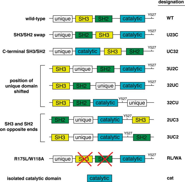
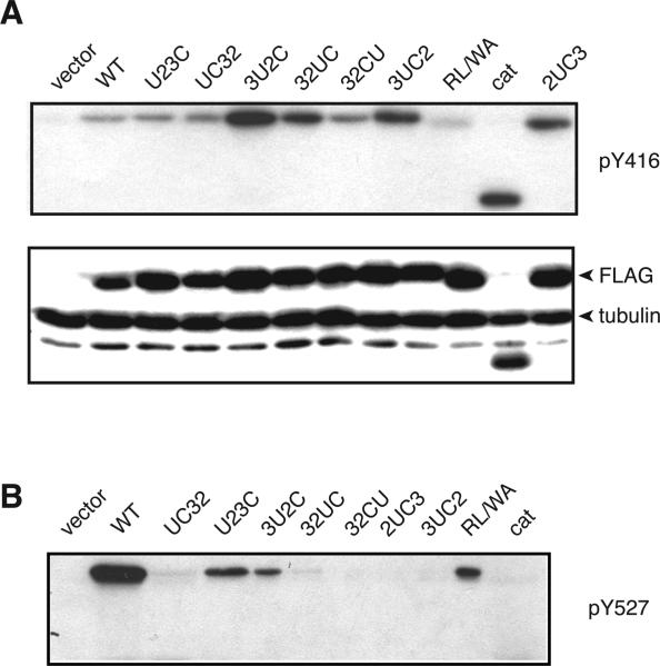
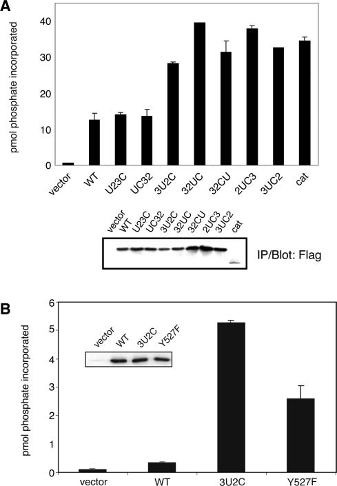
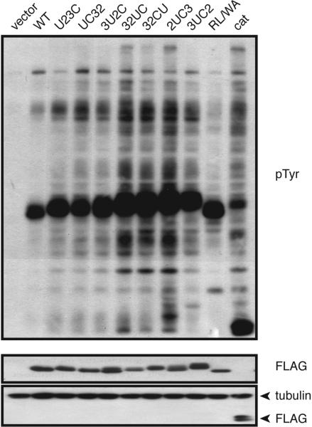
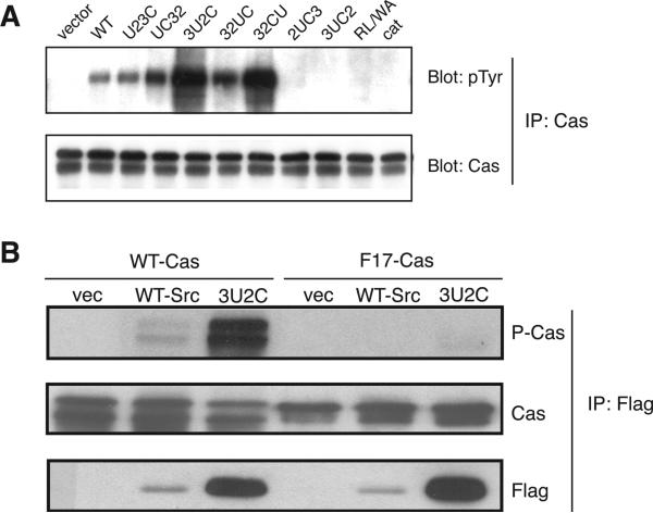
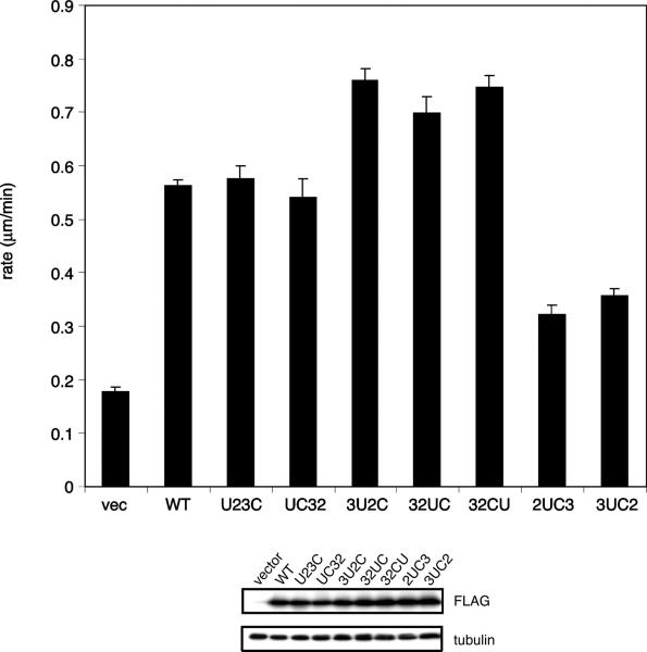
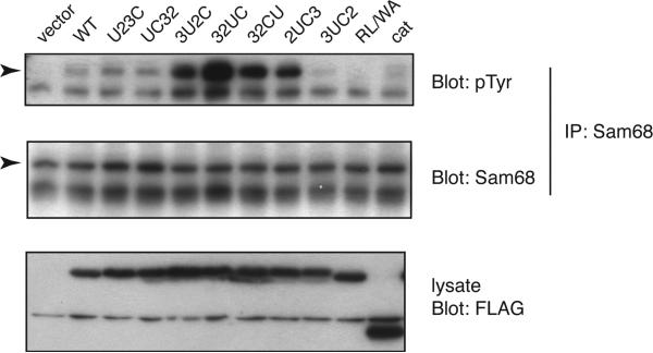
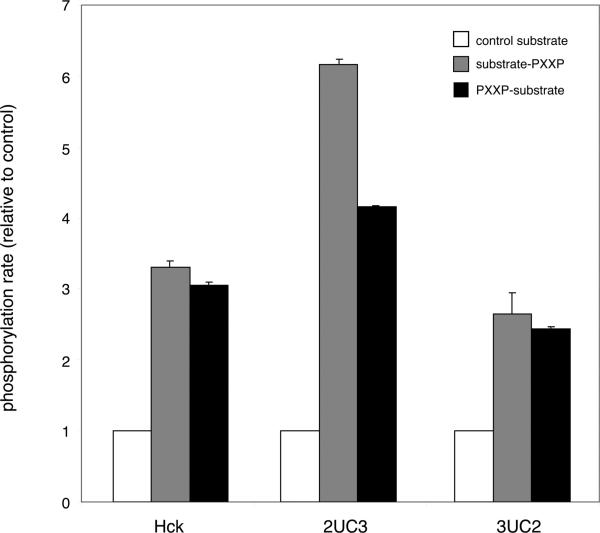
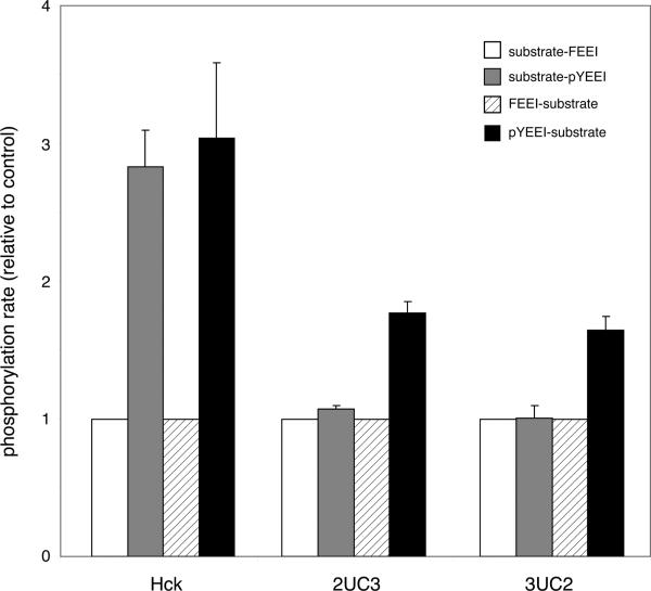
The SH3-SH2-kinase domain arrangement in nonreceptor tyrosine kinases has been conserved throughout evolution. For Src family kinases, the relative positions of the domains are important for enzyme regulation; they permit the assembly of Src kinases into autoinhibited conformations. The SH3 and SH2 domains of Src family kinases have an additional role in determining the substrate specificity of the kinase. We addressed the question of whether the domain arrangement of Src family kinases has a role in substrate specificity by producing mutants with alternative arrangements. Our results suggest that changes in the positions of domains can lead to specific changes in the phosphorylation of Sam68 and Cas by Src. Phosphorylation of Cas by several mutants triggers downstream signaling leading to cell migration. The placement of the SH2 domain with respect to the catalytic domain of Src appears to be especially important for proper substrate recognition, while the placement of the SH3 domain is more flexible. The results suggest that the involvement of the SH3 and SH2 domains in substrate recognition is one reason for the strict conservation of the SH3-SH2-kinase architecture.
Figures
Similar articles
-
SH3-dependent stimulation of Src-family kinase autophosphorylation without tail release from the SH2 domain in vivo.Nat Struct Biol. 2002 May;9(5):365-9. doi: 10.1038/nsb782. Nat Struct Biol. 2002. PMID: 11976726
-
Cooperative activation of Src family kinases by SH3 and SH2 ligands.Cancer Lett. 2007 Nov 8;257(1):116-23. doi: 10.1016/j.canlet.2007.07.012. Epub 2007 Aug 24. Cancer Lett. 2007. PMID: 17719722 Free PMC article.
-
Novel mechanism of regulation of the non-receptor protein tyrosine kinase Csk: insights from NMR mapping studies and site-directed mutagenesis.J Mol Biol. 2001 Nov 16;314(1):129-38. doi: 10.1006/jmbi.2001.5126. J Mol Biol. 2001. PMID: 11724538
-
The Src module: an ancient scaffold in the evolution of cytoplasmic tyrosine kinases.Crit Rev Biochem Mol Biol. 2018 Oct;53(5):535-563. doi: 10.1080/10409238.2018.1495173. Epub 2018 Sep 5. Crit Rev Biochem Mol Biol. 2018. PMID: 30183386 Free PMC article. Review.
-
Src protein-tyrosine kinase structure and regulation.Biochem Biophys Res Commun. 2004 Nov 26;324(4):1155-64. doi: 10.1016/j.bbrc.2004.09.171. Biochem Biophys Res Commun. 2004. PMID: 15504335 Review.
Cited by
-
Structure, Function, and Regulation of the SRMS Tyrosine Kinase.Int J Mol Sci. 2020 Jun 14;21(12):4233. doi: 10.3390/ijms21124233. Int J Mol Sci. 2020. PMID: 32545875 Free PMC article. Review.
-
PTB domain-directed substrate targeting in a tyrosine kinase from the unicellular choanoflagellate Monosiga brevicollis.PLoS One. 2011 Apr 26;6(4):e19296. doi: 10.1371/journal.pone.0019296. PLoS One. 2011. PMID: 21541291 Free PMC article.
-
Constitutive Activity in an Ancestral Form of Abl Tyrosine Kinase.PLoS One. 2015 Jun 19;10(6):e0131062. doi: 10.1371/journal.pone.0131062. eCollection 2015. PLoS One. 2015. PMID: 26090675 Free PMC article.
-
Bivalent inhibitors of protein kinases.Crit Rev Biochem Mol Biol. 2014 Mar-Apr;49(2):102-15. doi: 10.3109/10409238.2013.875513. Epub 2014 Feb 25. Crit Rev Biochem Mol Biol. 2014. PMID: 24564382 Free PMC article. Review.
-
Temperature sensitivities of metazoan and pre-metazoan Src kinases.Biochem Biophys Rep. 2020 Jun 10;23:100775. doi: 10.1016/j.bbrep.2020.100775. eCollection 2020 Sep. Biochem Biophys Rep. 2020. PMID: 32566764 Free PMC article.
References
-
- Robinson DR, Wu YM, Lin SF. The protein tyrosine kinase family of the human genome. Oncogene. 2000;19:5548–5557. - PubMed
-
- Brown MT, Cooper JA. Regulation, substrates and functions of src. Biochim. Biophys. Acta. 1996;1287:121–149. - PubMed
-
- Shiu SH, Li WH. Origins, lineage-specific expansions, and multiple losses of tyrosine kinases in eukaryotes. Mol. Biol. Evol. 2004;21:828–840. - PubMed
-
- Hubbard SR, Till JH. Protein Tyrosine Kinase Structure and Function. Annu. Rev. Biochem. 2000;69:373–398. - PubMed
-
- Manning G, Whyte DB, Martinez R, Hunter T, Sudarsanam S. The protein kinase complement of the human genome. Science. 2002;298:1912–1934. - PubMed
Publication types
MeSH terms
Substances
Grants and funding
LinkOut - more resources
Full Text Sources
Miscellaneous
