Multicenter Study
. 2008 Sep;153(3):360-8.
doi: 10.1111/j.1365-2249.2008.03720.x.

Impaired CD4 and CD8 T cell phenotype and reduced chemokine secretion in recent-onset type 1 diabetic children

Affiliations
Multicenter Study

Impaired CD4 and CD8 T cell phenotype and reduced chemokine secretion in recent-onset type 1 diabetic children

M Hedman et al. Clin Exp Immunol. 2008 Sep.

Abstract

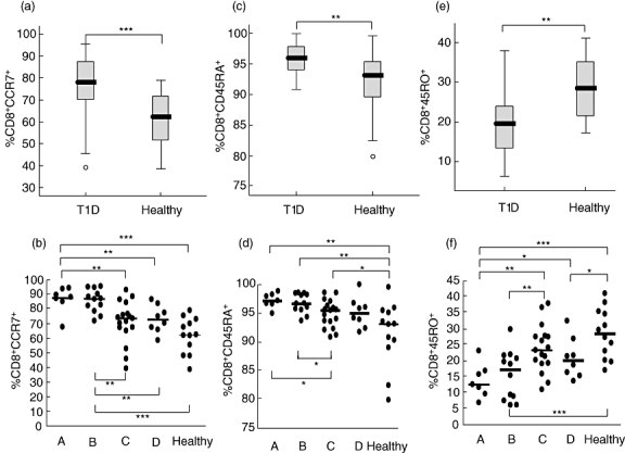
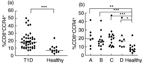
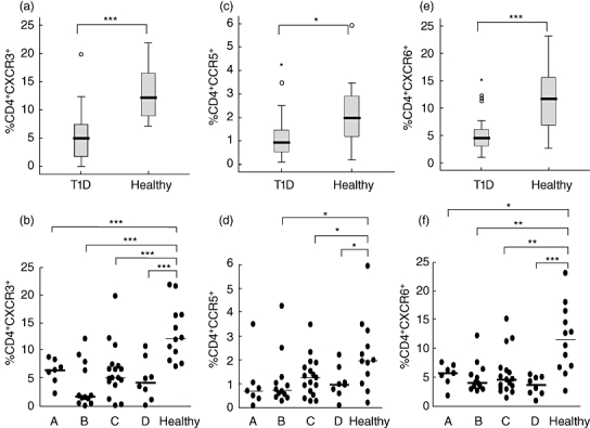
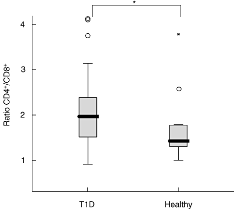
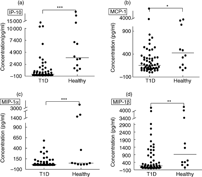
Although the role of the T cell-mediated autoimmune reaction in type 1 diabetes (T1D) is conclusive, studies including data from human circulating CD4(+) and CD8(+) lymphocytes subsets during the disease onset and posterior development are scarce. Further, chemokines and chemokine receptors are key players in the migration of pathogenic T cells into the islets of non-obese diabetic mice developing T1D, but few studies have investigated these markers in human T1D patients. We studied the expression of T helper 1 (Th1)- and Th2-associated chemokine receptors, and the two isoforms of CD45 leucocyte antigen on CD4(+) and CD8(+) lymphocytes from T1D and healthy children, as well as the secretion of chemokines in cell supernatants in peripheral blood mononuclear cells. Our results showed increased expression of CCR7 and CD45RA and reduced CD45RO on CD8(+) cells among recent-onset T1D patients. The percentages of CD4(+) cells expressing CXC chemokine receptor 3 (CXCR3), CXCR6 and CCR5, and the secretion of interferon-gamma-induced protein-10, monocyte chemoattractant protein-1, macrophage inflammatory protein (MIP)-1alpha and MIP-1beta was lower among diabetics. Low expression of Th1-associated receptors and secretion of chemokines, together with an increased amount of CD8(+) cells expressing CD45RA and CCR7 in T1D patients therefore might represent suboptimal Th function in T1D, leading to impaired T cytotoxic responses or alternatively reflect a selective recruitment of Th1 cells into the pancreas.


Figures

Fig. 1
Fig. 1
Expression of CCR7, CD45RA and CD45RO receptors on CD8+ cells was determined by flow cytometry. The percentages (%) of cells expressing each receptor in the diabetic (T1D) and healthy groups is shown in box-plots (a,c,e). Horizontal lines represent the median, the box comprises the 25th and 75th percentiles and the error bars the 10th and 90th percentiles. Outliers are indicated. CCR7 (b), CD45RA (d) and CD45RO (f) expression of the receptors on CD8+ cells in relation to duration of T1D in comparison with healthy controls is shown. Patients were grouped according to duration of disease: 0–4 months (A, n = 7); 5–9 months (B, n = 12); 10–14 months (C, n = 17) and 15–18 months (D, n = 8); healthy (n = 12). Asterisks indicate the level of significance: *P < 0·05; **P < 0·01, ***P < 0·001.
Fig. 2
Fig. 2
Expression of CCR4 receptors on CD8+ cells was determined by flow cytometry. The percentages (%) of cells expressing CCR4 is shown for each individual in the diabetic (T1D) and healthy groups (a). Patients were grouped according to duration of disease: 0–4 months (A, n = 7); 5–9 months (B, n = 12); 10–14 months (C, n = 17) and 15–18 months (D, n = 8); healthy (n = 12) (b). Horizontal lines represent the median and asterisks indicate the level of significance: *P < 0·05; **P < 0·01, ***P < 0·001.
Fig. 3
Fig. 3
Expression of CXC chemokine receptor 3 (CXCR3), CCR5 and CXCR6 chemokine receptors on CD4+ cells was determined by flow cytometry. The percentages (%) of cells expressing each receptor in the diabetic (T1D) and healthy groups is shown in box-plots (a,c,e). Horizontal lines represent the median, the box comprises the 25th and 75th percentiles and the error bars the 10th and 90th percentiles. Outliers are indicated as CXCR3 (b), CCR5 (d) and CXCR6 (f) expression of the receptors on CD8+ cells in relation to duration of T1D in comparison with healthy controls is shown. Patients were grouped according to duration of disease: 0–4 months (A, n = 7); 5–9 months (B, n = 12); 10–14 months (C, n = 17) and 15–18 months (D, n = 8); healthy (n = 12). Asterisks indicate the level of significance: *P < 0·05; **P < 0·01, ***P < 0·001.
Fig. 4
Fig. 4
CD4+ and CD8+ cells were analysed by flow cytometry. The ratio of the percentages of CD4+/CD8+ was calculated for each individual. Horizontal lines represent the median, the box comprises the 25th and 75th percentiles and the error bars the 10th and 90th percentiles and outliers are indicated. Asterisk indicates the level of significance: *P < 0·05.
Fig. 5
Fig. 5
Spontaneous secretion of the chemokines induced protein (IP)-10 (a), monocyte chemoattractant protein (MCP)-1 (b), macrophage inflammatory protein (MIP)-1α (c) and MIP-1β (d) in cell culture supernatant was determined by multiplex fluorochrome technique (Luminex). The chemokine concentrations are expressed as pg/ml. Horizontal lines indicate median values. Asterisks indicate the level of significance: *P < 0·05; **P < 0·01, ***P < 0·001.

Similar articles

Cited by

References

    1. Eisenbarth GS. Type I diabetes mellitus. A chronic autoimmune disease. N Engl J Med. 1986;314:1360–8. - PubMed
    1. Liblau RS, Wong FS, Mars LT, Santamaria P. Autoreactive CD8 T cells in organ-specific autoimmunity: emerging targets for therapeutic intervention. Immunity. 2002;17:1–6. - PubMed
    1. Loetscher P, Pellegrino A, Gong JH, et al. The ligands of CXC chemokine receptor 3, I-TAC, MIG, and IP-10, are natural antagonists for CCR3. J Biol Chem. 2001;276:2986–91. - PubMed
    1. Schrum S, Probst P, Fleischer B, Zipfel PF. Synthesis of the CC-chemokines MIP-1alpha, MIP-1beta, and RANTES is associated with a type 1 immune response. J Immunol. 1996;157:3598–604. - PubMed
    1. Taub DD, Lloyd AR, Conlon K, et al. Recombinant human interferon-inducible protein 10 is a chemoattractant for human monocytes and T lymphocytes and promotes T cell adhesion to endothelial cells. J Exp Med. 1993;177:1809–14. - PMC - PubMed

Publication types

MeSH terms