

Transcriptional regulation of mouse alpha A-crystallin gene in a 148kb Cryaa BAC and its derivates
- PMID: 18803847
- PMCID: PMC2567317
- DOI: 10.1186/1471-213X-8-88
Transcriptional regulation of mouse alpha A-crystallin gene in a 148kb Cryaa BAC and its derivates
Abstract
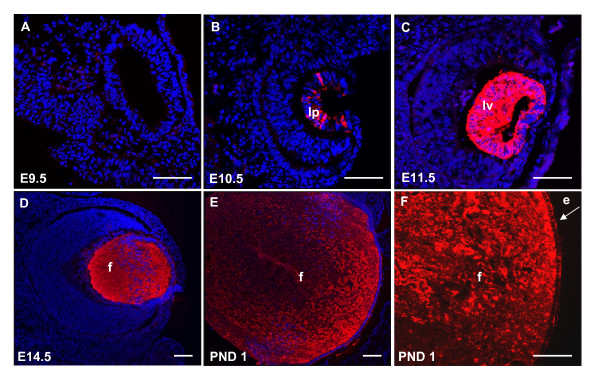
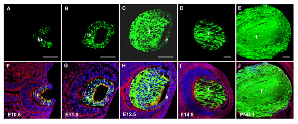
Background: alphaA-crystallin is highly expressed in the embryonic, neonatal and adult mouse lens. Previously, we identified two novel distal control regions, DCR1 and DCR3. DCR1 was required for transgenic expression of enhanced green fluorescent protein, EGFP, in lens epithelium, whereas DCR3 was active during "late" stages of lens primary fiber cell differentiation. However, the onset of transgenic EGFP expression was delayed by 12-24 hours, compared to the expression of the endogenous Cryaa gene.
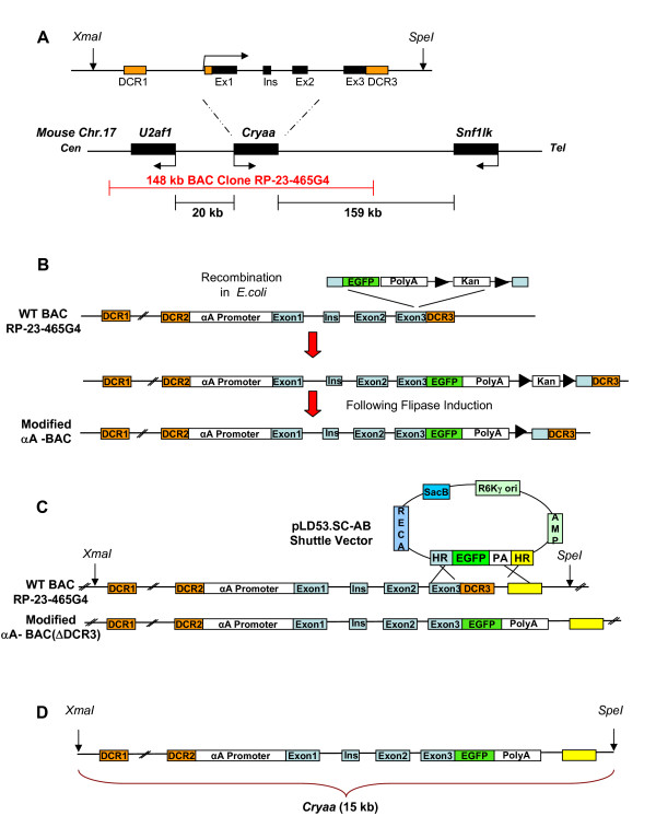
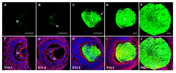
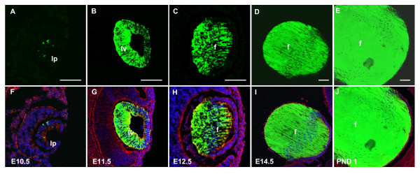
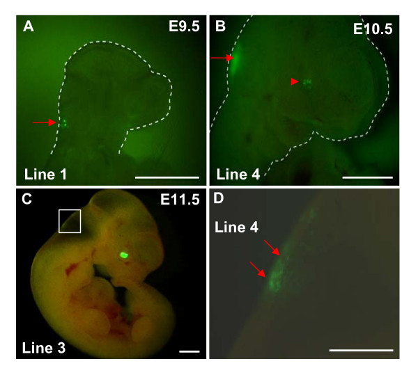
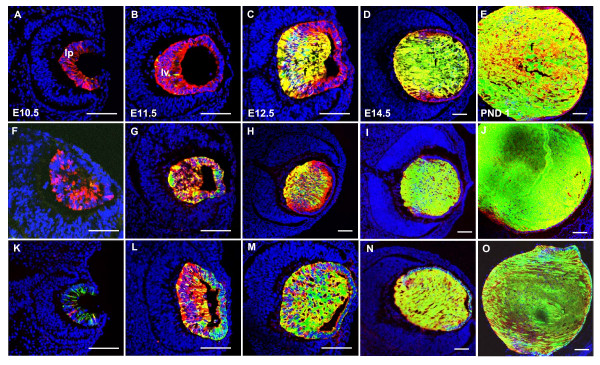
Results: Here, we used bacterial artificial chromosome (BAC) and standard transgenic approaches to examine temporal and spatial regulation of the mouse Cryaa gene. Two BAC transgenes, with EGFP insertions into the third coding exon of Cryaa gene, were created: the intact alphaA-crystallin 148 kb BAC (alphaA-BAC) and alphaA-BAC(DeltaDCR3), which lacks approximately 1.0 kb of genomic DNA including DCR3. Expression of EGFP in the majority of both BAC transgenics nearly recapitulated the endogenous expression pattern of the Cryaa gene in lens, but not outside of the lens. The number of cells expressing alphaA-crystallin in the lens pit was higher compared to the number of cells expressing EGFP. Next, we generated additional lines using a 15 kb fragment of alphaA-crystallin locus derived from alphaA-BAC(DeltaDCR3), 15 kb Cryaa/EGFP. A 15 kb region of Cryaa/EGFP supported the expression pattern of EGFP also in the lens pit. However, co-localization studies of alphaA-crystallin and EGFP indicated that the number of cells that showed transgenic expression was higher compared to cells expressing alphaA-crystallin in the lens pit.
Conclusion: We conclude that a 148 kb alphaA-BAC likely contains all of the regulatory regions required for alphaA-crystallin expression in the lens, but not in retina, spleen and thymus. In addition, while the 15 kb Cryaa/EGFP region also supported the expression of EGFP in the lens pit, expression in regions such as the hindbrain, indicate that additional genomic regions may play modulatory functions in regulating extralenticular alphaA-crystallin expression. Finally, deletion of DCR3 in either alphaA-BAC(DeltaDCR3) or Cryaa (15 kb) transgenic mice result in EGFP expression patterns that are consistent with DCR's previously established role as a distal enhancer active in "late" primary lens fiber cells.
Figures
Similar articles
-
Chick delta1-crystallin enhancer influences mouse alphaA-crystallin promoter activity in transgenic mice.Invest Ophthalmol Vis Sci. 2004 Nov;45(11):4083-90. doi: 10.1167/iovs.03-1270. Invest Ophthalmol Vis Sci. 2004. PMID: 15505059
-
Regulation of alphaA-crystallin via Pax6, c-Maf, CREB and a broad domain of lens-specific chromatin.EMBO J. 2006 May 17;25(10):2107-18. doi: 10.1038/sj.emboj.7601114. Epub 2006 May 4. EMBO J. 2006. PMID: 16675956 Free PMC article.
-
Insertion of a Pax6 consensus binding site into the alphaA-crystallin promoter acts as a lens epithelial cell enhancer in transgenic mice.Invest Ophthalmol Vis Sci. 2004 Jun;45(6):1930-9. doi: 10.1167/iovs.03-0856. Invest Ophthalmol Vis Sci. 2004. PMID: 15161860
-
Genetics of crystallins: cataract and beyond.Exp Eye Res. 2009 Feb;88(2):173-89. doi: 10.1016/j.exer.2008.10.011. Epub 2008 Nov 1. Exp Eye Res. 2009. PMID: 19007775 Review.
-
Developmental genetics in ophthalmology.Ophthalmic Genet. 2003 Mar;24(1):1-33. doi: 10.1076/opge.24.1.1.13888. Ophthalmic Genet. 2003. PMID: 12660863 Review.
Cited by
-
Crystallins in retinal ganglion cell survival and regeneration.Mol Neurobiol. 2013 Dec;48(3):819-28. doi: 10.1007/s12035-013-8470-2. Epub 2013 May 25. Mol Neurobiol. 2013. PMID: 23709342 Free PMC article. Review.
-
Differentiation of Induced Pluripotent Stem Cells to Lentoid Bodies Expressing a Lens Cell-Specific Fluorescent Reporter.PLoS One. 2016 Jun 20;11(6):e0157570. doi: 10.1371/journal.pone.0157570. eCollection 2016. PLoS One. 2016. PMID: 27322380 Free PMC article.
-
Promoter-enhancer looping and shadow enhancers of the mouse αA-crystallin locus.Biol Open. 2018 Dec 7;7(12):bio036897. doi: 10.1242/bio.036897. Biol Open. 2018. PMID: 30404901 Free PMC article.
-
Signaling and Gene Regulatory Networks in Mammalian Lens Development.Trends Genet. 2017 Oct;33(10):677-702. doi: 10.1016/j.tig.2017.08.001. Epub 2017 Aug 31. Trends Genet. 2017. PMID: 28867048 Free PMC article. Review.
-
Considerations for the use of Cre recombinase for conditional gene deletion in the mouse lens.Hum Genomics. 2019 Feb 15;13(1):10. doi: 10.1186/s40246-019-0192-8. Hum Genomics. 2019. PMID: 30770771 Free PMC article.
References
Publication types
MeSH terms
Substances
Grants and funding
LinkOut - more resources
Full Text Sources
Molecular Biology Databases
