

Lack of genomic evidence of AI-2 receptors suggests a non-quorum sensing role for luxS in most bacteria
- PMID: 18803868
- PMCID: PMC2561040
- DOI: 10.1186/1471-2180-8-154
Lack of genomic evidence of AI-2 receptors suggests a non-quorum sensing role for luxS in most bacteria
Abstract
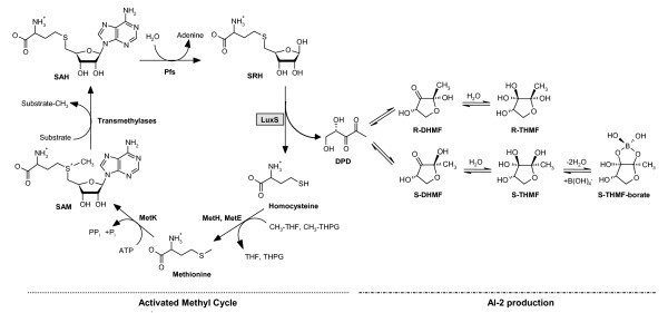
Background: Great excitement accompanied discoveries over the last decade in several Gram-negative and Gram-positive bacteria of the LuxS protein, which catalyzes production of the AI-2 autoinducer molecule for a second quorum sensing system (QS-2). Since the luxS gene was found to be widespread among the most diverse bacterial taxa, it was hypothesized that AI-2 may constitute the basis of a universal microbial language, a kind of bacterial Esperanto. Many of the studies published in this field have drawn a direct correlation between the occurrence of the luxS gene in a given organism and the presence and functionality of a QS-2 therein. However, rarely hathe existence of potential AI-2 receptors been examined. This is important, since it is now well recognized that LuxS also holds a central role as a metabolic enzyme in the activated methyl cycle which is responsible for the generation of S-adenosyl-L-methionine, the major methyl donor in the cell.
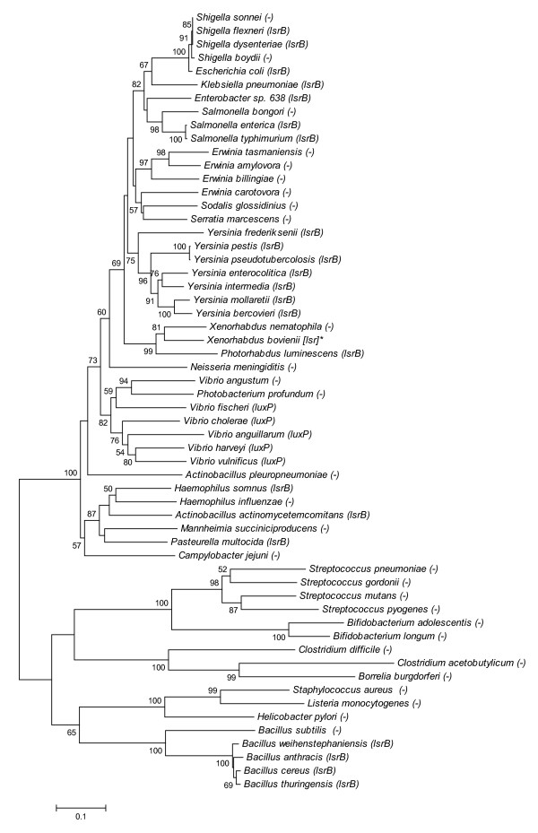
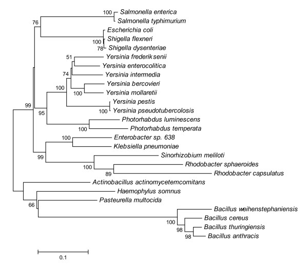
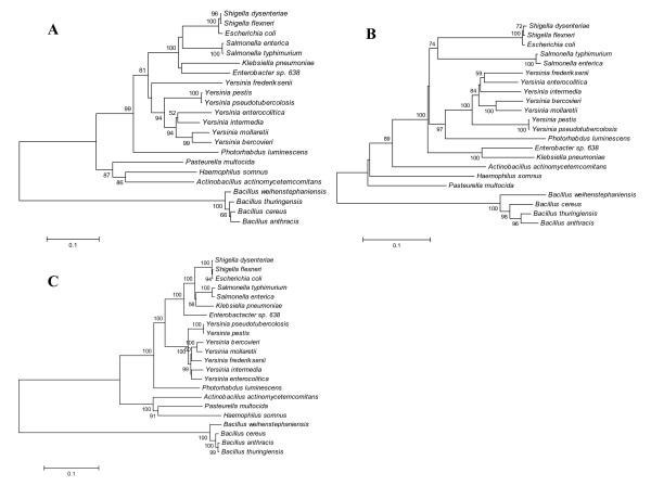
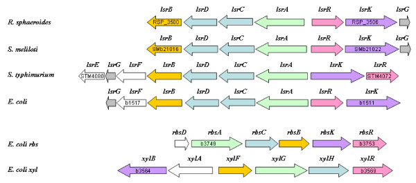
Results: In order to assess whether the role of LuxS in these bacteria is indeed related to AI-2 mediated quorum sensing we analyzed genomic databases searching for established AI-2 receptors (i.e., LuxPQ-receptor of Vibrio harveyi and Lsr ABC-transporter of Salmonella typhimurium) and other presumed QS-related proteins and compared the outcome with published results about the role of QS-2 in these organisms. An unequivocal AI-2 related behavior was restricted primarily to organisms bearing known AI-2 receptor genes, while phenotypes of luxS mutant bacteria lacking these genes could often be explained simply by assuming deficiencies in sulfur metabolism.
Conclusion: Genomic analysis shows that while LuxPQ is restricted to Vibrionales, the Lsr-receptor complex is mainly present in pathogenic bacteria associated with endotherms. This suggests that QS-2 may play an important role in interactions with animal hosts. In most other species, however, the role of LuxS appears to be limited to metabolism, although in a few cases the presence of yet unknown receptors or the adaptation of pre-existent effectors to QS-2 must be postulated.
Figures
Similar articles
-
Let LuxS speak up in AI-2 signaling.Trends Microbiol. 2006 Mar;14(3):114-9. doi: 10.1016/j.tim.2006.01.003. Epub 2006 Feb 3. Trends Microbiol. 2006. PMID: 16459080
-
The LuxS/AI-2 system of Streptococcus suis.Appl Microbiol Biotechnol. 2018 Sep;102(17):7231-7238. doi: 10.1007/s00253-018-9170-7. Epub 2018 Jun 25. Appl Microbiol Biotechnol. 2018. PMID: 29938319 Review.
-
Detection of autoinducer-2 and analysis of the profile of luxS and pfs transcription in Streptococcus suis serotype 2.Curr Microbiol. 2009 Feb;58(2):146-52. doi: 10.1007/s00284-008-9291-9. Epub 2008 Oct 28. Curr Microbiol. 2009. PMID: 18956225
-
Bifidobacteria exhibit LuxS-dependent autoinducer 2 activity and biofilm formation.PLoS One. 2014 Feb 5;9(2):e88260. doi: 10.1371/journal.pone.0088260. eCollection 2014. PLoS One. 2014. PMID: 24505453 Free PMC article.
-
Detection of AI-2 receptors in genomes of Enterobacteriaceae suggests a role of type-2 quorum sensing in closed ecosystems.Sensors (Basel). 2012;12(5):6645-65. doi: 10.3390/s120506645. Epub 2012 May 21. Sensors (Basel). 2012. PMID: 22778662 Free PMC article. Review.
Cited by
-
Heterologous expression of sahH reveals that biofilm formation is autoinducer-2-independent in Streptococcus sanguinis but is associated with an intact activated methionine cycle.J Biol Chem. 2012 Oct 19;287(43):36111-22. doi: 10.1074/jbc.M112.379230. Epub 2012 Aug 31. J Biol Chem. 2012. PMID: 22942290 Free PMC article.
-
Complete genome sequence of the fire blight pathogen Erwinia pyrifoliae DSM 12163T and comparative genomic insights into plant pathogenicity.BMC Genomics. 2010 Jan 4;11:2. doi: 10.1186/1471-2164-11-2. BMC Genomics. 2010. PMID: 20047678 Free PMC article.
-
Communication and autoinduction in the species Listeria monocytogenes: A central role for the agr system.Commun Integr Biol. 2009 Jul;2(4):371-4. doi: 10.4161/cib.2.4.8610. Commun Integr Biol. 2009. PMID: 19721895 Free PMC article.
-
Communication between Bacteria and Their Hosts.Scientifica (Cairo). 2013;2013:361073. doi: 10.1155/2013/361073. Epub 2013 Dec 8. Scientifica (Cairo). 2013. PMID: 24381789 Free PMC article. Review.
-
luxS in bacteria isolated from 25- to 40-million-year-old amber.FEMS Microbiol Lett. 2014 Jan;350(1):117-24. doi: 10.1111/1574-6968.12275. Epub 2013 Oct 7. FEMS Microbiol Lett. 2014. PMID: 24102660 Free PMC article.
References
-
- Whitehead NA, Barnard AM, Slater H, Simpson NJ, Salmond GP. Quorum-sensing in Gram-negative bacteria. FEMS Microbiol Rev. 2001;25:365–404. - PubMed
-
- Cao JG, Meighen EA. Purification and structural identification of an autoinducer for the luminescence system of Vibrio harveyi. J Biol Chem. 1989;264:21670–21676. - PubMed
-
- Engebrecht J, Nealson K, Silverman M. Bacterial bioluminescence: isolation and genetic analysis of functions from Vibrio fischeri. Cell. 1983;32:773–781. - PubMed
-
- Steidle A, Sigl K, Schuhegger R, Ihring A, Schmid M, Gantner S, Stoffels M, Riedel K, Givskov M, Hartmann A, Langebartels C, Eberl L. Visualization of N-acylhomoserine lactone-mediated cell-cell communication between bacteria colonizing the tomato rhizosphere. Appl Environ Microbiol. 2001;67:5761–5770. - PMC - PubMed
Publication types
MeSH terms
Substances
LinkOut - more resources
Full Text Sources
Molecular Biology Databases
