. 2008 Sep 22;15(9):908-19.
doi: 10.1016/j.chembiol.2008.07.008.

A heterospecific leucine zipper tetramer

Affiliations

A heterospecific leucine zipper tetramer

Yiqun Deng et al. Chem Biol. .

Abstract

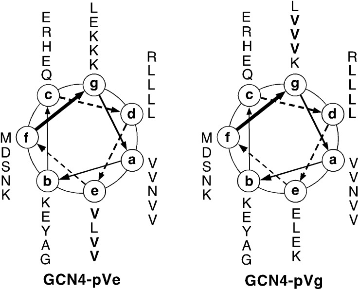
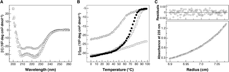
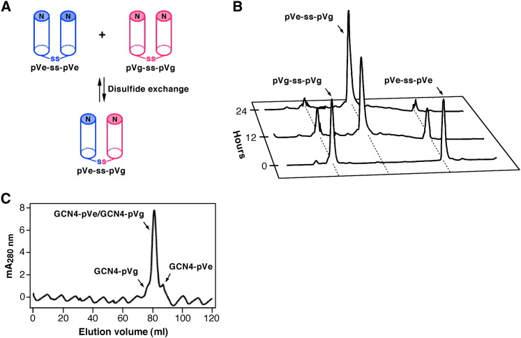
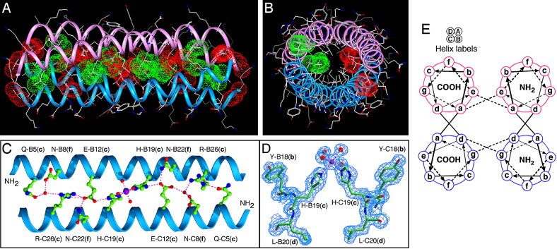
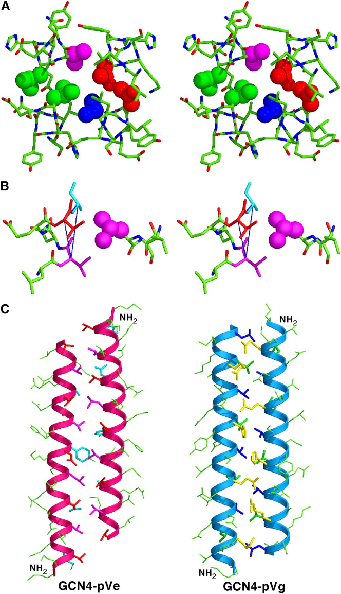
Protein-protein interactions play an essential role in the assembly of the macromolecular complexes that form functional networks and control cellular behavior. Elucidating principles of molecular recognition governing potentially complex interfaces is a challenging goal for structural and systems biology. Extensive studies of alpha-helical coiled coils have provided fundamental insight into the determinants of one seemingly tractable class of oligomeric protein interfaces. We report here that two different valine-containing mutants of the GCN4 leucine zipper that fold individually as four-stranded coiled coils associate preferentially in mixtures to form an antiparallel, heterotetrameric structure. X-ray crystallographic analysis reveals that the coinciding hydrophobic interfaces of the hetero- and homotetramers differ in detail, thereby controlling their partnering and structural specificity. Equilibrium disulfide exchange and thermal denaturation experiments show that the 50-fold preference for heterospecificity is determined by interfacial van der Waals interactions and hydrophobicity. Parallel studies of two alanine-containing variants confirm the above-mentioned interpretation of the basis and mechanism of this heterospecificity. Our results suggest that coiled-coil recognition is an inherently geometric process in which heterotypic interaction specificity derives from a complementarity of both shape and chemistry.


Figures

Figure 1
Figure 1
Axial Helical Projection of the GCN4-pVe and GCN4-pVg Sequences GCN4-pVe differs from GCN4-pVg only at the three charged e and g positions (bold). The view is from the N termini. Heptad-repeat positions are labeled ag. The sequence of GCN4-pVe is MK VKQLVDK VEELLSK NYHLVNE VARLVKL VGER; the sequence of GCN4-pVg is MK VKQLEDV VEELLSV NYHLENV VARLKKL VGER.
Figure 2
Figure 2
The GCN4-pVe and GCN4-pVg Peptides Associate to Form an Extremely Stable, α-Helical Dimer of Heterodimers (A) CD spectra of GCN4-pVe (open triangles), GCN4-pVg (open squares), and the GCN4-pVe/GCN4-pVg complex (open circles) at 4°C in TBS at a total peptide concentration of 10 μM. (B) Thermal melts monitored by the CD signal at 222 nm. The filled circles show thermal unfolding data of the GCN4-pVe/GCN4-pVg complex in the presence of 2 M GuHCl. (C) Equilibrium sedimentation data (27,000 rpm) of a mixture of GCN4-pVe (75 μM) and GCN4-pVg (75 μM) at 20°C in TBS. The data fit closely to a tetrameric complex. The deviation in the data from the linear fit for a tetrameric model is plotted.
Figure 3
Figure 3
Specificity of the Heterotypic Interaction between the GCN4-pVe and GCN4-pVg Peptides (A) Preferential formation of a heterodisulfide bond. Assuming that the glycyl linkers allow for random sorting of the C-terminal cysteine residues in a mixture of the pVe-SH and pVg-SH peptides (the variants of GCN4-pVe and GCN4-pVg that have a C-terminal Gly-Gly-Cys sequence), the thermodynamically preferred heterotetramer conformation should favor oxidative heterodisulfide formation. (B) HPLC analyses of disulfide rearrangement during the course of the equilibration under redox conditions. Disulfide exchange reactions were initiated from the disulfide-bonded pVe-ss-pVe and pVg-ss-pVg homodimers. (C) Size-exclusion chromatography profile of the refolded GCN4-pVe/GCN4-pVg sample. An equilmolar mixture of the two peptides was refolded by renaturation from GuHCl and was analyzed by size exclusion on a Superdex 75 column equilibrated with TBS at 4°C. Fractions were analyzed by reverse-phase HPLC. Relative concentrations of the GCN4-pVe/GCN4-pVg complex and the combined GCN4-pVg and GCN4-pVe homotetramers is ∼50:1, as calculated from the peak absorbance at 280 nm.
Figure 4
Figure 4
GCN4-pVe and GCN4-pVg Associate to Form an Antiparallel, Four-Stranded Coiled Coil (A) Lateral view of the antiparallel heterotetramer (residues 2–31). The Cα backbones of GCN4-pVe (magenta) and GCN4-pVg (blue) are depicted. Red van der Waals surfaces identify residues at the a positions, and green van der Waals surfaces identify residues at the d positions. (B) Axial view of the antiparallel heterotetramer. The view is from the N termini of helices A and B, looking down the superhelical axis. The van der Waals surfaces are colored red for Val10(a) of helices A and B and green for Leu27(d) of helices C and D. (C) The interhelical hydrogen-bonding network formed by a Mg2+ ion, structured waters, and the c and f residues of helices B and C. The Mg2+ ion is represented as a purple ball; water molecules are represented as small, red spheres; and hydrogen bonds are represented by dotted lines. (D) A portion of the 2Fo − Fc electron density map (contoured at 1.5σ) showing the coordination geometry of the Mg2+ ion (purple ball) connecting the helices B and C. Water molecules are shown as red spheres, and magnesium coordinations are denoted by dotted lines. (E) Helical wheel representation of the antiparallel heterotetramer. Heptad positions are labeled ag.
Figure 5
Figure 5
Extended Knobs-into-Holes Packing in the Antiparallel GCN4-pVe/GCN4-pVg Hetrotetramer (A) Packing of core residues in the antiparallel heterotetramer. Stereo helix cross-sectional layers centered on positions a, d, and e of GCN4-pVe and on positions a, d, and g of GCN4-pVg are shown in a space-filling representation. The view is from the N termini of helices A and B, looking down the superhelical axis. Residues at positions a (red) and d (green) are packed in register at the interfaces of the parallel A/B and C/D helices with classical coiled-coil interactions. Side chains at positions e of GCN4-pVe (magenta) and g of GCN4-pVg (blue) fit into triangular spaces on the opposite antiparallel helix to form interlocking hydrophobic seams between the antiparallel A/D and B/C helices, respectively. (B) Stereo view of knobs-into-triangle contacts in the antiparallel heterotetramer. The triangle of the Val21(e) (magenta), Val24(a) (red), and Ala25(b) (cyan) residues on the opposite antiparallel helix, into which the Leu14 side chain nestles, is indicated. (C) Coiled-coil packing in the antiparallel heterotetramer. The left panel shows the Cα backbones of GCN4-pVe (residues 1–31), with side chains at positions e (magenta) that pack inside triangles of residues at positions a (red), b (cyan), and e of the opposite helix. The right panel shows the Cα backbones of GCN4-pVg (residues 2–31), with side chains at positions g (blue) that pack inside triangles of residues at positions d (green), c (yellow), and g of the opposite helix. Contacting helical turns interdigitate; the vertical offset of heptads is half of a helix turn.
Figure 6
Figure 6
Heterotetramer Formation by the GCN4-pAe and GCN4-pAg Peptides (A) Coiled-coil helical wheel representation of the GCN4-pAe and GCN4-pAg sequences. They differ from the dimeric GCN4 leucine zipper by alanine substitutions (bold) at three e and three g positions, respectively. The sequence of GCN4-pAe is MK VKQLADK VEELLSK NYHLANE VARLAKL VGER; the sequence of GCN4-pAg is MK VKQLEDA VEELLSA NYHLENA VARLKKL VGER. (B) Equilibrium sedimentation data (27,000 rpm) of the GCN4-pAe/GCN4-pAg mixture (150 μM) at 20°C in TBS. The data fit closely to a tetrameric complex. The deviation in the data from the linear fit for a tetrameric model is plotted. (C) Thermal melts of the GCN4-pAe/GCN4-pAg mixture (circles) and GCN4-pAg (triangles) monitored by the CD signal at 222 nm at a total protein concentration of 10 μM. The insert shows the CD spectra of the GCN4-pAe/GCN4-pAg mixture (circles), GCN4-pAg (triangles), and GCN4-pAe (squares) at 4°C. (D) Size-exclusion chromatography profile of the refolded GCN4-pAe/GCN4-pAg sample. An equilmolar mixture of the two peptides was refolded by renaturation from GuHCl solution and was analyzed by size exclusion on a Superdex 75 column equilibrated with TBS at 4°C. Fractions were analyzed by reverse-phase HLPC and equilibrium sedimentation. Relative concentrations of the GCN4-pVe/GCN4-pVg complex and the GCN4-pVg tetramer were calculated from the peak absorbance at 280 nm. (E) Crystal structure of the GCN4-pAe/GCN4-pAg complex. The 1.70 Å 2Fo − Fc electron density map at 1.5σ contour shows a cross-section of the antiparallel heterotetramer. (F) Lateral view of the antiparallel heterotetramer. The Cα backbones of GCN4-pAe (magenta) and GCN4-pAg (blue) are depicted. Red van der Waals surfaces identify residues at the a positions, and green van der Waals surfaces identify residues at the d positions. (G) Axial view of the antiparallel heterotetramer. The view is from the N termini of helices A and B, looking down the superhelical axis. The van der Waals surfaces are colored red for Val10(a) of helices A and B and green for Leu27(d) of helices C and D.

Similar articles

Cited by

References

    1. Acharya A., Ruvinov S.B., Gal J., Moll J.R., Vinson C. A heterodimerizing leucine zipper coiled coil system for examining the specificity of a position interactions: amino acids I, V, L, N, A, and K. Biochemistry. 2002;41:14122–14131. - PubMed
    1. Adamson J.G., Zhou N.E., Hodges R.S. Structure, function and application of the coiled-coil protein folding motif. Curr. Opin. Biotechnol. 1993;4:428–437. - PubMed
    1. Behe M.J., Lattman E.E., Rose G.D. The protein-folding problem: the native fold determines packing, but does packing determine the native fold? Proc. Natl. Acad. Sci. USA. 1991;88:4195–4199. - PMC - PubMed
    1. Brunger A.T., Adams P.D., Clore G.M., DeLano W.L., Gros P., Grosse-Kunstleve R.W., Jiang J.S., Kuszewski J., Nilges M., Pannu N.S. Crystallography & NMR system: a new software suite for macromolecular structure determination. Acta Crystallogr. D Biol. Crystallogr. 1998;54:905–921. - PubMed
    1. Bryson J.W., Betz S.F., Lu H.S., Suich D.J., Zhou H.X., O'Neil K.T., DeGrado W.F. Protein design: a hierarchic approach. Science. 1995;270:935–941. - PubMed

LinkOut - more resources