. 2008 Dec 1;324(1):10-7.
doi: 10.1016/j.ydbio.2008.08.018. Epub 2008 Aug 29.

WNT5A selectively inhibits mouse ventral prostate development

Affiliations

WNT5A selectively inhibits mouse ventral prostate development

Sarah Hicks Allgeier et al. Dev Biol. .

Abstract

The establishment of prostatic budding patterns occurs early in prostate development but mechanisms responsible for this event are poorly understood. We investigated the role of WNT5A in patterning prostatic buds as they emerge from the fetal mouse urogenital sinus (UGS). Wnt5a mRNA was expressed in UGS mesenchyme during budding and was focally up-regulated as buds emerged from the anterior, dorsolateral, and ventral UGS regions. We observed abnormal UGS morphology and prostatic bud patterns in Wnt5a null male fetuses, demonstrated that prostatic bud number was decreased by recombinant mouse WNT5A protein during wild type UGS morphogenesis in vitro, and showed that ventral prostate development was selectively impaired when these WNT5A-treated UGSs were grafted under under kidney capsules of immunodeficient mice and grown for 28 d. Moreover, a WNT5A inhibitory antibody, added to UGS organ culture media, rescued prostatic budding from inhibition by a ventral prostatic bud inhibitor, 2,3,8,7-tetrachlorodibenzo-p-dioxin, and restored ventral prostate morphogenesis when these tissues were grafted under immunodeficient mouse kidney capsules and grown for 28 d. These results suggest that WNT5A participates in prostatic bud patterning by restricting mouse ventral prostate development.


Figures

Figure 1
Figure 1. Wnt5a transcripts are expressed in male mouse UGS mesenchyme (UGM) during prostatic bud formation and are increased focally where buds emerge from each UGS zone
Wnt5a mRNA distribution was determined by in-situ hybridization (ISH) of mid-sagittal male UGS tissue sections at E15.5, 16.5, and 17.5. Results are representative of three UGSs from each day. Colored dashed lines demarcate the UGE-UGM interface in the anterior (red), dorsolateral (green), and ventral (yellow) prostatic budding zones, respectively. Wnt5a-positive staining is dark blue.
Figure 2
Figure 2. E18.5 Wnt5a null male fetuses exhibit aberrant hindgut and urogenital morphology
Images of whole UGSs and hematoxylin and eosin stained sagittal UGS sections are shown for representative wild-type and Wnt5a null male mouse fetuses. Results are representative of ten fetuses from each genotype. Distal UGS boundaries are indicated by dashed white lines. The E18.5 Wnt5a null UGSs shown in panels C and D feature a fistulous hindgut (HG)-UGS connection (arrowhead) and agenic pelvic urethra (PU), compared to the normal anatomy of wild-type UGSs in panels A and B. The other structure shown is bladder (BL).
Figure 3
Figure 3. E18.5 Wnt5a null male fetuses exhibit an aberrant prostatic bud pattern
UGS mesenchyme was removed from wild type and Wnt5a null UGSs and the underlying UGS epithelium (UGE) was visualized by scanning electron microscopy. Results are representative of three UGSs per genotype. The Wnt5a null UGE shown above exhibits a shortened and distended bladder neck (BN), is devoid of anterior (red) and ventral (yellow) prostatic buds, and exhibits fewer dorsolateral buds (green). Also evident is a fistulous connection (arrowhead) between UGS and hindgut (HG). HG is distinguished from seminal vesicle (SV) by its numerous epithelial protrusions. The other structure shown is the pelvic urethra (PU).
Figure 4
Figure 4. Recombinant mouse WNT5A (rmWNT5A) restricts prostatic budding in mouse UGS organ culture
E14.5 UGSs were cultured for 3 d in serum-free media containing either 10 nM DHT and 0.1% DMSO (vehicle control, open bar) or DHT and graded concentrations of rmWNT5A (solid bars). Prostatic bud number (all lobes) was determined after the 3 d culture period by collecting each UGS, removing UGM as described previously, and then imaging the underlying UGE by scanning electron microscopy (SEM). A representative micrograph of a cultured UGS, after the mesenchyme was removed to reveal the underlying prostatic buds, is shown for each group. Prostatic buds are pseudocolored yellow. Results are mean ± SEM of five UGSs per group. Differences between groups were determined by ANOVA followed by Fisher’s least-significant difference test. “*” indicates significantly different from the vehicle control (0 µg/ml rmWNT5A), p < 0.05.
Figure 5
Figure 5. Recombinant mouse WNT5A restricts ventral prostate development
E14.5 male UGSs were cultured for 3 d with 10 nM DHT, 0.1% DMSO and either no rmWNT5A (control, open bar) or 2 µg/ml rmWNT5A (solid bar) then grafted under the kidney capsule of adult male nu/nu mice and grown for 28 d. Real-time RT-PCR was used to measure abundance of a gene selectively expressed in ventral prostate, spermine binding protein (Sbp), as an index of ventral prostate development. The abundance of Sbp mRNA was normalized to peptidylprolyl isomerase A abundance, and results are presented as mean ± SEM of five grafted UGSs per in vitro treatment. Differences between groups were determined by Student’s T test. “*” indicates significantly different from vehicle control.
Figure 6
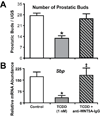
Figure 6. A WNT5A inhibitory antibody (anti-WNT5A-IgG) counteracts the effects of a selective ventral prostate inhibitor, TCDD, during mouse UGS development in vitro
E14.5 UGSs were cultured for 3 d in serum-free media containing 10 nM DHT and 0.1% DMSO (vehicle control, open bar) or these same two constituents and either TCDD (1 nM, gray-shaded bar) or TCDD + anti-WNT5A-IgG (gray-shaded, diagonally slashed bar). (A) Some cultured UGSs were imaged by scanning electron microscopy to assess total bud count. (B) Other cultured UGSs were grafted under the kidney capsule of nu/nu mice and grown for 28 d. Ventral prostate development was assessed by using real-time, RT-PCR to measure abundance of the ventral prostate-selective transcript, spermine binding protein (Sbp), in grafted tissues. Sbp abundance was normalized to peptidylprolyl isomerase A. Results are mean ± SEM of five UGS per treatment group. Differences between groups were determined by ANOVA followed by Fisher’s least significant difference test. The presence of “*” indicates significantly different from vehicle control and “‡” indicates significantly different from TCDD (p < 0.05).

References

    1. Benedict JC, Lin TM, Loeffler IK, Peterson RE, Flaws JA. Physiological role of the aryl hydrocarbon receptor in mouse ovary development. Toxicol Sci. 2000;56:382–388. - PubMed
    1. Castelo-Branco G, Sousa KM, Bryja V, Pinto L, Wagner J, Arenas E. Ventral midbrain glia express region-specific transcription factors and regulate dopaminergic neurogenesis through Wnt-5a secretion. Mol Cell Neurosci. 2006;31:251–262. - PubMed
    1. Cheng CW, Yeh JC, Fan TP, Smith SK, Charnock-Jones DS. Wnt5a-mediated non-canonical Wnt signalling regulates human endothelial cell proliferation and migration. Biochem Biophys Res Commun. 2008;365:285–290. - PubMed
    1. Cook C, Vezina CM, Allgeier SH, Shaw A, Yu M, Peterson RE, Bushman W. Noggin is required for normal lobe patterning and ductal budding in the mouse prostate. Dev Biol. 2007;312:217–230. - PMC - PubMed
    1. Cunha GR, Chung LW. Stromal-epithelial interactions--I. Induction of prostatic phenotype in urothelium of testicular feminized (Tfm/y) mice. J Steroid Biochem. 1981;14:1317–1324. - PubMed

Publication types