. 2008 Dec;55(8):1329-39.
doi: 10.1016/j.neuropharm.2008.08.028. Epub 2008 Aug 30.

Excitatory actions of peptide histidine isoleucine on thalamic relay neurons

Affiliations

Excitatory actions of peptide histidine isoleucine on thalamic relay neurons

Sang-Hun Lee et al. Neuropharmacology. 2008 Dec.

Abstract

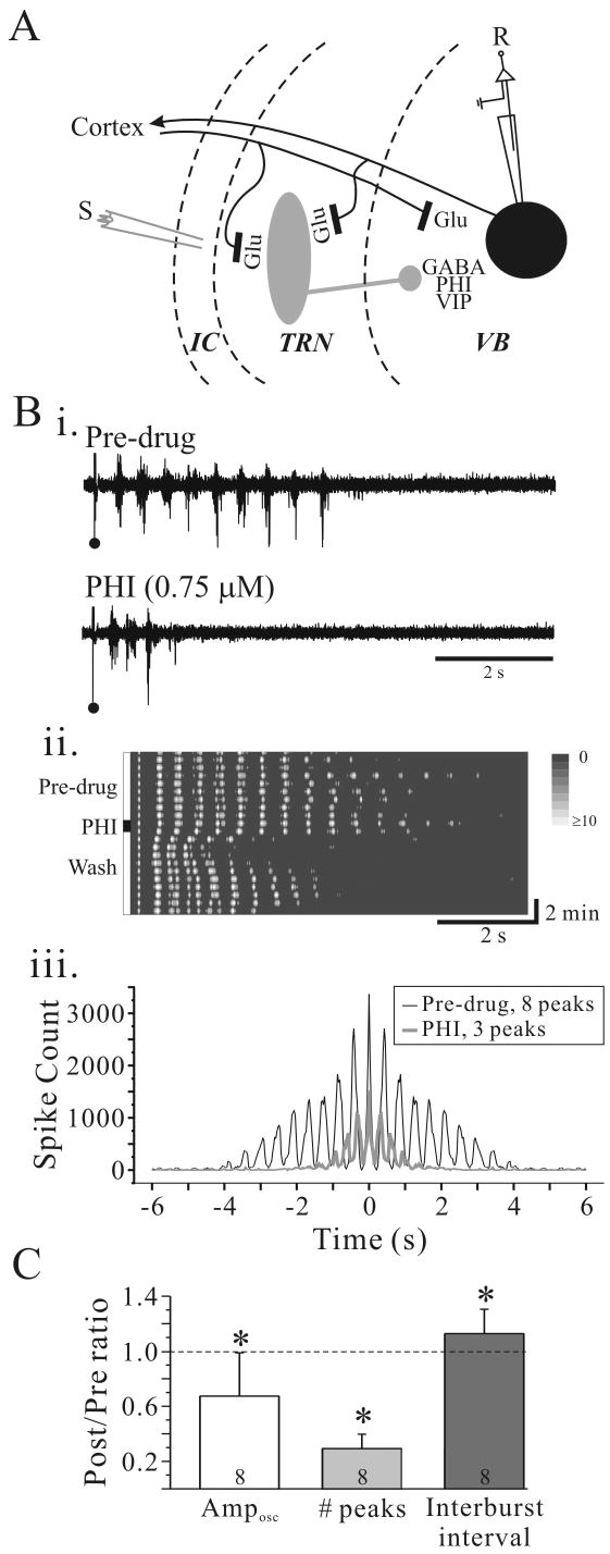
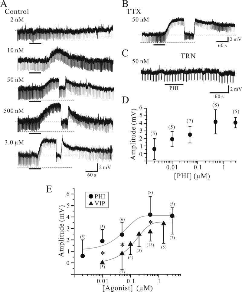
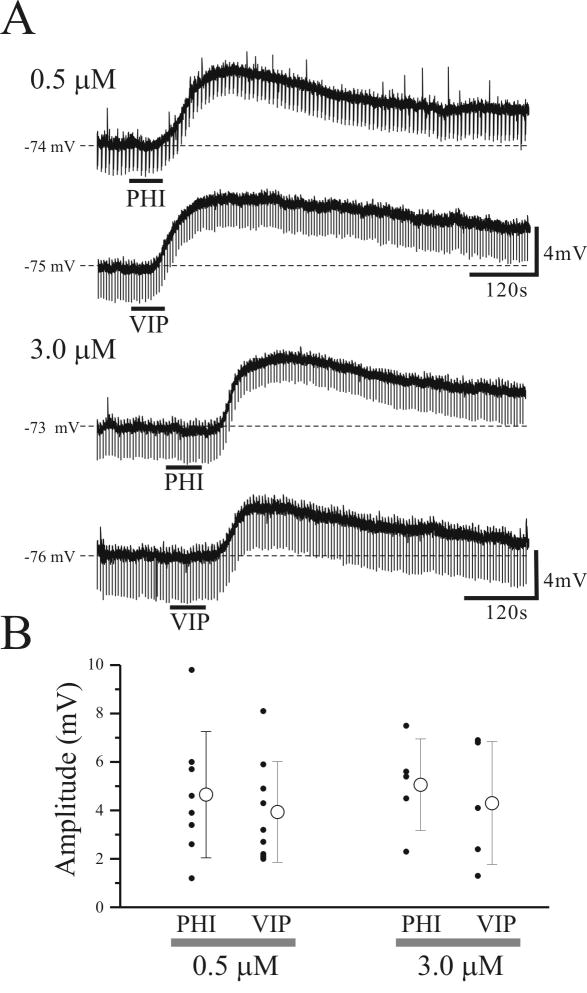
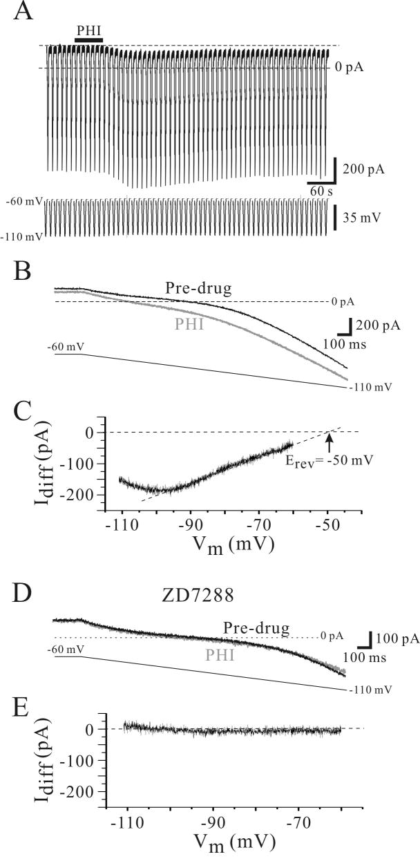
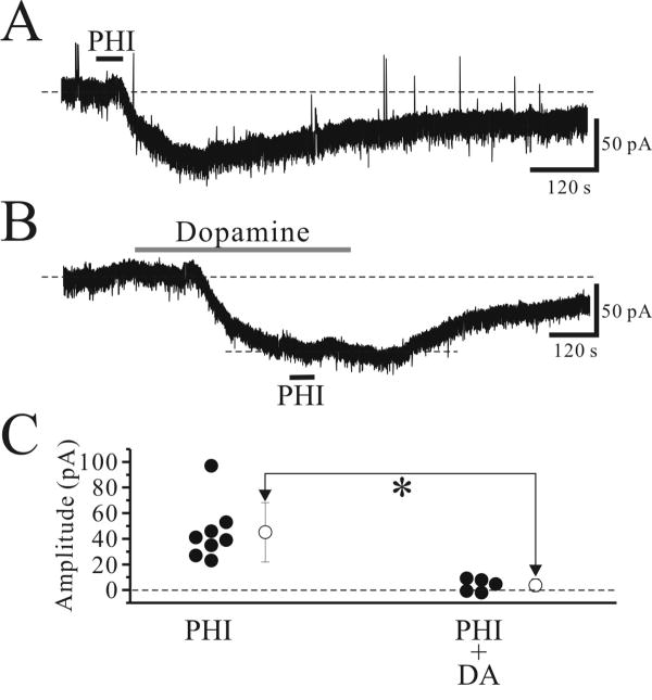
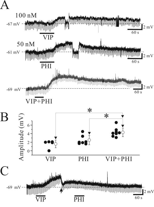
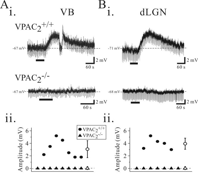
Peptide histidine isoleucine (PHI) and vasoactive intestinal peptide (VIP) are neuropeptides synthesized from a common precursor, prepro-VIP, and share structural similarity and biological functions in many systems. Within the central nervous system and peripheral tissues, PHI and VIP have overlapping distribution. PHI-mediated functions are generally via activation of VIP receptors; however, the potency and affinity of PHI for VIP receptors are significantly lower than VIP. In addition, several studies suggest distinct PHI receptors that are independent of VIP receptors. PHI receptors have been cloned and characterized in fish, but their existence in mammals is still unknown. This study focuses on the functional role of PHI in the thalamus because of the localization of both PHI and VIP receptors in this brain region. Using extracellular multiple-unit recording techniques, we found that PHI strongly attenuated the slow intrathalamic rhythmic activity. Using intracellular recording techniques, we found that PHI selectively depolarized thalamic relay neurons via an enhancement of the hyperpolarization-activated mixed cation current, Ih. Further, the actions of PHI were occluded by VIP and dopamine, indicating these modulators converge onto a common mechanism. In contrast to previous work, we found that PHI was more potent than VIP in producing excitatory actions on thalamic neurons. We next used the transgenic mice lacking a specific VIP receptor, VPAC2, to identify its possible role in PHI-mediated actions in the thalamus. PHI depolarized all relay neurons tested from wild-type mice (VPAC2(+/+)); however, in knockout mice (VPAC2(-/-)), PHI produced no change in membrane potential in all neurons tested. Our findings indicate that excitatory actions of PHI are mediated by VPAC2 receptors, not by its own PHI receptors and the excitatory actions of PHI clearly attenuate intrathalamic rhythmic activities, and likely influence information transfer through thalamocortical circuits.


Figures

Figure 1
Figure 1
PHI attenuates intrathalamic rhythmic activity. A. Simplified schematic illustrating thalamic circuitry with putative localization of PHI. Abbreviations: S, stimulus electrode; R, recording electrode; GABA, γ-aminobutyric acid; Glu, glutamate; PHI, peptide histidine isoleucine; VB, ventrobasal nucleus; TRN, thalamic reticular nucleus; IC, internal capsule. Bi. Extracellular multiple-unit recording from VB in a rat thalamic slice. In BMI (10 μM; Pre-drug), a single stimulus (•) in TRN evokes rhythmic discharge in VB. PHI (0.75 μM, 60 seconds) dramatically suppresses the rhythmic activity. Bii. Contour plot of experiment in Bi illustrates the time course of PHI effect on intrathalamic rhythmic activity. Prior to PHI application the rhythmic activity is very stable and lasts for many cycles. After PHI application, the rhythmic activity is dramatically attenuated, but returns near control levels within 5 minutes. Biii. Autocorrelogram of experiment in Bi illustrates a highly synchronized response that lasts nearly four seconds in control conditions (black trace). PHI (gray trace) reduces the numbers of peaks from 8 to 3. C. Summary of effects of PHI on oscillation amplitude (Amposc), number of peaks, and oscillation frequency. * p<0.05, **p<0.01.
Figure 2
Figure 2
PHI selectively depolarizes relay neurons. A. Intracellular recording from thalamic relay neurons reveal that PHI produces a concentration-dependent depolarization. Lowest PHI concentration (2 nM) produced no membrane depolarization. In a different relay neuron, higher PHI concentration (500 nM) produced a larger depolarization (4.2 mV). The depolarization is briefly interrupted by current injection to test for alteration in apparent input resistance. The downward deflections are voltage responses to hyperpolarizing current steps (10 pA, 500 ms, 0.2 Hz), and in this neuron PHI decreased input resistance by 12 %. Dotted lines indicate resting membrane potentials ranging -68 mV to -72 mV. B. In TTX (1 μM), PHI (500 nM) produces a similar membrane depolarization (6.2 mV). Upper dotted line indicates -71 mV. C. PHI (500 nM) produces no detectable changes in membrane potential or input resistance in a TRN neuron. Dotted line indicates -74 mV. D. Summary of all PHI-mediated membrane depolarizations of VB neurons. Cell counts for each concentration are listed in parentheses. E. Summary of PHI-mediated (●) and VIP-mediated (▲) membrane depolarizations of VB neurons. Solid gray lines indicate the sigmoidal function of each agonoist-mediated depolarizations. Cell counts for each concentration are listed in parentheses. *, p<0.05.
Figure 3
Figure 3
PHI and VIP depolarize relay neurons from adult rats (postnatal age: 40-55 days). A. Intracellular recording from thalamic relay neurons reveal that PHI (≥0.5μM) and VIP (≥0.5μM) produce a similar membrane depolarization in all VB relay neurons tested (n=27). B. Summary of all PHI- and VIP- mediated membrane depolarizations of VB neurons. Open circle symbol in the right indicates the mean ± SD of membrane depolarizations evoked by PHI and VIP.
Figure 4
Figure 4
PHI enhances Ih in a VB neuron. A. In voltage clamp recordings from a VB neuron, PHI (500 nM) produces an inward current associated with increase in membrane conductance. Slow ramped voltage commands (-60 to -110 mV, 4 s duration) are used to measure conductance before and after agonist application. B. Expanded traces of the membrane response to the ramped voltage command reveal not only the inward current, but the conductance increase by PHI (gray trace). Each trace consists of an average of 3 subsequent responses prior to and at the peak of the PHI-mediated inward current. C. The difference between the PHI (B, gray trace) and Pre-drug (B, black trace) is indicative of the PHI-sensitive current (Idiff). Extrapolation of the linear portion of this current (dotted line) indicates that the PHI-mediated current has a reversal potential (Erev) of -50 mV. D. The Ih blocker ZD7288 attenuates the PHI receptor-mediated inward current in the same neuron. In TTX (1.0 μM) and ZD7288 (100 μM), PHI does not produce an inward current or alter membrane conductance. E. The difference between the PHI and pre-drug clearly indicates the lack of PHI effect in ZD7288.
Figure 5
Figure 5
Excitatory actions of PHI are occluded by dopamine. A. In a voltage clamp recording of relay neurons (Vhold=-60mV), PHI (0.5 μM) produces an inward current. B. In a different relay neuron, DA (50 μM) produces an inward current that reaches a steady state, and at this point, subsequent PHI application produces a significantly smaller inward current. C. Summary of PHI-mediated inward currents in the presence and absence of DA. Open circle symbol in the right indicate the mean ± standard deviation of inward currents evoked by PHI. *, p<0.05.
Figure 6
Figure 6
Additive effects of submaximal responses of VIP and PHI in thalamic relay neurons. A. PHI (0.05 μM) and VIP (0.1 μM) produced small membrane depolarizations when applied separately. In another cell, combined application of PHI (0.05 μM) and VIP (0.1 μM) produced a significantly larger depolarization than the individual agonists, and is similar to that the sum of each agonists-induced membrane depolarizations. B. Population data illustrating individual and combined agonist applications. Open circle symbol in the right indicates the mean ± standard deviation of membrane depolarizations evoked by agonists. *, p<0.05. C. High VIP concentration (1 μM) occludes PHI-mediated membrane depolarizations. At the peak of VIP-mediated depolarization (arrowhead), the membrane potential was manually adjusted to resting levels, and PHI (0.5 μM) was applied. Under these conditions, PHI produced no changes in membrane potential.
Figure 7
Figure 7
PHI-mediated depolarizations are dependent on VPAC2 receptor activation in the mouse thalamus. Ai. Intracellular recording from a VB neuron in a slice from a wildtype mouse (VPAC2+/+) reveals that PHI (500 nM) evokes a long-lasting, membrane depolarization associated with a decrease in input resistance (top trace). In a slice from a knock-out (VPAC2-/-) animal, PHI does not alter membrane potential (bottom trace). Aii. Population data illustrating membrane depolarizations produced by PHI in VB neurons from VPAC2+/+ (n = 7, solid circles) and VPAC2-/- (n = 8, solid triangles) animals. Bi. Intracellular recordings from VPAC2+/+ dLGN relay neurons reveals that PHI (500 nM) evokes membrane depolarizations (top trace). In dLGN neurons from VPAC2-/- animal, PHI do not alter membrane potentials (bottom trace). Bii. Population data illustrating membrane depolarizations produced in dLGN neurons from VPAC2+/+ (n = 5) and VPAC2-/- (n = 7) slices. Open circle symbol in the right indicates the mean ± standard deviation of membrane depolarizations evoked in VPAC2+/+ mice.

Similar articles

Cited by

References

    1. Anagnostides AA, Manolas K, Christofides ND, Yiangou Y, Welbourn RB, Bloom SR, Chadwick VS. Peptide histidine isoleucine (PHI). A secretagogue in porcine intestine. Dig Dis Sci. 1983;28:893–896. - PubMed
    1. Bailey CJ, Wilkes LC, Conlon JM, Armstrong PH, Buchanan KD. Effects of gastric inhibitory polypeptide, vasoactive intestinal polypeptide and peptide histidine isoleucine on the secretion of hormones by isolated mouse pancreatic islets. J Endocrinol. 1990;125:375–379. - PubMed
    1. Bal T, Debay D, Destexhe A. Cortical feedback controls the frequency and synchrony of oscillations in the visual thalamus. J Neurosci. 2000;20:7478–7488. - PMC - PubMed
    1. Bal T, von Krosigk M, McCormick DA. Role of the ferret perigeniculate nucleus in the generation of synchronized oscillations in vitro. J Physiol (Lond) 1995a;483:665–685. - PMC - PubMed
    1. Bal T, von Krosigk M, McCormick DA. Synaptic and membrane mechanisms underlying synchronized oscillations in the ferret lateral geniculate nucleus in vitro. J Physiol (Lond) 1995b;483:641–663. - PMC - PubMed

Publication types

MeSH terms

LinkOut - more resources