. 2008 Sep;15(3):416-425.
doi: 10.1016/j.devcel.2008.07.010.

A subcortical maternal complex essential for preimplantation mouse embryogenesis

Affiliations

A subcortical maternal complex essential for preimplantation mouse embryogenesis

Lei Li et al. Dev Cell. 2008 Sep.

Abstract

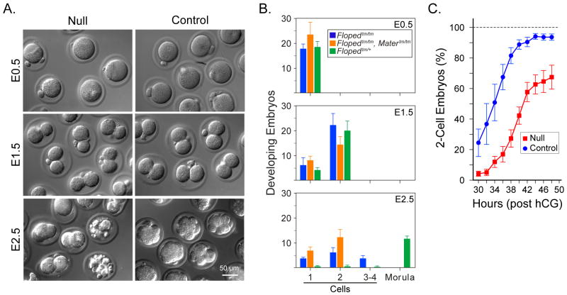
We have identified a subcortical maternal complex (SCMC) that assembles during oocyte growth and is essential for zygotes to progress beyond the first embryonic cell divisions. At least four maternally encoded proteins contribute to this MDa complex: FLOPED, MATER, and TLE6 interact with each other while Filia binds independently to MATER. Although the transcripts encoding these proteins are degraded during meiotic maturation and ovulation, the SCMC proteins persist in the early embryo. The SCMC, located in the subcortex of eggs, is excluded from regions of cell-cell contact in the cleavage-stage embryo and segregates to the outer cells of the morulae and blastocyst. Floped(tm/tm) and/or Mater(tm/tm) eggs lack the SCMC but can be fertilized. However, these embryos do not progress beyond cleavage stage development and female mice are sterile. The proteins are conserved in humans, and similar maternal effect mutations may result in recurrent embryonic loss.


Figures

Figure 1
Figure 1. Expression of Floped and Flopedtm/tm mouse lines
(A). Quantitative reverse-transcription, polymerase chain reaction (qRT-PCR) with total RNA extracted from newborn brain (Br), heart (He), intestines (In), kidney (Ki), liver (Li), lung (Lu), muscle (Mu), ovary (Ov), spleen (Sp), testis (Te), uterus (Ut) and pancreas (Pa) expressed as a percent of GAPDH. (B). In situ hybridization of fixed, paraffin-embedded 4 μm ovarian sections probed with DIG-labeled antisense (left) or sense (right) synthetic Floped oligonucleotides. Scale bar, 50 μm. (C). qRT-PCR of Floped (blue bars) and Figla expression (grey background) using total RNA isolated at embryonic day 12.5 (E12.5) to E19.5, newborn (NB), 1–7 days post-partum (dpp) and at six weeks (6wk). (D). Eggs and two-cell embryos were isolated, fixed and stained with peptide-purified antibodies to FLOPED or with Hoechst and phalloidin which bind to DNA and F-actin, respectively. Morphology of eggs and early embryos was observed with differential interference contrast (DIC). (E). Total ovarian RNA was primed with oligo dT and PCR with P1 and P2 primers (Supplemental Data) produced a 229 bp band in normal (+/+) and heterozygote (+/−), but not in Floped null (−/−) mice (left). RT-PCR with P1 and P3 primers produced a 361 bp band in null (−/−) and in heterozygote (+/−), but not in normal mice (right). M, molecular mass markers. (F). Immunoblots of total ovarian extract (20 μg) and 10 ovulated eggs from heterozygous (+/−) or homozygous (−/−) Floped null mice were probed with anti-FLOPED antibody. (G). Plastic embedded ovarian sections from homozygous (top) and control heterozygous (bottom) Floped null mice. (H). Ovulated eggs from hormonally stimulated homozygous (top) and control heterozygous (bottom,) Floped null mice were imaged by DIC.
Figure 2
Figure 2. Phenotype of embryos derived from Flopedtm/tm female mice
(A). Embryos were flushed from the oviducts of Floped null and heterozygous controls at E0.5, E1.5 and E2.5. (B). In vivo progression of pre-implantation embryos from (A) was quantified from at least five females and expressed as the average ±s.e.m. Embryos derived from nine Flopedtm/tm, Matertm/tm and double mutant females were isolated at E0.5 and cultured in vitro for one (E1.5) or two (E2.5) days. (C). Homozygous and heterozygous (control) Floped null females were mated with normal males to isolate one-cell embryos 30 hours after hCG administration. Embryos (174 from Floped+/tm and 132 from Flopedtm/tm females) were cultured an additional 18 hours and progression to two-cell embryos was assessed morphologically. Data is the percent of two-cell embryos (average ± s.e.m.) observed at two hour intervals.
Figure 3
Figure 3. Identification of FLOPED binding partners
(A). Ovarian lysates from normal and Floped null mice were precipitated with peptide-purified, rabbit anti-FLOPED antibody. Immunoprecipitates were separated by SDS-PAGE and stained with colloidal blue. Arrows indicate protein bands present in normal, but not null extracts and represent the relative mobility of MATER (1), TLE6 (2), Filia (3) and FLOPED (4). Molecular mass (kDa) indicated on left. (B). Tissue-specific expression of Tle6 determined by qRT-PCR of total RNA using TLE6 specific primers and probes. Inset, in situ hybridization with DIG-labeled antisense synthetic Tle6 oligonucleotides. (C). Developmental expression profile of Tle6. (D). Poly(A)+ RNA was isolated from oocytes/eggs/embryos, reversed transcribed with oligo dT and aliquots were analyzed by qRT-PCR using synthetic oligonucleotide primers and TaqMan® probes specific to Floped, Tle6, and Mater transcripts. Results were normalized to the abundance in full grown (80 μm) oocytes. (E). Immunoblot of lysates isolated from 10 growing oocytes, eggs and pre-implantation embryos were probed with antibodies specific to FLOPED, MATER, TLE6 or β-actin as a load control.
Figure 4
Figure 4. Interactions of FLOPED, MATER, TLE6 and Filia
(A). Ovarian lysates, before (Input) or after immunoprecipitation with antibodies to FLOPED (α-FLP) or TLE6 (α-TLE) were immunoblotted and probed with antibodies to FLOPED, MATER, TLE6 and Filia. Abbreviations include: Norm (normal) and Null (Flopedtm/tm) ovarian lysates; α-FLP (rabbit anti-FLOPED antibody); α-TLE (rabbit anti-TLE6 antibody); IgG (normal rabbit immunoglobulin), as a negative control. (B). FLOPED-EGFP and either MATER-myc, TLE6 or Filia-HA expression vectors were co-transfected (Co-Txfect) into COS cells. Lysates before (Input) or after immunoprecipitation with control immunoglobulin (IgG) an antibody to EGFP (α-EGFP) were used to isolate FLOPED and associated proteins. Immunoblots were probed with antibodies to myc, TLE6 and HA (hemaglutinin) to detect MATER, TLE6 and Filia, respectively. (C). Same as (B) except that MATER with either FLOPED, TLE6 or Filia were co-transfected into COS cells; antibodies to myc (α-myc) were used to immunoprecipitate MATER and associated proteins. Immunoblots were probed with appropriate antibodies to detect FLOPED, TLE6 and Filia. (D). Same as (B) except that TLE6 with either FLOPED, MATER or Filia were co-transfected into COS cells; antibodies to TLE6 (α-TLE6) were used to immunoprecipitate TLE and associated proteins. Immunoblots were probed with appropriate antibodies to detect FLOPED, MATER and Filia. (E). Same as (B) except that Filia with either FLOPED, MATER or TLE6 were co-transfected into COS cells; antibodies to HA (α-HA) were used to immunoprecipitate Filia and associated proteins. Immunoblots were probed with appropriate antibodies to detect FLOPED, MATER and TLE6. (F). Normal egg lysates (150) were chromatographed by FPLC on a Superose 6 10/300 GL column with a void volume (V0) of ~2 × 10⁶ Da (upper). Immunoblots of individual 1 ml fractions (lower) detected FLOPED (fractions 8–16), MATER (fractions 8–14), TLE6 (fractions 8–14) and Filia (fractions 8–16). The data (average of two experiments) was quantified by fluorescent image analysis and the peak values in fraction 9 were set at a relative intensity of 100%. Elution of protein standards are indicated by arrows.
Figure 5
Figure 5. Localization of the SCMC in eggs and pre-implantation embryos
(A). Confocal microscopic images of eggs and pre-implantation embryos after permeabilization and incubation with antibodies to FLOPED, MATER, and TLE6. Co-localization of FLOPED, MATER and TLE6 was observed in the merge. (B). At E3.5, control (Tead4+/tm) and Tead4tm/tm embryos were isolated from the oviduct of mated mice, permeabilized and incubated with antibodies to TLE6 and OCT4 (epiblast marker) or (C) with antibodies to FLOPED and OCT4. In all eggs and embryos, DNA was visualized with Hoechst and morphology was determined by DIC.
Figure 6
Figure 6. Formation of the SCMC depends on FLOPED and MATER
(A). Eggs isolated from normal, Flopedtm/tm or Matertm/tm mice were fixed, permeabilized and incubated with antibodies to FLOPED and phalloidin to detect F-actin. (B) Same as (A) except with antibodies to MATER. (C) Same as (A) except with antibodies to TLE6. (D) Same as (A) except with antibodies to Filia. (E). Egg lysates (10) from control (normal), Flopedtm/tm or Matertm/tm mice were immunoblotted and probed with antibodies specific to FLOPED, MATER, TLE6, Filia and β-actin. (F). Flopedtm/tm oocytes were injected with a plasmid independently expressing FLOPED and GFP (positive transcription/translation control). After ~2 days in culture, oocytes were fixed and imaged by confocal microscopy for FLOPED, Filia and GFP as well as by DIC. (G). A model of the subcortical maternal complex (SCMC) formed during oogenesis that is required for cleavage stage embryogenesis. The SCMC is composed of FLOPED, MATER, TLE6 and Filia. The first three proteins (FLOPED, TLE6, MATER) interact directly with one another and Filia interacts with MATER, but not FLOPED or TLE6. The size of each protein corresponds to its predicted molecular mass.

References

    1. Aronson J, Solter D. Developmental potency of gametic and embryonic genomes revealed by nuclear transfer. Curr Top Dev Biol. 1987;23:55–71. - PubMed
    1. Bajoghli B. Evolution of the Groucho/Tle gene family: gene organization and duplication events. Dev Genes Evol. 2007;217:613–618. - PubMed
    1. Bischoff M, Parfitt DE, Zernicka-Goetz M. Formation of the embryonic-abembryonic axis of the mouse blastocyst: relationships between orientation of early cleavage divisions and pattern of symmetric/asymmetric divisions. Development. 2008;135:953–962. - PMC - PubMed
    1. Bolton VN, Oades PJ, Johnson MH. The relationship between cleavage, DNA replication, and gene expression in the mouse 2-cell embryo. J Embryol Exp Morphol. 1984;79:139–163. - PubMed
    1. Bultman SJ, Gebuhr TC, Pan H, Svoboda P, Schultz RM, Magnuson T. Maternal BRG1 regulates zygotic genome activation in the mouse. Genes Dev. 2006;20:1744–1754. - PMC - PubMed

Publication types

MeSH terms