. 2008 Nov 28;383(5):1181-94.
doi: 10.1016/j.jmb.2008.09.006. Epub 2008 Sep 12.

Reconstituting initial events during the assembly of apolipoprotein B-containing lipoproteins in a cell-free system

Affiliations

Reconstituting initial events during the assembly of apolipoprotein B-containing lipoproteins in a cell-free system

Z Gordon Jiang et al. J Mol Biol. .

Abstract

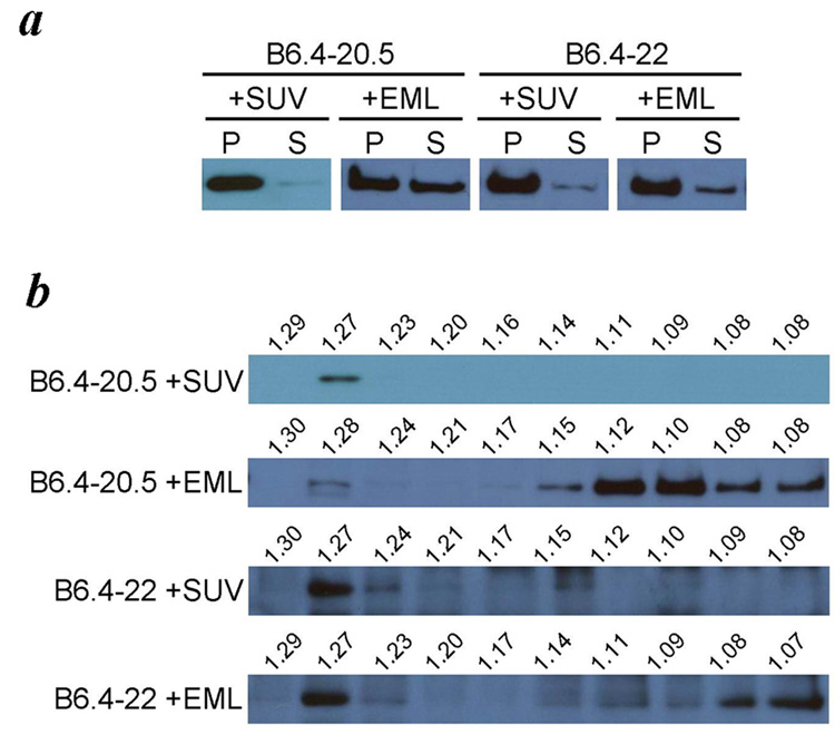
The synthesis of apolipoprotein B (apoB) dictates the formation of chylomicrons and very low-density lipoproteins, two major lipoprotein precursors in the human plasma. Despite its biological significance, the mechanism of the assembly of these apoB-containing lipoproteins remains elusive. An essential obstacle is the lack of systems that allow fine dissection of key components during assembly, including nascent apoB peptide, lipids in defined forms, chaperones, and microsomal triglyceride transfer protein (MTP). In this study, we used a prokaryotic cell-free expression system to reconstitute early events in the assembly of apoB-containing lipoprotein that involve the N-terminal domains of apoB. Our study shows that N-terminal domains larger than 20.5% of apoB (B20.5) have an intrinsic ability to remodel vesicular phospholipid bilayers into discrete protein-lipid complexes. The presence of appropriate lipid substrates during apoB translation plays a pivotal role for successful lipid recruitment, and similar lipid recruitment fails to occur if the lipids are added posttranslationally. Cotranslational presence of MTP can dramatically promote the folding of B6.4-20.5 and B6.4-22. Furthermore, apoB translated in the presence of MTP retains its phospholipid recruitment capability posttranslationally. Our data suggest that during the synthesis of apoB, the N-terminal domain has a short window for intrinsic phospholipid recruitment, the time frame of which is predetermined by the environment where apoB synthesis occurs. The presence of MTP prolongs this window of time by acting as a chaperone. The absence of either proper lipid substrate or MTP may result in the improper folding of apoB and, consequently, its degradation.


Figures

Fig. 1
Fig. 1. Lipid analysis of two cell-free expression systems
Lipids extracted from the cell lysate for one reaction of the Expressway (Invitrogen) and EasyXpress systems (Qiagen) were compared with thin layer chromatography, as described in the experimental procedures section. A lipid marker containing common lipid species is shown on the left.
Fig. 2
Fig. 2. Domain diagram of apoB
The proposed domain structure of the N-terminal region of apoB is compared with that of lipovitellin. Sequences missing in the lipovitellin crystal structure are shown in white boxes. In the diagram of apoB, disulfide bond linkages are shown with orange bridges, and N-linked glycosylation sites are shown with green hexagons. A scale with both percentage of B100 and amino acid number is shown below the diagram of apoB. Also listed are the three constructs used in this study – B6.4–17, B6.4–20.5 and B6.4–22.
Fig. 3
Fig. 3. Cotranslational cell-free apoB expression
a. Comparison of the solubility of B6.4–17, B6.4–20.5 and B6.4–22. Each construct was expressed in the cell-free system in the absence of additional lipids. The cell pellet (P) and supernatant (S) were separated by centrifugation and analyzed by western blot using anti-His antibody. The expression of B6.4–20.5 in the presence of RNase A was included as a negative control. b. Density of apoB expressed with lipids. Supernatant of the ApoB expressed without lipids, with SUV or with emulsion (EML) were further separated by potassium bromide density gradient centrifugation and analyzed by western blot using anti-His antibody. The density of each fraction was determined by measuring its refractory index, and the calculated density is labeled above each lane.
Fig. 4
Fig. 4. Posttranslational incubation with lipids
B6.4–20.5 and B6.4–22 were incubated with SUV (0.6 mg/ml) or emulsion (EML) (1.8 mg/ml) after their cell free expression. The supernatants were separated by potassium bromide density gradient centrifugation and analyzed by western blot using anti-His antibody. The density of each fraction was determined by measuring its refractory index, and the calculated density is labeled above each lane.
Fig. 5
Fig. 5. Refolding of B6.4–20.5 and B6.4–22 in the presence of lipids
B6.4–20.5 and B6.4–22 were refolded in the presence of SUV or emulsion at 8 or 24 times the protein weight concentration, respectively. Arginine and glutathione that had assisted in the protein refolding were removed by dialysis, and the products were separated on a potassium bromide gradient. Density separated fractions were analyzed on a dot blot using anti-His antibody. The density of each fraction was determined by measuring its refractory index, and the calculated density is labeled above each lane.
Fig. 6
Fig. 6. Electron microscopy imaging of lipids and protein lipid complexes
a. Egg PC SUVs. A high density ring with a thickness of 5 nm is characteristic for unilamellar vesicles. b. Egg PC / Triolein emulsions. Emulsion particles have a homogenous density. In this view, many emulsion particles are adsorbed on the grid (top third of the image), and only a few were trapped in the vitreous ice. Egg PC vesicles are sometimes found in the emulsion sample. c. B6.4–20.5 refolded with SUV. Large unilamellar vesicles are observed in this sample, likely due to the fusion of unstable small unilamellar vesicles overtime. d. B6.4–20.5 refolded with emulsions. The appearance of most particles is similar to pure emulsion (b). It is noteworthy that samples in c and d had been purified by Ni-NTA resin, thus proteins were present on all lipid particles. e. and f. B6.4–20.5 expressed with SUV in the cell-free system. The sample was analyzed by negative staining electron microscopy in e. Samples imaged by cryo-electron microscopy in f. is less concentrated on the grid and often contains impurities from liquid ethane. This contamination can be differentiated by a characteristic white ring on the edge, suggesting their presence on the surface of the vitreous ice. Circled particles are the identified complex containing B6.4–20.5 and egg PC. The arrow points to a vesicle with additional densities on its surface, probably representing B6.4–20.5 particle. g. A collage of 18 cropped images containing B6.4–20.5 translated in the presence of egg PC particles from 8 negative films. These particles adopt relatively uniform shape and dimension. Preparation of samples for electron microscopy is described in the experimental procedures section. All grids were prepared with cryo method, except for the sample in e, which was stained by 1% phosphotunstate. Images were acquired at different magnifications, and scale bars are shown in each image.
Fig. 7
Fig. 7. Role of MTP for protein folding and lipid binding
a. The effect of MTP on B6.4–20.5 expression. MTP was added either cotranslationally or posttranslationally during B6.4–20.5 expression. RNase inhibitor increases the expression yield when MTP was present cotranslationally. b. The effect of MTP on the lipid binding of B6.4–20.5. Four combinations of MTP and SUV during B6.4–20.5 translation were tested, and their respective outcome on protein density were examined with potassium bromide density gradient and western blot using anti-His antibody: cotranslational presence of MTP (B6.4–20.5 w/MTP), posttranslational incubation of SUV (B6.4–20.5 + SUV), posttranslational incubation of both MTP and SUV (B6.4–20.5 + MTP & SUV) and cotranslational presence of MTP and posttranslational incubation of SUV (B6.4–20.5 w/MTP + SUV). The density of each fraction was determined by measuring its refractory index, and the calculated density is labeled above each lane.
Fig. 8
Fig. 8. Model of B20.5
a. A homology model of B20.5. The homology model of B20.5 was built upon the crystal structure of lipovitellin as described in the experimental procedures section. The β-barrel domain is shown in green, α-helical in cyan, C-sheet in red, and A-sheet in blue. b. A simplified cartoon for the folding topology of B20.5, with a proposed lipid binding site shown as an orange sphere. c. A proposed mechanism during the initiation of lipid binding. The nascent apoB polypeptide tested in our system beginning from the α-helical domain has an intrinsic lipid binding and remodeling property, which can break the appropriate lipid bilayer substrates, such as egg PC SUVs into discrete protein/lipid complexes. However, in the absence of such lipid substrates, the protein misfolds and irreversibly aggregates. MTP during apoB expression facilitates apoB folding even in the absence of appropriate lipid substrate, and retains the lipid binding capability of apoB.

References

    1. Knott TJ, Rall SC, Jr, Innerarity TL, Jacobson SF, Urdea MS, Levy-Wilson B, Powell LM, Pease RJ, Eddy R, Nakai H, et al. Human apolipoprotein B: structure of carboxyl-terminal domains, sites of gene expression, and chromosomal localization. Science. 1985;230:37–43. - PubMed
    1. Chen SH, Habib G, Yang CY, Gu ZW, Lee BR, Weng SA, Silberman SR, Cai SJ, Deslypere JP, Rosseneu M, et al. Apolipoprotein B-48 is the product of a messenger RNA with an organ-specific in-frame stop codon. Science. 1987;238:363–366. - PubMed
    1. Powell LM, Wallis SC, Pease RJ, Edwards YH, Knott TJ, Scott J. A novel form of tissue-specific RNA processing produces apolipoprotein-B48 in intestine. Cell. 1987;50:831–840. - PubMed
    1. Fisher EA, Ginsberg HN. Complexity in the secretory pathway: the assembly and secretion of apolipoprotein B-containing lipoproteins. J Biol Chem. 2002;277:17377–17380. - PubMed
    1. Yao Z, Tran K, McLeod RS. Intracellular degradation of newly synthesized apolipoprotein B. J Lipid Res. 1997;38:1937–1953. - PubMed

Publication types

Substances

LinkOut - more resources