. 2008 Nov 25;26(50):6405-14.
doi: 10.1016/j.vaccine.2008.08.069. Epub 2008 Sep 18.

Immune modulating effect by a phosphoprotein-deleted rabies virus vaccine vector expressing two copies of the rabies virus glycoprotein gene

Affiliations

Immune modulating effect by a phosphoprotein-deleted rabies virus vaccine vector expressing two copies of the rabies virus glycoprotein gene

Jonathan Cenna et al. Vaccine. .

Abstract

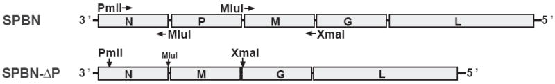
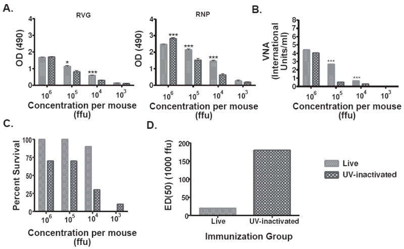
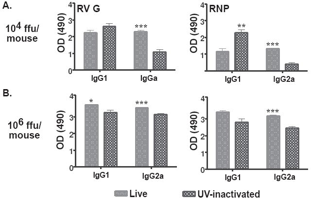
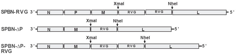
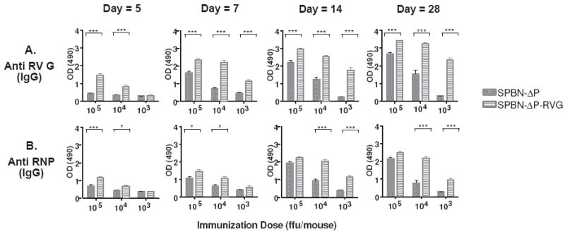
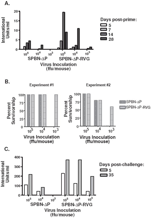
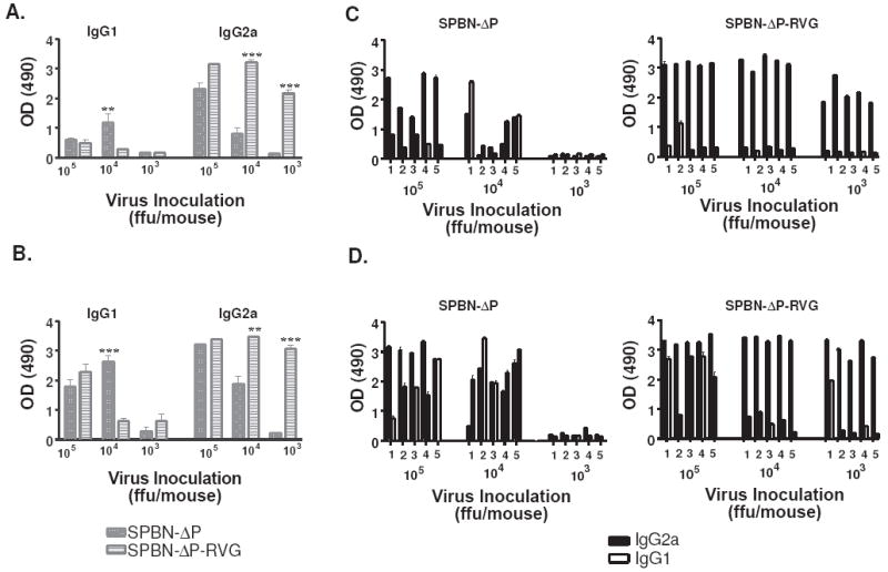
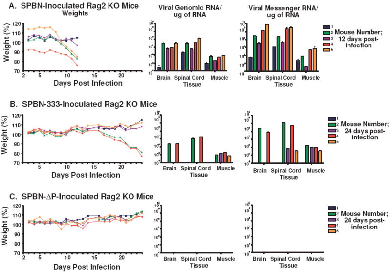
The type of immune response induced by a vaccine is a critical factor that determines its effectiveness in preventing infection or disease. Inactivated and live rabies virus (RV) vaccine strains elicit an IgG1-biased and IgG1/IgG2a-balanced antibody response, respectively. However, IgG2a antibodies are potent inducers of anti-viral effector functions, and therefore, a viral vaccine vector that can elicit an IgG2a-biased antibody response may be more effective against RV infection. Here we describe the humoral immune response of a live replication-deficient phosphoprotein (P)-deleted RV vector (SPBN-DeltaP), or a recombinant P-deleted virus that expresses two copies of the RV glycoprotein (G) gene (SPBN-DeltaP-RVG), and compare it to a UV-inactivated RV. Mice inoculated with UV-inactivated RV induced predominantly an IgG1-specific antibody response, while live recombinant SPBN-DeltaP exhibited a mixed IgG1/IgG2a antibody response, which is consistent with the isotype profiles from the replication-competent parental viruses. Survivorship in mice after pathogenic RV challenge indicates a 10-fold higher efficiency of live SPBN-DeltaP compared to UV-inactivated SPBN-DeltaP. In addition, SPBN-DeltaP-RVG induced a more rapid and robust IgG2a response that protected mice more effectively than SPBN-DeltaP. Of note, 10(3)ffu of SPBN-DeltaP-RVG-induced anti-RV antibodies that were 100% protective in mice against pathogenic RV challenge. The increased immune response was directed not only against RV G but also against the ribonucleoprotein (RNP), indicating that the expression of two RV G genes from SPBN-DeltaP-RVG enhances the immune response to other RV antigens as well. In addition, Rag2 mice inoculated intramuscularly with 10(5)ffu/mouse of SPBN-DeltaP showed no clinical signs of rabies, and no viral RNA was detected in the spinal cord or brain of inoculated mice. Therefore, the safety of the P-deleted vectors along with the onset and magnitude of the IgG2a-induced immune response by SPBN-DeltaP-RVG indicate that this vector holds great promise as either a therapeutic or preventative vaccine against RV or other infectious diseases.


Figures

Figure 1
Figure 1
Construction of SPBN-ΔP. The P gene was deleted from SPBN (28) (top), which is based on the attenuated SAD-B19 RV vaccine strain, using a PCR strategy and standard cloning techniques. Infectious virus was recovered and propagated on BSR cells stably expressing RV P, as described in Materials and Methods
Figure 2
Figure 2
Live SPBN-ΔP induces high levels of anti-RV G and anti-RNP antibodies, and protects mice more effectively against pathogenic RV challenge than UV-inactivated RV. Groups of 10 Balb/c mice were immunized intramuscularly (i.m.) with the indicated doses of live or UV-inactivated SPBN-ΔP and sera from mice 14 days post-immunization were tested for total IgG antibodies against RV G or RNP by ELISA at a 1:50 sera dilution (A), or virus neutralizing antibodies (VNA) expressed in international units/ml (IU/ml) (B). The data represents the average and standard error of the mean (SEM) of n=10 completed in duplicate and analyzed using the unpaired t-test, 95% confidence (see the text for p values). Four weeks post-immunization, the mice were challenged i.m. with 100 LD50 of pathogenic Challenge Virus Strain (CVS)-N2c and observed for three weeks and their survivorship recorded (C). The ED50 was calculated using the survivorship rates between the two groups of immunized animals (D) and indicates a 10-fold higher efficiency of live SPBN-ΔP versus UV-inactivated SPBN-ΔP. (***p<0.001; **p=0.001-0.01; *p=0.01-0.05)
Figure 3
Figure 3
Live SPBN-ΔP induces a mixed IgG1/IgG2a antibody titer compared with an IgG1-biased response from UV-inactivated RV. Fourteen days post-immunization, sera from mice that received either 104 (A) or 106 ffu (B) of live SPBN-ΔP or UV-inactivated RV, as described in Figure 2, were tested by ELISA for anti-RV G (left) or anti-RNP (right) antibodies at a 1:50 dilution. The data shows an increase in IgG2a-specific antibody titers against both RV G and RNP at both doses used of live SPBN-ΔP compared with UV-inactivated RV. The data represents the average and SEM of n=10 completed in duplicate and analyzed using the unpaired t-test, 95% confidence (p values provided in the text; ***p<0.001; **p=0.001-0.01; *p=0.01-0.05).
Figure 4
Figure 4
Construction of SPBN-ΔP-RVG. Two copies of the RV G gene were digested from SPBN-RVG (top) using the two unique restriction sites, XmaI and NheI. The two RV G genes were inserted into SPBN-ΔP (middle) also digested with XmaI and NheI, resulting in SPBN-ΔP-RVG (bottom).
Figure 5
Figure 5
SPBN-ΔP-RVG induces rapid and potent anti-RV G and anti-RNP antibodies. Groups of five Balb/c mice were immunized i.m. with 105, 104,, or 103 ffu of SPBN-ΔP or SPBN-ΔP-RVG and sera was collected on days 5, 7, and 14 post-immunization (Experiment #1). The experiment was repeated with an additional five mice, except in this experiment, sera were collected on days 5, 7, 14 and 28 post-immunization. At each time point, sera were tested for total IgG by ELISA for anti-RV G (A) or anti-RV RNP (B) antibodies. The data for days 5, 7, and 14 represent the average and SEM for both Experiments #1 and #2 (n=10) completed in duplicate and for day 28 from Experiment #2 (n=5) also completed in duplicate. The data were analyzed using the unpaired t-test, 95% confidence (see the text for p values; ***p<0.001; **p=0.001-0.01; *p=0.01-0.05).
Figure 6
Figure 6
SPBN-ΔP-RVG induces strong virus neutralization antibody titers and provides protection against pathogenic RV challenge with as little as 103 ffu/mouse. The mice described in Figure 5 were challenged i.m. 4 (Experiment #1) or 6 (Experiment #2) weeks post-immunization with 100 LD50 pathogenic CVS-N2c. Pre-challenge virus neutralizing antibody titers, expressed in international units/ml, are shown (A), and survivorship was recorded (B). Virus neutralizing antibodies were also determined from all mice on day 5 post-challenge and from mice that survived at day 35 post-challenge.
Figure 7
Figure 7
SPBN-ΔP-RVG induces an IgG2a-biased antibody response, which is amplified post-challenge. Sera from mice collected 14 days post-immunization (A) or 5 days post-challenge (B) were analyzed by ELISA for IgG1- or IgG2a-specific antibodies against RV G at a sera dilution of 1:50. The average and SEM antibody results from each group of mice (n=5) (A and B) or from individual mice (C and D) are shown. Data were analyzed using the unpaired t-test, 95% confidence (see the text for p values; (***p<0.001; **p=0.001-0.01; *p=0.01-0.05).
Figure 8
Figure 8
Input RV P is sufficient for cell surface expression of RV G. NA cells were infected with SPBN (dotted line), SPBN-ΔP (solid line) or SPBN-ΔP-RVG (dashed line) at an moi of 3, or left uninfected (solid histogram). At 24 (top) and 48 h p.i. (bottom), cells were incubated with a polyclonal rabbit anti-RV G antiserum, followed by a Cy-2-conjugated anti-rabbit antibody. Surface expression was determined by flow cytometry, and fluorescence intensity was plotted.
Figure 9
Figure 9
SPBN-ΔP does not spread to the spinal cord or brain of infected RAG2 KO mice. Groups of 5 RAG2 KO mice were inoculated i.m. with 105 ffu of either SPBN (A), SPBN-333 (B) or SPBN-ΔP (C) and their weights were recorded (left panel). Once one or more animal(s) in a group became moribund due to rabies infection, all mice in that group were euthanized. Mice inoculated with the highly attenuated SPBN-333 vector served as a guide for the time point to collect samples from mice inoculated with SPBN-ΔP since it was not known whether SPBN-ΔP would induce visible signs of rabies infection. Total RNA was isolated from muscle, brain and spinal cord and viral RT-PCR and quantitative real-time PCR was used to amplify and quantify viral genomic (middle panels) or messenger (right panels) RNA from individual samples.

References

    1. Knobel DL, Cleaveland S, Coleman PG, et al. Re-evaluating the burden of rabies in Africa and Asia. Bull World Health Organ. 2005;83(5):360–8. - PMC - PubMed
    1. McGettigan JP, Pomerantz RJ, Siler CA, et al. Second-generation rabies virus-based vaccine vectors expressing human immunodeficiency virus type 1 gag have greatly reduced pathogenicity but are highly immunogenic. Journal of Virology. 2003;77(1):237–44. - PMC - PubMed
    1. McKenna PM, Koser ML, Carlson KR, et al. Highly attenuated rabies virus-based vaccine vectors expressing simian-human immunodeficiency virus89.6P Env and simian immunodeficiency virusmac239 Gag are safe in rhesus macaques and protect from an AIDS-like disease. Journal of Infectious Diseases. 2007;195(7):980–8. - PubMed
    1. Pirofski LA, Casadevall A. Use of licensed vaccines for active immunization of the immunocompromised host. Clinical Microbiology Reviews. 1998;11(1):1–26. - PMC - PubMed
    1. Conzelmann KK, Cox JH, Schneider LG, Thiel HJ. Molecular cloning and complete nucleotide sequence of the attenuated rabies virus SAD B19. Virology. 1990;175(2):485–99. - PubMed

Publication types

MeSH terms