

Structure and function of the reduced folate carrier a paradigm of a major facilitator superfamily mammalian nutrient transporter
- PMID: 18804694
- PMCID: PMC3806226
- DOI: 10.1016/S0083-6729(08)00405-6
Structure and function of the reduced folate carrier a paradigm of a major facilitator superfamily mammalian nutrient transporter
Abstract
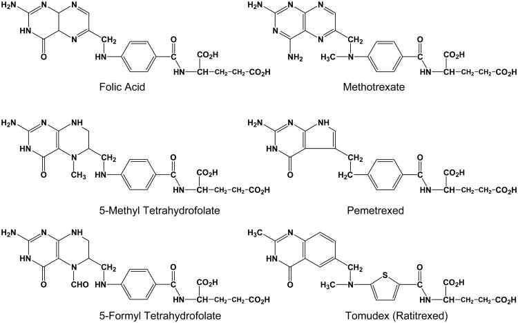
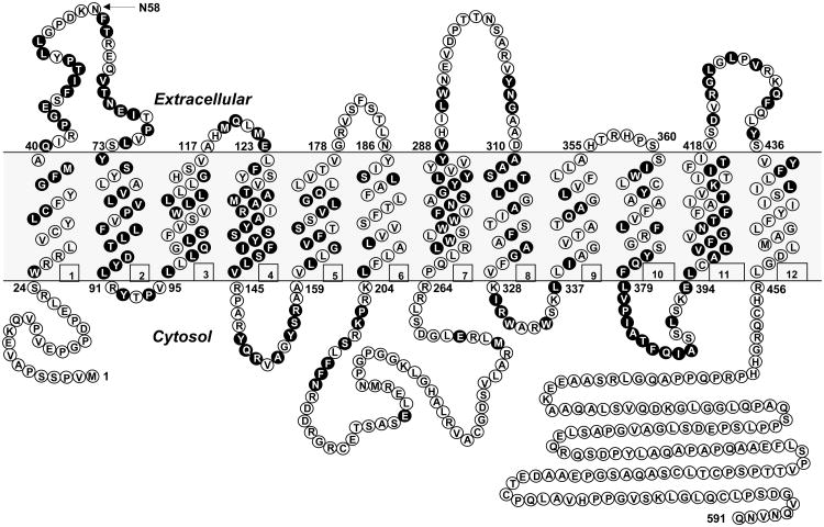
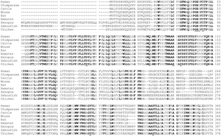
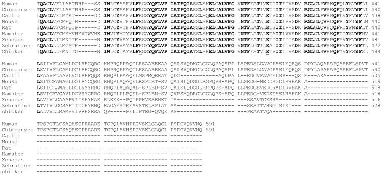
Folates are essential for life and folate deficiency contributes to a host of health problems including cardiovascular disease, fetal abnormalities, neurological disorders, and cancer. Antifolates, represented by methotrexate, continue to occupy a unique niche among the modern day pharmacopoeia for cancer along with other pathological conditions. This article focuses on the biology of the membrane transport system termed the "reduced folate carrier" or RFC with a particular emphasis on RFC structure and function. The ubiquitously expressed RFC is the major transporter for folates in mammalian cells and tissues. Loss of RFC expression or function portends potentially profound physiological or developmental consequences. For chemotherapeutic antifolates used for cancer, loss of RFC expression or synthesis of mutant RFC protein with impaired function results in antifolate resistance due to incomplete inhibition of cellular enzyme targets and low levels of substrate for polyglutamate synthesis. The functional properties for RFC were first documented nearly 40 years ago in murine leukemia cells. Since 1994, when RFC was first cloned, tremendous advances in the molecular biology of RFC and biochemical approaches for studying the structure of polytopic membrane proteins have led to an increasingly detailed picture of the molecular structure of the carrier, including its membrane topology, its N-glycosylation, identification of functionally and structurally important domains and amino acids, and helix packing associations. Although no crystal structure for RFC is yet available, biochemical and molecular studies, combined with homology modeling, based on homologous bacterial major facilitator superfamily transporters such as LacY, now permit the development of experimentally testable hypotheses designed to establish RFC structure and mechanism.
Figures
References
-
- Abramson J, Smirnova I, Kasho V, Verner G, Kaback HR, Iwata S. Structure and mechanism of the lactose permease of Escherichia coli. Science. 2003;301:610–615. - PubMed
-
- Almqvist J, Huang Y, Hovmoller S, Wang DN. Homology modeling of the human microsomal glucose 6-phosphate transporter explains the mutations that cause the glycogen storage disease type Ib. Biochemistry. 2004;43:9289–9297. - PubMed
-
- Balamurugan K, Said HM. Role of reduced folate carrier in intestinal folate uptake. Am J Physiol Cell Physiol. 2006;291:C189–193. - PubMed
-
- Barber RC, Bennett GD, Greer KA, Finnell RH. Expression of patterns of folate binding proteins one and two in the developing mouse embryo. Mol Genet Metab. 1999;66:31–39. - PubMed
-
- Barril X, Aleman C, Orozco M, Luque FJ. Salt bridge interactions: stability of the ionic and neutral complexes in the gas phase, in solution, and in proteins. Proteins. 1998;32:67–79. - PubMed
Publication types
MeSH terms
Substances
Grants and funding
LinkOut - more resources
Full Text Sources
Medical
Molecular Biology Databases
