Review
. 2008 Nov;24(11):564-73.
doi: 10.1016/j.tig.2008.08.006. Epub 2008 Sep 18.

Changing partners: moving from non-homologous to homologous centromere pairing in meiosis

Affiliations
Review

Changing partners: moving from non-homologous to homologous centromere pairing in meiosis

Mara N Stewart et al. Trends Genet. 2008 Nov.

Abstract

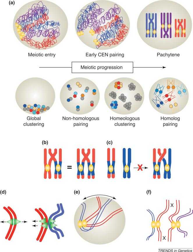
Reports of centromere pairing in early meiotic cells have appeared sporadically over the past thirty years. Recent experiments demonstrate that early centromere pairing occurs between non-homologous centromeres. As meiosis proceeds, centromeres change partners, becoming arranged in homologous pairs. Investigations of these later centromere pairs indicate that paired homologous centromeres are actively associated rather than positioned passively, side-by-side. Meiotic centromere pairing has been observed in organisms as diverse as mice, wheat and yeast, indicating that non-homologous centromere pairing in early meiosis and active homologous centromere pairing in later meiosis might be themes in meiotic chromosome behavior. Moreover, such pairing could have previously unrecognized roles in mediating chromosome organization or architecture that impact meiotic segregation fidelity.


Figures

Figure 1
Figure 1
Early centromere pairing. The arrangement of centromeres in pairs in early meiotic cells has been noted in many organisms. This pairing seems to be primarily between non-homologous centromeres. This non-homologous pairing gives way to the aligning of homologous centromeres as homologous chromosomes pair in later meiotic prophase. (a) Top row: in most organisms, cells enter meiosis with clustered centromeres (gold ovals) and decondensed chromosomes (homologous chromosomes are indicated by the same colors). It is not clear whether early centromere pairing occurs at exactly the same meiotic stage in all organisms in which it has been reported; furthermore, it has yet to be determined whether early pairing precedes or follows meiotic S phase and the early events of recombination initiation. In all cases, early pairing precedes alignment of homologous chromosomes and synaptonemal complex assembly. By pachytene, the stage of maximal homolog alignment and complete synaptonemal-complex formation (green), non-homologous centromere pairing has given way to the side-by-side arrangement of homologous centromeres. Bottom row: in wheat, early non-homologous pairing gives way to clustering of related (homeologous) centromeres (represented by dots of a similar shade). The organization of the chromosomes within the homeologous clusters is not known. By pachytene, centromeres have left the cluster and each is aligned with its true homologous partner. (b). Cytological experiments in wheat using wheat chromosomes (red) bearing rye centromere regions (blue) have addressed the roles of centromeres and chromosome arms in chromosome pairing [21]. Homologous chromosome pairs with either heterologous (one wheat centromere, one rye centromere) or homologous centromeres (both rye centromeres) show indistinguishable levels of pairing and chiasma formation, thereby demonstrating that centromere homology is not required for homologous chromosome pairing. (c) As a second test of the ability of homologous centromeres to drive chromosome pairing, heterologous chromosomes (one rye, blue; and one wheat, red) with homologous rye centromeres were examined. These showed no evidence of meiotic pairing or chiasma formation, demonstrating that homologous centromeres do not drive partner choice, a conclusion also supported by earlier genetic experiments in yeast [22]. (d) Mitotic kinetochores (green) are orientated towards opposite poles (red sister chromatids on the left). Early centromere pairing, shown for red and blue sister chromatid pairs on the right, might serve as a signal or provide a template for the assembly of sister kinetochores that orientate towards the same pole at meiosis I. (e) Experiments in budding yeast [45,46] and rat [47] show that meiotic telomeres move actively in early meiosis, probably to influence chromosome organization or interactions. Early centromere pairing might function as an anchor against telomere-led chromosome movements in early meiosis. (f) In most organisms tested, recombination is limited in centromere regions. Early non-homologous centromere pairing (different colored chromosomes are non-homologous) might sterically limit interactions of homologous centromere regions, thereby preventing homologous recombination near centromeres. (All chromosomes are drawn as condensed to simplify the illustration.)
Figure 2
Figure 2
Centromere pairing in late prophase. In late meiotic prophase, homologous chromosomes align end-to-end. At pachytene, the stage of maximal alignment, a proteinaceous structure runs the length of the aligned homologs. This alignment juxtaposes homologous centromeres. This late centromere pairing has a role in the proper attachment of microtubules during meiosis I. (a) Evidence for active association of late prophase centromeres comes from studies of mouse spermatocytes. In pachytene, chromosomes are tightly synapsed along their length (synaptonemal complex is shown in green). As cells move into diplotene, the synaptonemal complex is lost and homologs remain tethered to one another by chiasmata and the centromeres (yellow). (b) Late centromere pairing is clearly revealed in this micrograph of mouse spermatocyte diplotene chromosomes, linked at chiasmata and centromeres (image provided by Huiling Xu, Peter MacCallum Cancer Centre; http://www.petermac.org/). The chromosome arms are stained with antibodies against SCP3 (synaptonemal-complex protein 3; green); each green line represents one chromosome (two tightly associated sister chromatids). Chiasmata are revealed as the points where the green lines come together. Centromeres are stained with CREST (a human autoimmune syndrome) antibodies against kinetochore components (red) [71]. (c) Centromere pairing of achiasmate chromosomes. In Drosophila, budding yeast and fission yeast, chromosomes that are not tethered by crossovers (red) pair at their centromere regions (but not their arms) in late meiotic prophase (pairing symbolized by blue rectangle), whereas exchange chromosomes (purple) are linked by chiasmata. Centromere pairing promotes disjunction at meiosis I. (d) Centromere pairing between achiasmate chromosomes seems to promote attachment of the chromosomes to opposite poles of the spindle. Centromere pairing could fulfill the same role for exchange chromosomes. Centromere pairing (blue rectangle) might secure the homologous kinetochores (green triangles) such that they face in opposite directions before microtubule attachment. In the absence of centromere pairing, kinetochores might have rotational freedom, thereby enabling attachment of the kinetochores to the same pole. For simplicity, the kinetochores of each sister chromatid pair is represented as a single triangle. (e) Does centromere pairing help orientate sister kinetochores towards the same pole? In mouse oocytes, chromosomes that have failed to pair with a partner (univalents) frequently segregate their sister chromatids to opposite poles at meiosis I. This indicates that partnerless sister chromatid pairs orientate their kinetochores to opposite poles (sister pair on left). Centromere pairing (blue rectangle) might help co-orientate sister kinetochores to the same pole (right).
Figure I
Figure I
Kinetochore orientation and cohesin removal during mitosis and meiosis. (a) (i) Mitotic kinetochores (green triangles) are assembled in a back-to-back in a fashion. (ii) In meiosis, sister chromatids have kinetochores that function as a unit. (iii) Failure to adopt a meiosis-specific kinetochore assembly would result in precocious separation of the sister chromatids in meiosis I, rather than meiosis II. (b) (i) In mitosis, cohesins (orange lines) are all removed by anaphase, facilitating sister chromatid separation. In meiosis, cohesins are removed in two steps. (ii) Before anaphase I, cohesins distal to the chiasmata are removed, enabling homologs to separate. (iii) Before anaphase II, the remaining cohesins are removed, thereby facilitating separation of sister centromeres.
Figure I
Figure I
The stages of meiosis. Depicted is a diploid cell with only one chromosome; one homolog in blue and the other in purple. To simplify the illustration of meiotic events, the chromosomes are represented as condensed throughout the process (which is not actually the case). The time at which early centromere pairing initiates has not been determined, but could be before or during meiotic S phase (red dashed line). Early pairing continues into leptotene (red line). Known examples of late centromere pairing initiate in leptotene or zygotene and continue until chromosomes separate at metaphase or anaphase I.

Similar articles

Cited by

References

    1. Bennett MD. Centromere arrangements in Triticum aestivum and their relationship to synapsis. Heredity. 1979;43:157.
    1. Brinkley BR, et al. Arrangements of kinetochores in mouse cells during meiosis and spermiogenesis. Chromosoma. 1986;94:309–317. - PubMed
    1. Church K, Moens PB. Centromere behavior during interphase and meiotic prophase in Allium fistulosum from 3-D, E.M. reconstruction. Chromosoma. 1976;56:249–263.
    1. Kleckner N. Meiosis: how could it work? Proc. Natl. Acad. Sci. U. S. A. 1996;93:8167–8174. - PMC - PubMed
    1. Yunis JJ, Yasmineh WG. Model for mammalian constitutive heterocromatin. In: DuPraw FJ, editor. Advances in Cell and Molecular Biology. Academic Press; 1972. pp. 1–46.

Publication types