. 2009 Jan;17(1):73-82.
doi: 10.1016/j.joca.2008.05.019. Epub 2008 Sep 19.

Analysis of radial variations in material properties and matrix composition of chondrocyte-seeded agarose hydrogel constructs

Affiliations

Analysis of radial variations in material properties and matrix composition of chondrocyte-seeded agarose hydrogel constructs

T-A N Kelly et al. Osteoarthritis Cartilage. 2009 Jan.

Abstract

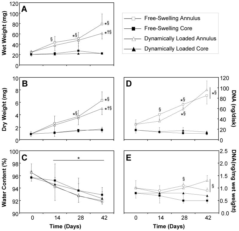
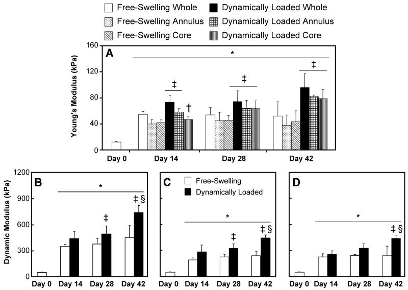
Objective: To examine the radial variations in engineered cartilage that may result due to radial fluid flow during dynamic compressive loading. This was done by evaluating the annuli and the central cores of the constructs separately.

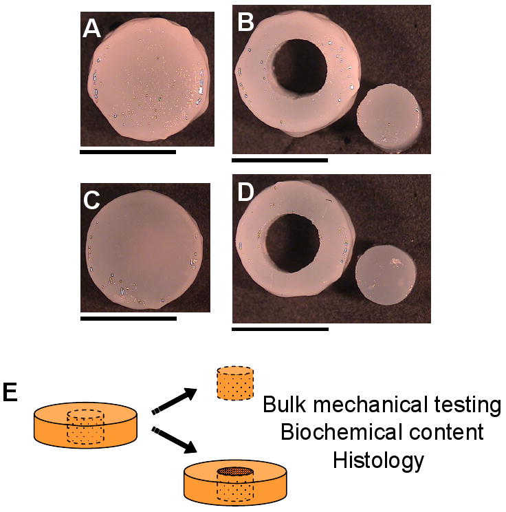
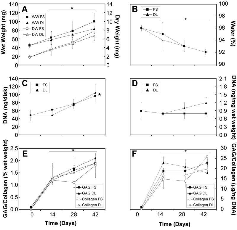
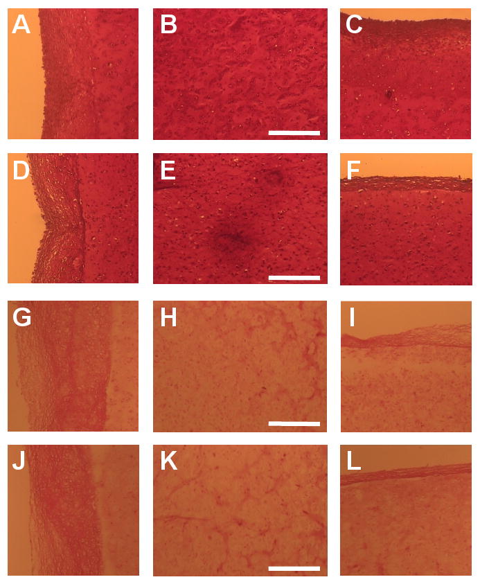
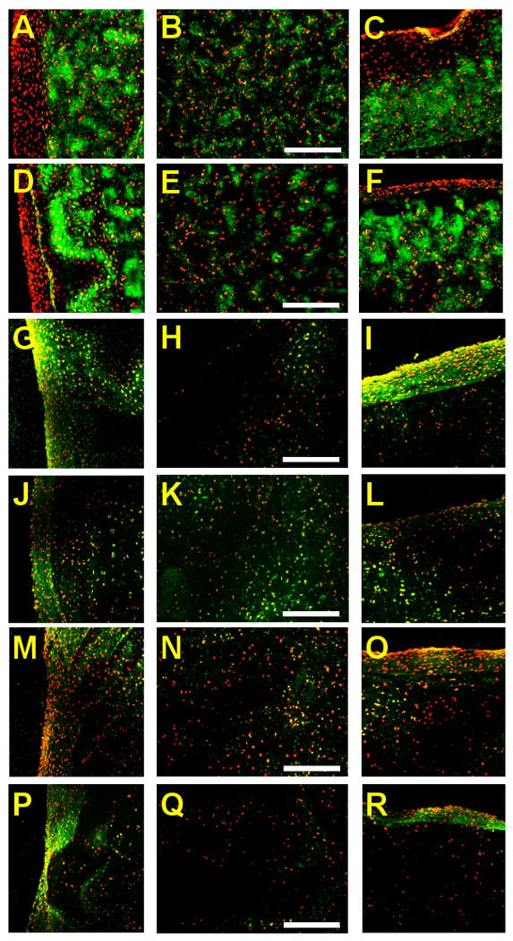
Method: Chondrocyte-seeded agarose hydrogels were grown in free-swelling and dynamic, unconfined loading cultures for 42 days. After mechanical testing, constructs were allowed to recover for 1-2h, the central 3mm cores removed, and the cores and annuli were retested separately. Histological and/or biochemical analyses for DNA, glycosaminoglycan (GAG), collagen, type I collagen, type II collagen, and elastin were performed. Multiple regression analysis was used to determine the correlation between the biochemical and material properties of the constructs.

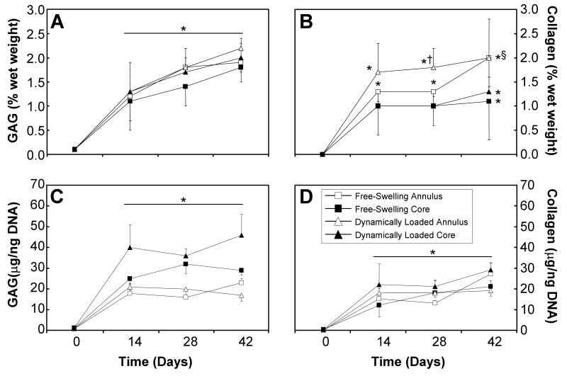
Results: The cores and annuli of chondrocyte-seeded constructs did not exhibit significant differences in material properties and GAG content. Annuli possessed greater DNA and collagen content over time in culture than cores. Dynamic loading enhanced the material properties and GAG content of cores, annuli, and whole constructs relative to free-swelling controls, but it did not alter the radial variations compared to free-swelling culture.

Conclusion: Surprisingly, the benefits of dynamic loading on tissue properties extended through the entire construct and did not result in radial variations as measured via the coring technique in this study. Nutrient transport limitations and the formation of a fibrous capsule on the periphery may explain the differences in DNA and collagen between cores and annuli. No differences in GAG distribution may be due to sufficient chemical signals and building blocks for GAG synthesis throughout the constructs.


Figures

Figure 1
Figure 1
Images of free-swelling (A, B) and dynamically loaded (C, D) constructs were acquired at day 42, before (A, C) and after (B, D) coring of the constructs. Scale bars equal 5 mm. For this study, samples were tested whole, allowed to recover and then the annuli and cores were tested separately (E).
Figure 2
Figure 2
Wet and dry weights (A), percent water (B), DNA content (C, D), and GAG and collagen contents (E, F) of intact free-swelling and dynamically loaded chondrocyte-seeded constructs. * represents significant differences versus day 0 constructs (ANOVA p<0.005; post hoc p<0.005).
Figure 3
Figure 3
Wet weight (A), dry weight (B), percent water (C), and DNA content (D, E), of the annuli (open) and cores (closed) of free-swelling (boxes) and dynamically loaded (triangles) constructs over the 6-week culture period (n=4-9). Significant effect of time was observed for these samples (ANOVA p<0.05). * represents significant differences versus day 0 constructs (ANOVA p<0.05; post hoc p<0.05); † represents significant differences versus free-swelling controls (ANOVA p<0.01; post hoc p<0.05); § represents significant differences versus the respective cores (ANOVA p<0.001; post hoc p<0.05).
Figure 4
Figure 4
Wet GAG (A, B) and collagen (C, D) contents of the annuli (open) and cores (closed) of free-swelling (boxes) and dynamically loaded (triangles) constructs over the 6-week culture period (n=4-9). Significant effect of time was observed for these samples (ANOVA p<0.05). * represents significant differences versus day 0 constructs (ANOVA p<0.05; post hoc p<0.05); † represents significant differences versus free-swelling controls (ANOVA p<0.05; post hoc p<0.05); § represents significant differences versus the respective annular samples (ANOVA p<0.05; post hoc p<0.05).
Figure 5
Figure 5
Young's (A) and dynamic (B, C, D; measured at 0.5 Hz) moduli of free-swelling and dynamically loaded constructs over the 42-day culture period (n=7). The constructs were allowed to recover for 1 hour before the annulus and the 3 mm central core were tested. As the dynamic modulus is a structural property, inter-group comparisons between intact constructs (B), annuli (C), and cores (D) were not performed. *represents significant differences compared to day 0 (ANOVA p<0.005; Tukey p<0.003); ‡represents significant differences compared to corresponding free-swelling controls (ANOVA p<0.0005; Tukey p<0.005); †represents significant differences compared to whole sample (ANOVA p<0.0005; Tukey p<0.02); §represents significant differences compared to day 28 constructs (p<0.05).
Figure 6
Figure 6
Safranin O (GAG; A-F) and picrosirius red staining (collagen; G-L) of free-swelling (A-C, G-I) and dynamically loaded (D- F, J-L) constructs on day 42. Images were acquired along the radial edge (first column), in the center (second column), and along the axial edge (third column) of the constructs. Scale bars equal 250 μm. Staining intensity distribution along the axial direction of the constructs for Safranin O (M; GAG) and Picrosirius Red (N; Collagen). Intensity is normalized by the intensity at the center region (x/h = 0.5), where x/h = 0 represents the top and bottom surfaces of the construct (n = 3).
Figure 7
Figure 7
Immunofluorescent labeling (green) for type II collagen (A-F), type I collagen (G-L), and elastin (M-R) of free-swelling (A-C, G-I, M-O) and dynamically loaded (D-F, J-L, P-R) chondrocyte-seeded constructs at day 42. Images were acquired along the radial edge (first column), in the center (second column), and along the axial edge (third column) of the constructs. Propidium iodide counter staining (red) was used tovisualize the cell nuclei. Scale bars equal 200 μm.

References

    1. Mauck RL, Soltz MA, Wang CC, Wong DD, Chao PH, Valhmu WB, et al. Functional tissue engineering of articular cartilage through dynamic loading of chondrocyte-seeded agarose gels. J Biomech Eng. 2000;122:252–260. - PubMed
    1. Kelly TA, Ng KW, Wang CC, Ateshian GA, Hung CT. Spatial and temporal development of chondrocyte-seeded agarose constructs in free-swelling and dynamically loaded cultures. J Biomech. 2005;39:1489–1497. - PubMed
    1. Ateshian GA, Wang H. A theoretical solution for the frictionless rolling contact of cylindrical biphasic articular cartilage layers. J Biomech. 1995;28:1341–1355. - PubMed
    1. Park S, Krishnan R, Nicoll SB, Ateshian GA. Cartilage interstitial fluid load support in unconfined compression. J Biomech. 2003;36:1785–1796. - PMC - PubMed
    1. Mauck RL, Hung CT, Ateshian GA. Modeling of neutral solute transport in a dynamically loaded porous permeable gel: implications for articular cartilage biosynthesis and tissue engineering. J Biomech Eng. 2003;125:602–614. - PMC - PubMed

Publication types

MeSH terms