

Review
doi: 10.1016/j.cell.2008.09.002.
Retinoic acid synthesis and signaling during early organogenesis
Affiliations
- PMID: 18805086
- PMCID: PMC2632951
- DOI: 10.1016/j.cell.2008.09.002
Item in Clipboard
Review
Retinoic acid synthesis and signaling during early organogenesis
Cell.
.
Abstract
Retinoic acid, a derivative of vitamin A, is an essential component of cell-cell signaling during vertebrate organogenesis. In early development, retinoic acid organizes the trunk by providing an instructive signal for posterior neuroectoderm and foregut endoderm and a permissive signal for trunk mesoderm differentiation. At later stages, retinoic acid contributes to the development of the eye and other organs. Recent studies suggest that retinoic acid may act primarily in a paracrine manner and provide insight into the cell-cell signaling networks that control differentiation of pluripotent cells.
Figures
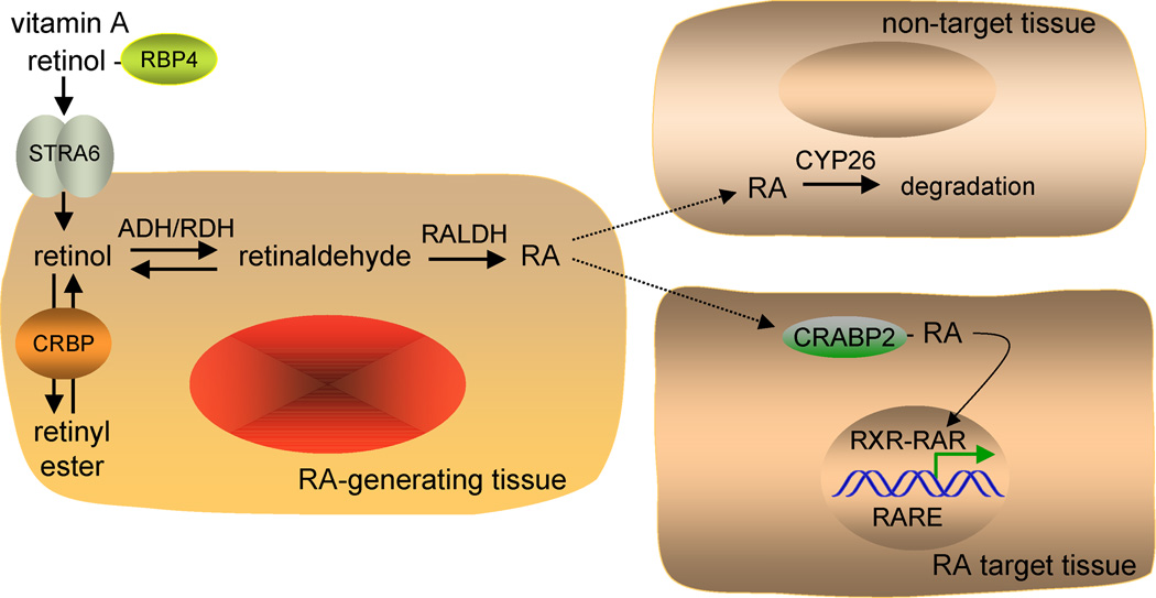
Depicted is the paracrine mechanism of retinoic acid (RA) signaling. Retinol is carried in the serum by retinol-binding protein (RBP4) secreted from the liver. Retinol enters cells via a specific receptor STRA6, and cellular retinol-binding protein (CRBP) facilitates conversion of retinol to retinyl esters for storage. In an RA-generating tissue, retinol is oxidized to retinaldehyde by either alcohol dehydrogenase (ADH) or retinol dehydrogenase (RDH), and retinaldehyde is oxidized to RA by retinaldehyde dehydrogenase (RALDH). RA is then released and taken up by surrounding cells. Cells that express cytochrome P450 (CYP26) initiate the further oxidation of RA for degradation and excretion and are not RA target cells. Some RA target cells express cellular-RA binding protein (CRABP) that facilitates uptake of RA and transport to the nucleus where RA binds the RA receptor (RAR). The ternary complex of ligand bound-RAR with RXR and a retinoic acid response element (RARE) regulates transcription of RA target genes by altering the binding of corepressors and coactivators.
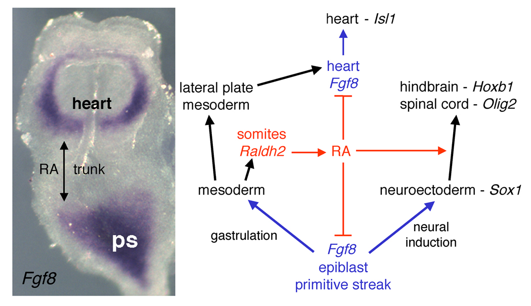
Shown on the left is an E8.0 mouse embryo stained for fibroblast growth factor 8 (Fgf8) mRNA by in situ hybridization. At this stage Fgf8 mRNA is expressed in an anterior cardiac domain where it induces Isl1, plus a posterior domain encompassing the epiblast and primitive streak (ps) where FGF8 is needed during gastrulation and neural induction; retinaldehyde dehydrogenase 2 (Raldh2) is expressed in between these two domains in the somitic mesoderm where the future trunk will form. The diagram depicts the function of retinoic acid (RA) as a repressor of both Fgf8 domains and as an inducer of neural posteriorization in the trunk to allow hindbrain and spinal cord differentiation.
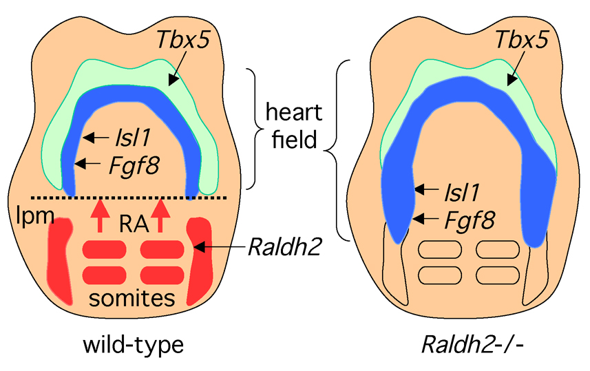
Retinoic acid (RA) generated by retinaldehyde dehydrogenase 2 (Raldh2) in the somites and lateral plate mesoderm (lpm) travels anteriorly where it provides a signal that helps establish the posterior border of the heart field. The lateral domain of the heart field expresses Tbx5 whereas the medial domain expresses Isl1 as well as Fgf8 which is required for cardiac Isl1 expression. Embryos from Raldh2 knockout mice exhibit posterior expansion of Fgf8 and Isl1 into lateral plate mesoderm that normally is not part of the heart field. As the Fgf8 promoter has been reported to contain an RA response element, RA may function as a repressor of cardiac Fgf8.
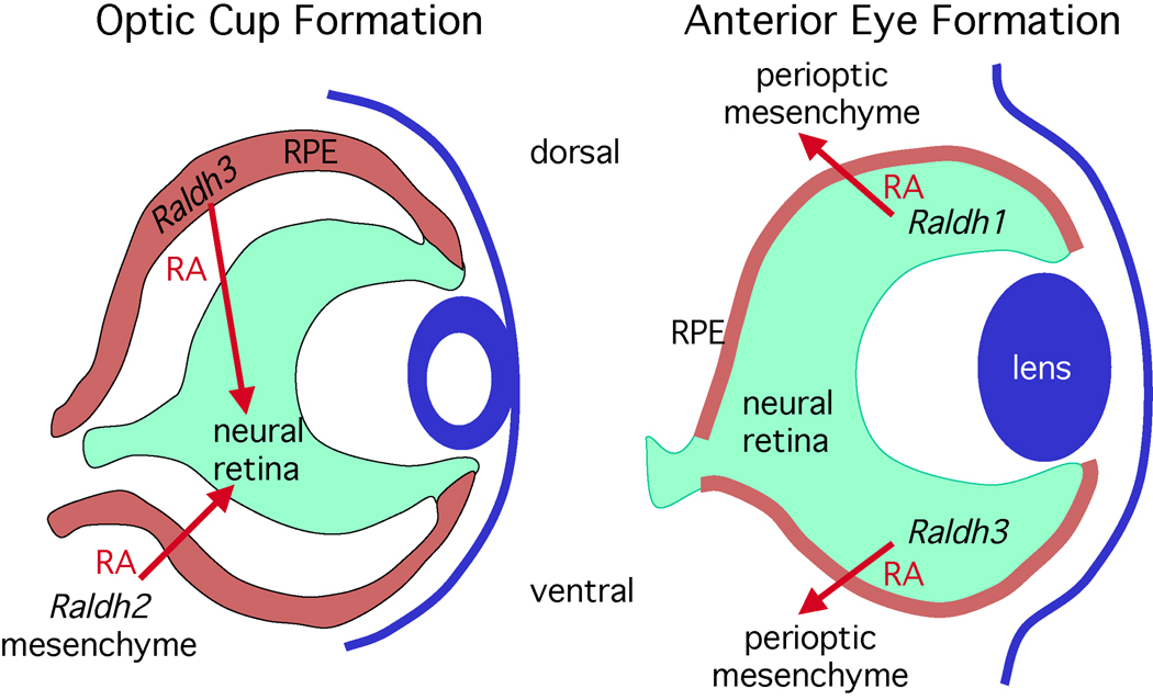
Paracrine retinoic acid (RA) signaling controls two distinct phases of eye morphogenetic movements and involves all three retinaldehyde dehydrogenase (Raldh) genes. During invagination of the optic vesicle to form an optic cup (E9.5–E10.5 in mouse), RA generated by Raldh2 in the perioptic mesenchyme as well as Raldh3 in tissue fated to become the dorsal retinal pigment epithelium (RPE) travels to the neural retina where it is required for ventral invagination during optic cup formation. After optic cup formation (E10.5-birth), Raldh2 is no longer expressed in the perioptic mesenchyme and Raldh3 expression ends in the RPE, but Raldh1 and Raldh3 are now expressed in the dorsal and ventral neural retina, respectively. RA generated by Raldh1 and Raldh3 is not required for patterning the neural retina, but this RA travels outside the retina where it limits invasion of perioptic mesenchyme during anterior eye development (cornea and eyelid formation).
References
-
- Ang HL, Deltour L, Hayamizu TF, Zgombic-Knight M, Duester G. Retinoic acid synthesis in mouse embryos during gastrulation and craniofacial development linked to class IV alcohol dehydrogenase gene expression. J. Biol. Chem. 1996;271:9526–9534. - PubMed
-
- Arnhold T, Tzimas G, Wittfoht W, Plonait S, Nau H. Identification of 9-cis-retinoic acid, 9,13-di-cis-retinoic acid, and 14-hydroxy-4,14-retro-retinol in human plasma after liver consumption. Life Sci. 1996;59:PL 169–PL 177. - PubMed
-
- Batourina E, Gim S, Bello N, Shy M, Clagett-Dame M, Srinivas S, Costantini F, Mendelsohn C. Vitamin A controls epithelial/mesenchymal interactions through Ret expression. Nature Genet. 2001;27:74–78. - PubMed
-
- Batourina E, Tsai S, Lambert S, Sprenkle P, Viana R, Dutta S, Hensle T, Wang FW, Niederreither K, McMahon AP, et al. Apoptosis induced by vitamin A signaling is crucial for connecting the ureters to the bladder. Nature Genet. 2005;37:1082–1089. - PubMed
Publication types
MeSH terms
Substances
Grants and funding
LinkOut - more resources
Full Text Sources
Other Literature Sources
Molecular Biology Databases
