. 2008 Sep 19;134(6):969-80.
doi: 10.1016/j.cell.2008.08.030.

Mechanism of replication-coupled DNA interstrand crosslink repair

Affiliations

Mechanism of replication-coupled DNA interstrand crosslink repair

Markus Räschle et al. Cell. .

Erratum in

  • Cell.2009 May 29;137(5):972. Knipsheer, Puck [corrected to Knipscheer, Puck]

Abstract

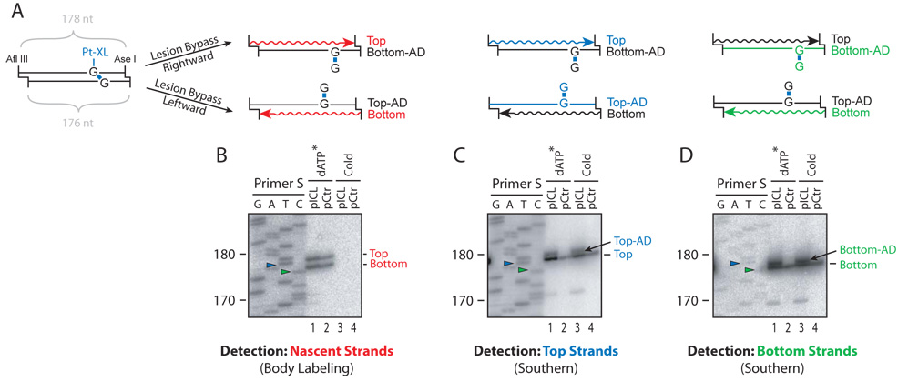
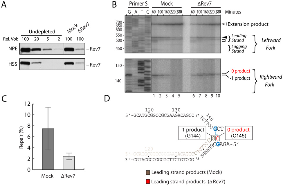
DNA interstrand crosslinks (ICLs) are toxic DNA lesions whose repair occurs in the S phase of metazoans via an unknown mechanism. Here, we describe a cell-free system based on Xenopus egg extracts that supports ICL repair. During DNA replication of a plasmid containing a site-specific ICL, two replication forks converge on the crosslink. Subsequent lesion bypass involves advance of a nascent leading strand to within one nucleotide of the ICL, followed by incisions, translesion DNA synthesis, and extension of the nascent strand beyond the lesion. Immunodepletion experiments suggest that extension requires DNA polymerase zeta. Ultimately, a significant portion of the input DNA is fully repaired, but not if DNA replication is blocked. Our experiments establish a mechanism for ICL repair that reveals how this process is coupled to DNA replication.


Figures

Figure 1
Figure 1. DNA replication forks converge on an interstrand cross-link
(A) Structure of nitrogen mustard-like (postulated) and (B) cisplatin [based on (Huang et al., 1995)] ICLs. (C) Replication of pICLNm in Xenopus egg extracts. pCtr or pICLNm was incubated sequentially with HSS and NPE/32P-α-dATP. At the indicated times after NPE addition, replication products were analyzed on a native agarose gel. (D) The average replication efficiency of three independent experiments was plotted with error bars. (E) Model for replication of pICL. (F) pICLNm was replicated as in (C). 30 minutes after NPE addition, DNA was analyzed by electron microscopy. The predominant species, a "Figure 8" structure, is shown.
Figure 2
Figure 2. Multi-step lesion bypass of an interstrand cross-link
(A) Structure of the replicated Afl III fragment which includes the ICL. S, primer used to generate the sequencing ladder shown in (B). (B) Mapping of nascent strands during replication of pICLNm or pCtr (final concentration 1.2 ng/µl). At the indicated times after NPE addition, reaction products were digested with Afl III and analyzed on a sequencing gel alongside a sequencing ladder derived from extension of primer S on pCtr (see A). Numbers to the left indicate the sizes of the sequencing products. Leading and lagging strands for the rightward and leftward forks are indicated. Square brackets show the positions of leading strands after the initial pausing, whereas red and green arrowheads show their location after advancing towards the ICL. Open arrowhead, extension product. (C) Enlarged and darker exposure of the bottom part of the autoradiogram shown in (B). The most prominent species observed at 45 minutes are indicated on the right, with the predicted last nucleotide denoted by a single letter. The exact sizes of species T147–A150 were confirmed in Supplementary Figure 9. (D) Cartoon-form depiction of the results in (C).
Figure 3
Figure 3. Detection of incisions near the ICL
(A) Expected intermediates resulting from single or dual incisions near the ICL (see text). Note that the first incision could also occur to the left of the ICL, giving rise to a short arm and large Y structure. (B) pICLNm replication products were digested with Hinc II, and separated on a native agarose gel (lanes 1–8). Replicated pCtr was digested with Hinc II to generate a 5.6 kb size marker (lane 9, only 20% of the reaction loaded). (C) Advance of the leading strand to the ICL precedes incisions. At each time point in (B), the relative abundance of X-shaped molecules was quantified using a phosphorimager (blue line). At 18 and 22 minutes, before replication was complete, the level was assigned a value of 100. The relative abundance of leading strand products from both forks at the −1 position (Figure 2B and Figure 2D) (grey dashed line) is plotted. The graph shows the average of four independent experiments.
Figure 4
Figure 4. Replication-dependent repair of an ICL
(A) An ICL blocks cleavage by Acc I. 15 ng of pICLNm or pCtr was digested with Acc I, separated on a native agarose gel, and stained with SYBR Gold. (B) ICL repair assay. At different times after addition of NPE/32P-α-dATP, pICLNm or pCtr was recovered and equal aliquots were digested with Hinc II (lanes 1–9) or Acc I (lanes 10–18). (Note: lanes 1–9 represent a darker exposure of panel 3B). 20% of the reaction was loaded in lanes 9 and 18. (C) At each time point shown in (B), the repair efficiency was calculated as explained in the text and graphed (Red line). Extension products from the same experiment shown in Figure 4B were plotted for comparison (grey dashed line). (D) pICLNm or pCtr was replicated using NPE lacking radioactivity and optionally supplemented with p27Kip. Plasmid was recovered, digested as indicated, and examined by Southern blotting using pCtr DNA as probe. 20% of the reaction was loaded in lane 13. Samples were supplemented with a 1.2 kb Hind III fragment of pCtr before extraction (loading control).
Figure 5
Figure 5. An adduct persists in the parental strand after lesion bypass
(A) Cartoon illustrating replication of an Afl III / Ase I restriction fragment harboring a cisplatin ICL. Due to the different overhangs generated by these enzymes, digestion of pICLPt yields Top and Bottom Strands of 178 and 176 nts, respectively. Lesion bypass by the rightward fork yields a radioactively labeled nascent Top Strand and an adducted, parental Bottom Strand (Bottom-AD), while lesion bypass by the leftward fork results in a labeled nascent Bottom Strand and an adducted parental Top Strand (Top-AD). Strand-specific Southern blotting was used to detect either the top strands (blue lines) or the bottom strands (green lines). (B) Detection of nascent strands. pICLPt or pCtr was replicated in the presence (lanes 1, 2) or absence (lanes 3, 4) of 32P-dATP. After 4 hours, replication products were digested with Afl III and Ase I, separated on a 5% denaturing polyacrylamide gel and transferred to a Nylon membrane. Radioactive products were visualized using a phophorimager. (C) Detection of the nascent Top Strand and the adducted parental Top Strand (Top-AD) on the membrane in (B) by Southern blotting using a bottom-strand probe. (D) Detection of the nascent Bottom Strand and the adducted parental Bottom Strand by stripping and reprobing the membrane in (C) using a top-strand probe. Primer S was used to generate a sequencing ladder from pCtr that serves as a size marker (see Figure 2). Green and Blue arrowheads indicate the 176 nt and 178 nt sequencing products, respectively (see Supplementary Figure 5C for sequence information and location of primer S). The migration of the digested DNA replication products is retarded by 1 nucleotide with respect to the sequencing products (See Supplementary Figure 9 for discussion of this effect). Adducted parental strands also persisted on pICLNm (data not shown).
Figure 6
Figure 6. Repair of pICL is defective in Rev7-depleted extracts
(A) Rev7 immunodepletion. Undepleted, mock-depleted, and Rev7-depleted HSS and NPE were analyzed by Western blotting using Rev7 antibody. A relative volume of 100 corresponds to 0.3 µl extract. (B) Accumulation of a new lesion bypass intermediate in Rev7-depleted extracts. pICLPt was replicated in mock-depleted or Rev7-depleted HSS and NPE (4 ng/µl final DNA concentration). At the indicated times, products were digested with Afl III and analyzed on a sequencing gel (as in Supplementary Figure 7B). Numbers to the left indicate the size of the sequencing products. The new replication intermediate is indicated ("0 product"). (C) The average repair efficiency in Mock- and Rev7-depleted extracts in 4 independent experiments is plotted with error bars. (D) Cartoon depicting the intermediate that accumulates in Rev7- depleted extracts. We infer that a C residue is inserted at position 145, since the translesion step is likely performed by the cytidyl transferase Rev1, and some of the products are digestible with Sap I.
Figure 7
Figure 7. Model for ICL repair in Xenopus egg extracts
See Discussion for details.

References

    1. Akkari YM, Bateman RL, Reifsteck CA, D'Andrea AD, Olson SB, Grompe M. The 4N cell cycle delay in Fanconi anemia reflects growth arrest in late S phase. Mol Genet Metab. 2001;74:403–412. - PubMed
    1. Akkari YM, Bateman RL, Reifsteck CA, Olson SB, Grompe M. DNA replication is required To elicit cellular responses to psoralen-induced DNA interstrand cross-links. Mol Cell Biol. 2000;20:8283–8289. - PMC - PubMed
    1. Berezney R, Dubey DD, Huberman JA. Heterogeneity of eukaryotic replicons, replicon clusters, and replication foci. Chromosoma. 2000;108:471–484. - PubMed
    1. Bessho T. Induction of DNA replication-mediated double strand breaks by psoralen DNA interstrand cross-links. J Biol Chem. 2003;278:5250–5254. - PubMed
    1. Byun TS, Pacek M, Yee MC, Walter JC, Cimprich KA. Functional uncoupling of MCM helicase and DNA polymerase activities activates the ATR-dependent checkpoint. Genes Dev. 2005;19:1040–1052. - PMC - PubMed

Publication types

LinkOut - more resources