. 2008 Sep 19;134(6):981-94.
doi: 10.1016/j.cell.2008.08.037.

Sgs1 helicase and two nucleases Dna2 and Exo1 resect DNA double-strand break ends

Affiliations

Sgs1 helicase and two nucleases Dna2 and Exo1 resect DNA double-strand break ends

Zhu Zhu et al. Cell. .

Abstract

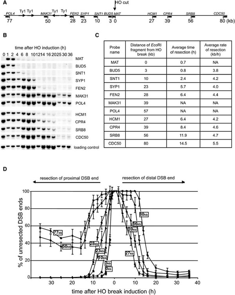
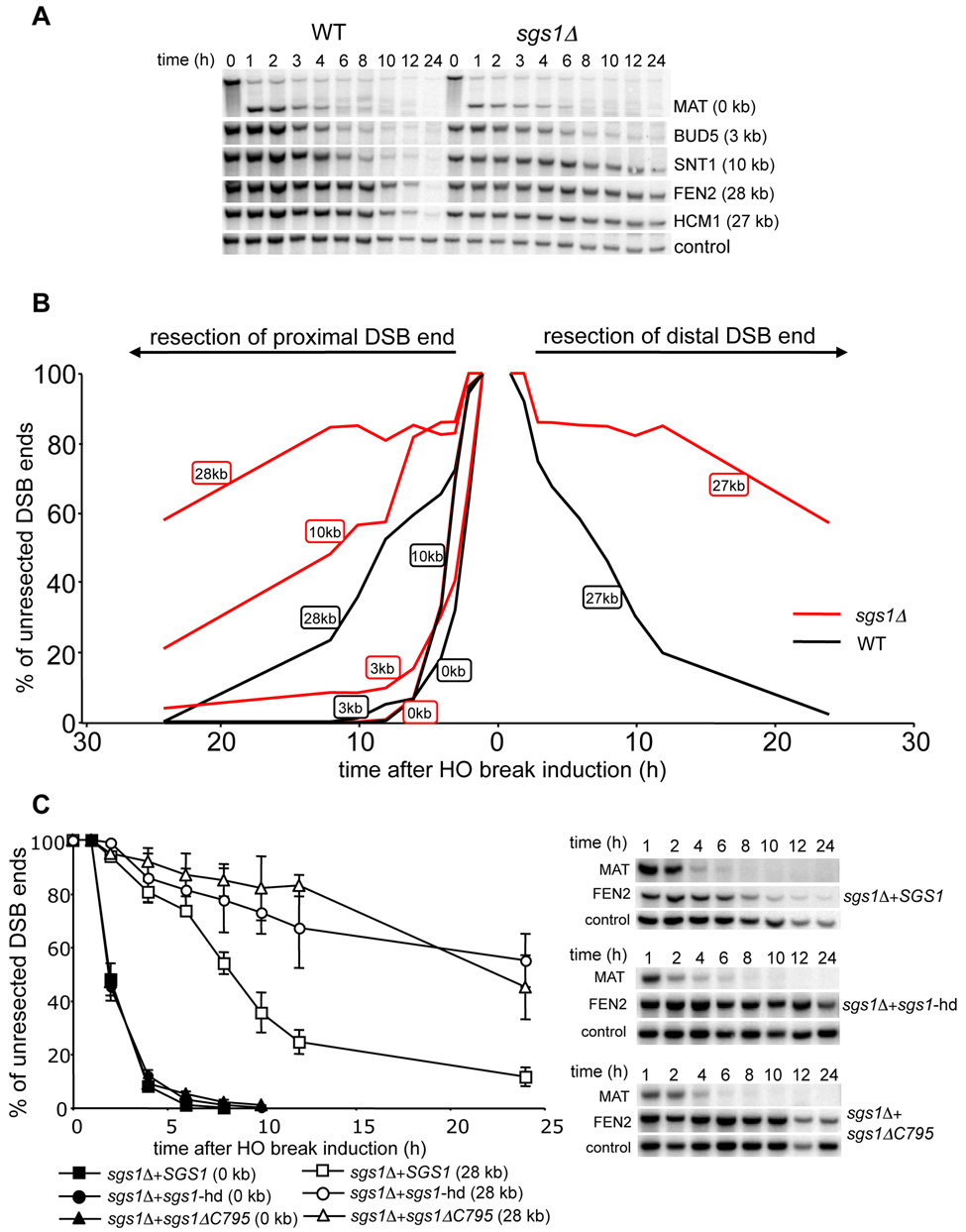
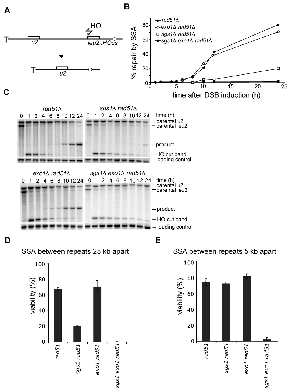
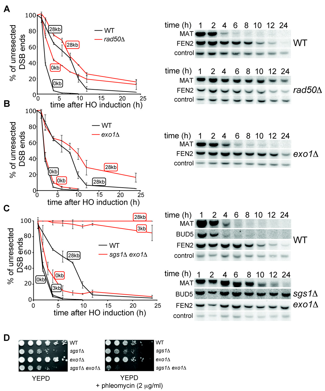
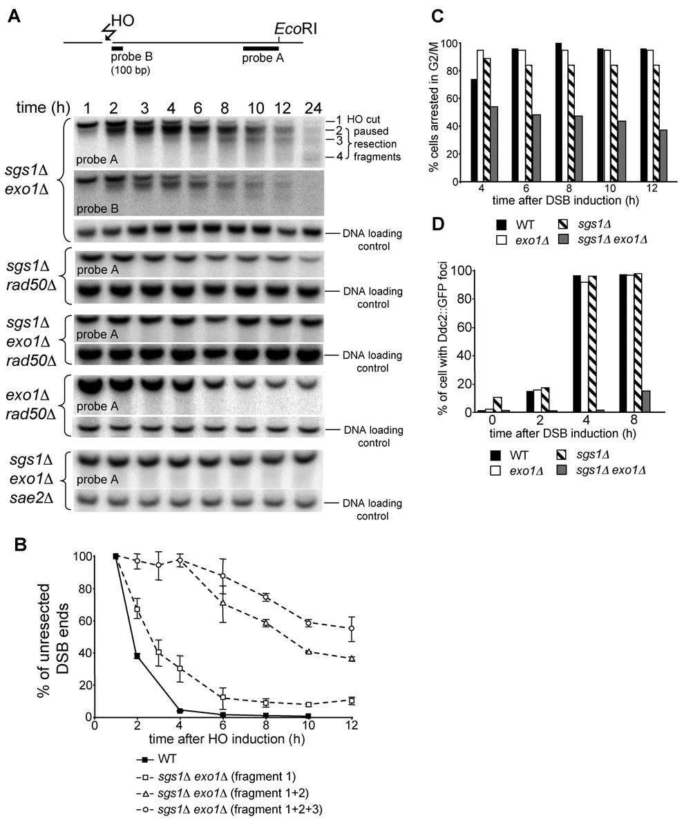
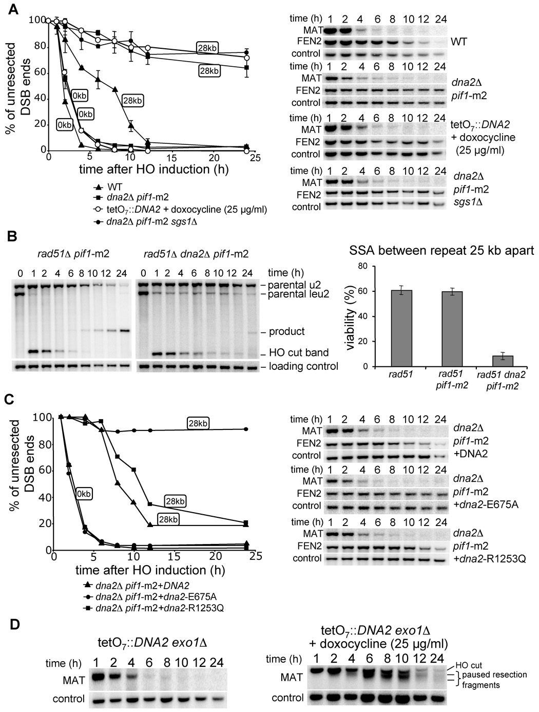
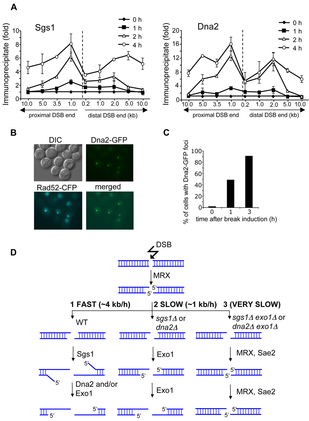
Formation of single-strand DNA (ssDNA) tails at a double-strand break (DSB) is a key step in homologous recombination and DNA-damage signaling. The enzyme(s) producing ssDNA at DSBs in eukaryotes remain unknown. We monitored 5'-strand resection at inducible DSB ends in yeast and identified proteins required for two stages of resection: initiation and long-range 5'-strand resection. We show that the Mre11-Rad50-Xrs2 complex (MRX) initiates 5' degradation, whereas Sgs1 and Dna2 degrade 5' strands exposing long 3' strands. Deletion of SGS1 or DNA2 reduces resection and DSB repair by single-strand annealing between distant repeats while the remaining long-range resection activity depends on the exonuclease Exo1. In exo1Deltasgs1Delta double mutants, the MRX complex together with Sae2 nuclease generate, in a stepwise manner, only few hundred nucleotides of ssDNA at the break, resulting in inefficient gene conversion and G2/M damage checkpoint arrest. These results provide important insights into the early steps of DSB repair in eukaryotes.


Figures

Figure 1
Figure 1. Analysis of 5’ strand resection in wild-type cells
(A) Position of EcoRI sites and DNA probes used to analyze 5’ strand processing with respect to the HO recognition site on chromosome III. (B) Southern blot analysis of 5’ strand resection in wild-type cells. Names of the probes are indicated. (C) Average rate of resection beyond each studied EcoRI site. NA – not applicable. (D) Plot demonstrating percentage of unprocessed 5’ strand for each studied EcoRI site.
Figure 2
Figure 2. Sgs1 helicase is required for normal rate of DSB end resection
(A) Southern blot analysis of 5’ strand resection in sgs1Δ cells. (B) Plot demonstrating percentage of unprocessed 5’ strand for each EcoRI site in wild-type (black line) and sgs1Δ cells (red line). (C) Analysis of resection in sgs1Δ cells carrying a centromeric plasmid with either wild-type or helicase mutant genes of SGS1.
Figure 3
Figure 3. Sgs1 promotes SSA between distant repeats
(A) Scheme representing SSA assay between partial LEU2 gene repeats (Vaze et al., 2002). (B) Kinetics of SSA product formation in wild-type and mutant cells lacking one or more genes. (C) Southern blot analysis of SSA in wild type and indicated mutants. (D–E) Viability of mutants on galactose-containing plates, where an HO break is repaired by SSA between repeats separated by 25 kb (D) or 5 kb (E).
Figure 4
Figure 4. Sgs1 and Exo1 can process 5’ strands independently
Kinetics of resection in rad50Δ (A), exo1Δ (B), and sgs1Δ exo1Δ mutant cells (C) compared to wild-type cells. Southern blot analysis is shown. (D) Sensitivity of wild-type, sgs1Δ, exo1Δ and sgs1Δ exo1Δ cells to phleomycin.
Figure 5
Figure 5. Analysis of resection and G2/M DNA damage checkpoint arrest in sgs1Δ exo1Δ cells
(A) Position of two probes with respect to the DSB and EcoRI sites used to analyze 5’ strand processing in sgs1Δ exo1Δ cells. Southern blot analysis of 5’ strand resection in indicated mutants. Position of HO cut band (DNA fragment 1) and additional bands (DNA fragments 2 to 4) observed in sgs1Δ exo1Δ cells is indicated. (B) Plot demonstrating kinetics of resection in wild-type and sgs1Δ exo1Δ cells. Pixel intensities of the signal corresponding to the unprocessed HO cut band separately or added to the signal of the band(s) corresponding to paused degradation are presented. (C) Analysis of G2/M arrest after induction of the HO break in indicated mutants. (D) Number of cells forming Ddc2 foci after DSB induction was analyzed in indicated mutant and wild-type cells.
Figure 6
Figure 6. Dna2 nuclease processes the 5’ strand at a DSB
(A) Southern blot analysis and kinetics of 5’ strand resection in pif1-m2 dna2Δ, pif1-m2 dna2Δ sgs1Δ and TetO7 ::TATA::DNA2 cells compared to wild-type cells. (B) Southern blot analysis and viability in the SSA assay in indicated mutants. (C) Southern blot analysis and kinetics of 5’ strand resection in pif1-m2 dna2Δ supplemented with a plasmid carrying either the wild-type DNA2 gene, or a point mutation eliminating nuclease (E675A) or helicase activity (R1253Q). (D) Southern blot analysis of 5’ strand resection in the absence of both Exo1 and Dna2 nucleases.
Figure 7
Figure 7. Recruitment of Dna2 and Sgs1 to a DSB and a model of 5’ strand resection at DSBs
(A) Localization of Sgs1 and Dna2 to DSBs at the MAT locus estimated by ChIP before and 1, 2 and 4 h after break induction. IP represents the ratio of the Sgs1p or Dna2p IP PCR signal before and after HO induction, normalized by the PCR signal of the PRE1 control. A dotted line indicates the location of the HO-induced break. (B) Dna2-GFP foci formed after HO break induction colocalize with Rad52-CFP foci. (C) Number of cells with Dna2-GFP foci before and 1 and 3 h after break induction. (D) Model representing three different 5’ strand resection pathways at a DSB with various processivity.

References

    1. Amundsen SK, Smith GR. Interchangeable parts of the Escherichia coli recombination machinery. Cell. 2003;112:741–744. - PubMed
    1. Aravind L, Makarova KS, Koonin EV. SURVEY AND SUMMARY: holliday junction resolvases and related nucleases: identification of new families, phyletic distribution and evolutionary trajectories. Nucleic Acids Res. 2000;28:3417–3432. - PMC - PubMed
    1. Bachrati CZ, Hickson ID. RecQ helicases: guardian angels of the DNA replication fork. Chromosoma. 2008;117:219–233. - PubMed
    1. Bae SH, Bae KH, Kim JA, Seo YS. RPA governs endonuclease switching during processing of Okazaki fragments in eukaryotes. Nature. 2001;412:456–461. - PubMed
    1. Bae SH, Choi E, Lee KH, Park JS, Lee SH, Seo YS. Dna2 of Saccharomyces cerevisiae possesses a single-stranded DNA-specific endonuclease activity that is able to act on double-stranded DNA in the presence of ATP. J. Biol. Chem. 1998;273:26880–26890. - PubMed

Publication types

MeSH terms