. 2008 Sep 19;134(6):1030-41.
doi: 10.1016/j.cell.2008.07.018.

Intramolecular strain coordinates kinesin stepping behavior along microtubules

Affiliations

Intramolecular strain coordinates kinesin stepping behavior along microtubules

Ahmet Yildiz et al. Cell. .

Abstract

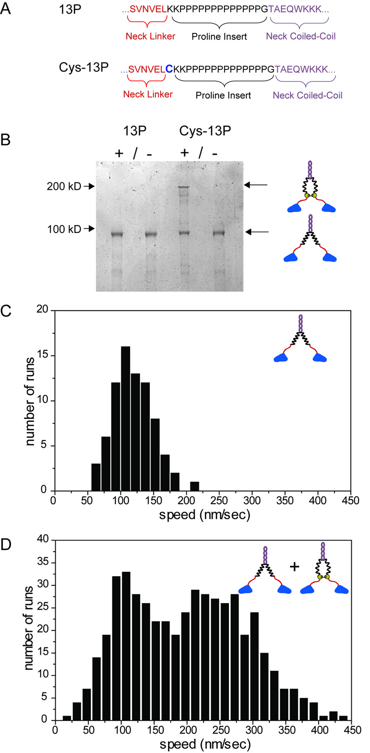
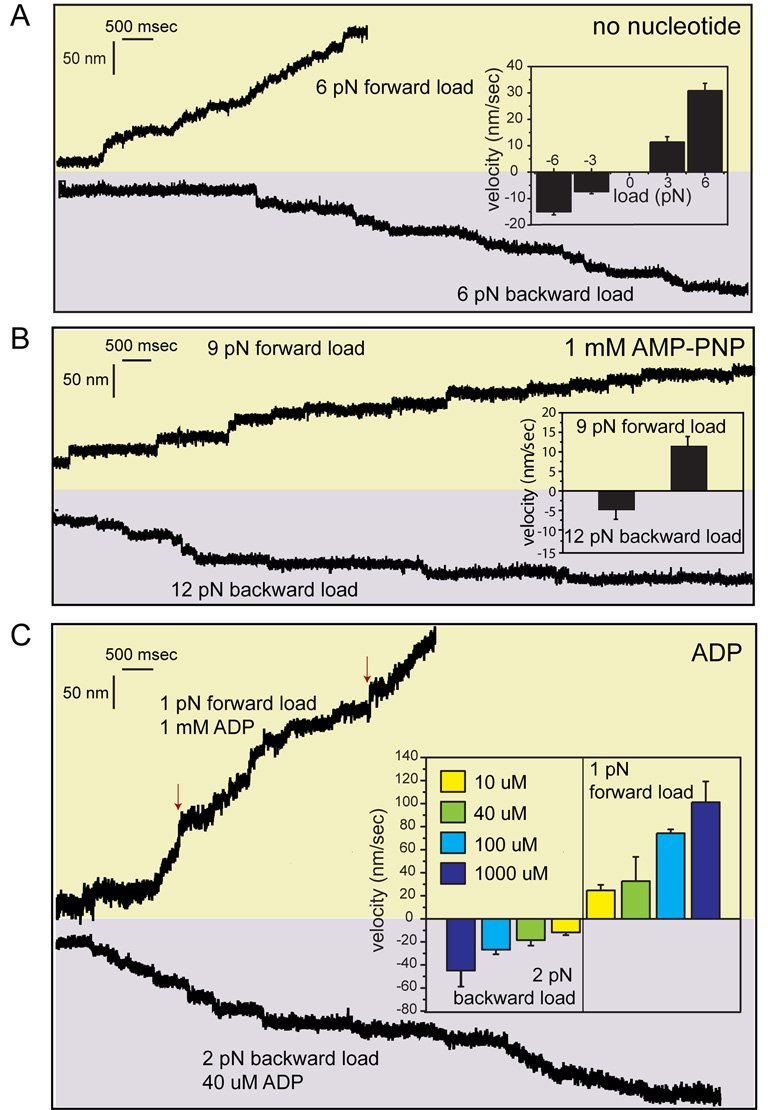
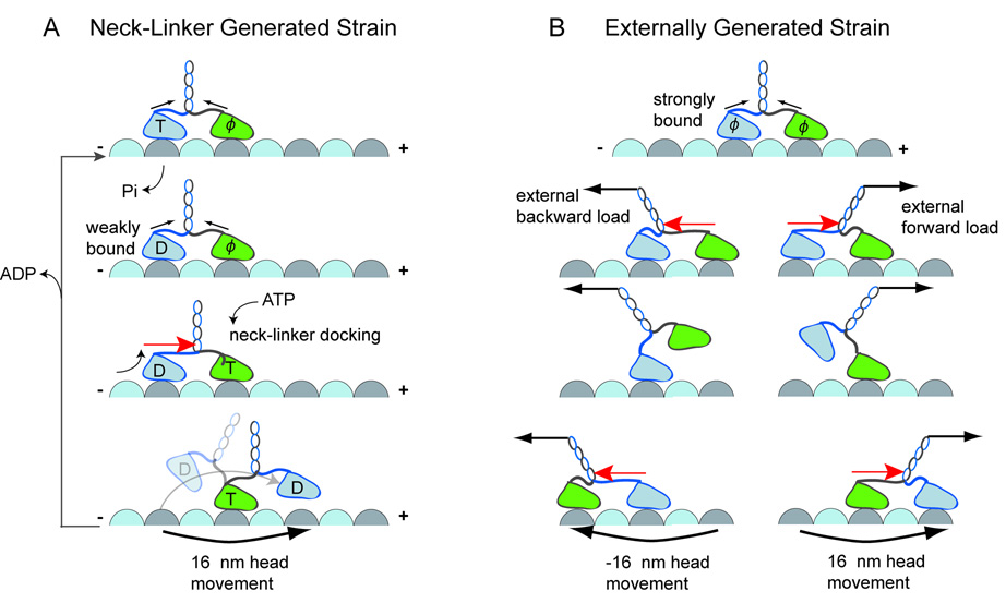
Kinesin advances 8 nm along a microtubule per ATP hydrolyzed, but the mechanism responsible for coordinating the enzymatic cycles of kinesin's two identical motor domains remains unresolved. Here, we have tested whether such coordination is mediated by intramolecular tension generated by the "neck linkers," mechanical elements that span between the motor domains. When tension is reduced by extending the neck linkers with artificial peptides, the coupling between ATP hydrolysis and forward stepping is impaired and motor's velocity decreases as a consequence. However, speed recovers to nearly normal levels when external tension is applied by an optical trap. Remarkably, external load also induces bidirectional stepping of an immotile kinesin that lacks its mechanical element (neck linker) and fuel (ATP). Our results indicate that the kinesin motor domain senses and responds to strain in a manner that facilitates its plus-end-directed stepping and communication between its two motor domains.


Figures

Figure 1
Figure 1. Motility Properties of Kinesins with Extended Neck Linkers
A. Various lengths of polyproline and glycine-serine repeats, preceded by two lysines and followed by a single glycine residue, were inserted between the neck linker (red) and the neck coiled-coil domains (gray). As examples, insertions (black) of 2, 4, 6, 13, 19, 26 prolines (2P, 4P, 6P, 13P, 19P, 26P) are expected to extend the neck linker (red) length to 4.7, 5.4, 6, 8, 10 and 12 nm, respectively. The insert of 7 repeats of glycine and serine (14GS) is depicted as a flexible 6 nm linker. B–C. Average run lengths and speeds of single GFP-tagged kinesin molecules at 1 mM ATP. D. Maximum ATP turnover rates of GFP-tagged kinesin dimers are similar at saturating microtubule concentrations. E. Calculated coupling ratio of mechanical stepping to ATP hydrolysis. See Supplemental Methods for details of these measurements.
Figure 2
Figure 2. Chemical Crosslinking of the Neck Linker in an Extended Kinesin Increases the Velocity of Movement
A. A unique cysteine residue was added to the N-terminus of the 13P insertion in a cys-light kinesin construct. B. Specific interchain crosslinking of Cys-13P, but not 13P, induced by a bifunctional crosslinking agent (Coomassie stained SDS-PAGE gel). C. Speed histogram of single Cys-13P molecules without crosslinking shows a peak at 110 nm/s. D. After crosslinking, Cys-13P shows a second peak at higher speeds centered around 250 nm. The bimodal distribution of steps agrees with mixed populations of crosslinked and noncrosslinked kinesin observed in SDS-PAGE.
Figure 3
Figure 3. Stepping Behavior of Extended Kinesin Mutants
A. Kinesin motors were labeled with a single quantum dot on one head and head movement was recorded with 70 ms integration time. Stepping traces of WT (blue), 6P (wine), 13P (black), 19P (cyan), 26P (navy), and 14GS (olive), fitted with a step detection program (red lines) (Kerssemakers et al., 2006). Motor velocity was kept in the range of 10–15 nm/s by adding different concentrations of ATP for each construct (1 µM for WT, 15 µM for 6P and 13P, 25 µM for 19P, 40 µM for 26P, 100 µM for 14GS)(see Experimental Procedures for details). B. A histogram of analyzed step sizes along the microtubule axis shows that the WT head moves with a consistent 16 nm step whereas the head of the neck mutants takes highly variable steps that show peaks at multiples of ~8 nm. The number of analyzed steps (Nstep) and probability for each construct to take a backward step (pBW) are indicated in each panel. C. Available binding sites (black circles) for the rear head in the microtubule plus end direction. D–F. Traces showing motor head displacement parallel (black trace) and perpendicular (blue trace) to the long microtubule axis for wild type (WT), 14GS and 26P, respectively. Vertical dotted lines indicate diagonal steps. The inserts show histograms of sideways step sizes, separated into left and right directions. The overall probability of taking a sideways step (psw) is shown on each panel. G. Randomness parameter (r) is derived from the dwell time measurements and suggests an uncoupling defect for extended kinesins (see Results for details).
Figure 4
Figure 4. Extended Kinesin Velocity can be Rescued by an Assisting Load
A. Schematic representation of a kinesin motor attached to a bead (green, not to scale) and trapped with a stationary laser beam (left panel). Displacement records of WT, 13P, 14GS kinesin motors at 1 mM ATP show successive runs, stalling, and motor detachment events. The stall force distributions (mean ± s.d.) are shown on the right. B. Kinesin movement under a constant assisting load applied by a force-feedback controlled trap (diagram). Individual traces of WT, 13P, 14GS and 26P under 3 pN (blue), 6 pN (green) and 9 pN (red) forward load at 1 mM ATP show stepwise movement (inserts). C. Under forward load, the velocity of WT shows a very small increase, while 13P, 14GS and 26P mutants speed up considerably with increasing load. D. Histograms of center-of-mass steps at a 9 pN assisting load show a larger average step size for 26P (11.51 ± 4.89 nm (mean ± s.d.), N = 305) than WT (7.98 ± 2.89 nm (mean ± s.d), N = 250)).
Figure 5
Figure 5. Load-Induced Kinesin Movement under Different Nucleotide Conditions
A. In the absence of a nucleotide, WT kinesin motors displayed bidirectional movement under a 6 pN forward (plus-end-directed) or backward load. Motors moved faster toward the plus-end under 6 pN load (30.8 ± 2.8 nm/s (mean ± s.e.m.) compared to 3 pN load (11.3 ± 2.1 nm/s). Under 6 pN, higher velocities were recorded toward the plus end compared to the minus end (8.5 ± 1.3 nm/sec). B. In the presence of 1 mM AMP-PNP, higher loads were required to induce stepping of single kinesin motors. Under 9 pN forward load, WT kinesin took 8 nm center-of-mass steps along the microtubule with a velocity of 12 nm/sec. 12 pN backward load was required to induce efficient movement at 5 nm/sec. C. In the presence of ADP, kinesin displayed bidirectional movement along microtubules under forces as low as 1 pN in forward or 2 pN in backward direction. The inset shows the average velocities recorded at different ADP concentrations. The red arrows show a rare slippage (<3% of stepping) along the microtubule axis during processive movement.
Figure 6
Figure 6. Role of Neck linker in Kinesin Stepping
A. Kinesin’s native neck linker sequence was replaced by 19 proline residues (19P-NL). B. A kymograph shows that the fluorescently-tagged 19P-NL construct does not move along axonemes with 1 mM ATP. C. Under 6 pN external load, 19P-NL showed processive movement in both directions. The insert shows that the force-induced kinesin movement occurs by consecutive 8 nm displacements and not by slippages along the microtubule axis. The motor moved faster under 1 mM ATP (black traces) compared to no nucleotide condition (red traces). Bar graph shows the measured velocities (N=40–70).
Figure 7
Figure 7. Internal or External Strain Facilitates Kinesin Stepping
A. Model for ATP-dependent kinesin stepping by neck linker -generated strain. ATP binding to the front head causes neck linker docking, which increases intramolecular strain (red arrow); forward strain on the rear head favors its dissociation after it transits to a weakly-bound ADP state (D). After its dissociation, the head shifts forward, biasing its attachment to the next available forward binding site. Note, however, that intramolecular strain from the stretched neck linkers (Figure S1) may suffice to release the rear head in its ADP state, without neck linker docking. B. Strain on kinesin heads can be generated by external load (black arrows). Forward load increases the strain on the rear head by pulling the neck linker. This favors the rear head detachment and displacement toward the microtubule plus end (right). Similarly, a backward load pulls the front head backward due to strain-dependent detachment (left). This type of stepping can occur in the absence of ATP and the neck linker.

Comment in

Similar articles

Cited by

References

    1. Alonso MC, Drummond DR, Kain S, Hoeng J, Amos L, Cross RA. An ATP gate controls tubulin binding by the tethered head of kinesin-1. Science. 2007;316:120–123. - PMC - PubMed
    1. Asbury CL, Fehr AN, Block SM. Kinesin moves by an asymmetric hand-over-hand mechanism. Science. 2003;302:2130–2134. - PMC - PubMed
    1. Block SM. Kinesin motor mechanics: binding, stepping, tracking, gating, and limping. Biophys J. 2007;92:2986–2995. - PMC - PubMed
    1. Block SM, Asbury CL, Shaevitz JW, Lang MJ. Probing the kinesin reaction cycle with a 2D optical force clamp. Proc Natl Acad Sci U S A. 2003;100:2351–2356. - PMC - PubMed
    1. Carter NJ, Cross RA. Mechanics of the kinesin step. Nature. 2005;435:308–312. - PubMed

Publication types

LinkOut - more resources