

Cyclosporine preserves mitochondrial morphology after myocardial ischemia/reperfusion independent of calcineurin inhibition
- PMID: 18805178
- PMCID: PMC4041111
- DOI: 10.1016/j.athoracsur.2008.06.033
Cyclosporine preserves mitochondrial morphology after myocardial ischemia/reperfusion independent of calcineurin inhibition
Abstract
Background: Opening of the mitochondrial permeability transition pore (MPTP) has been shown to contribute to myocardial ischemia/reperfusion injury. We sought to demonstrate that the myocardial protective effect of inhibiting MPTP opening with cyclosporine A (CsA) results in stabilization of mitochondrial morphology and is independent of CsA-induced calcineurin inhibition.
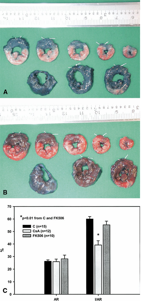
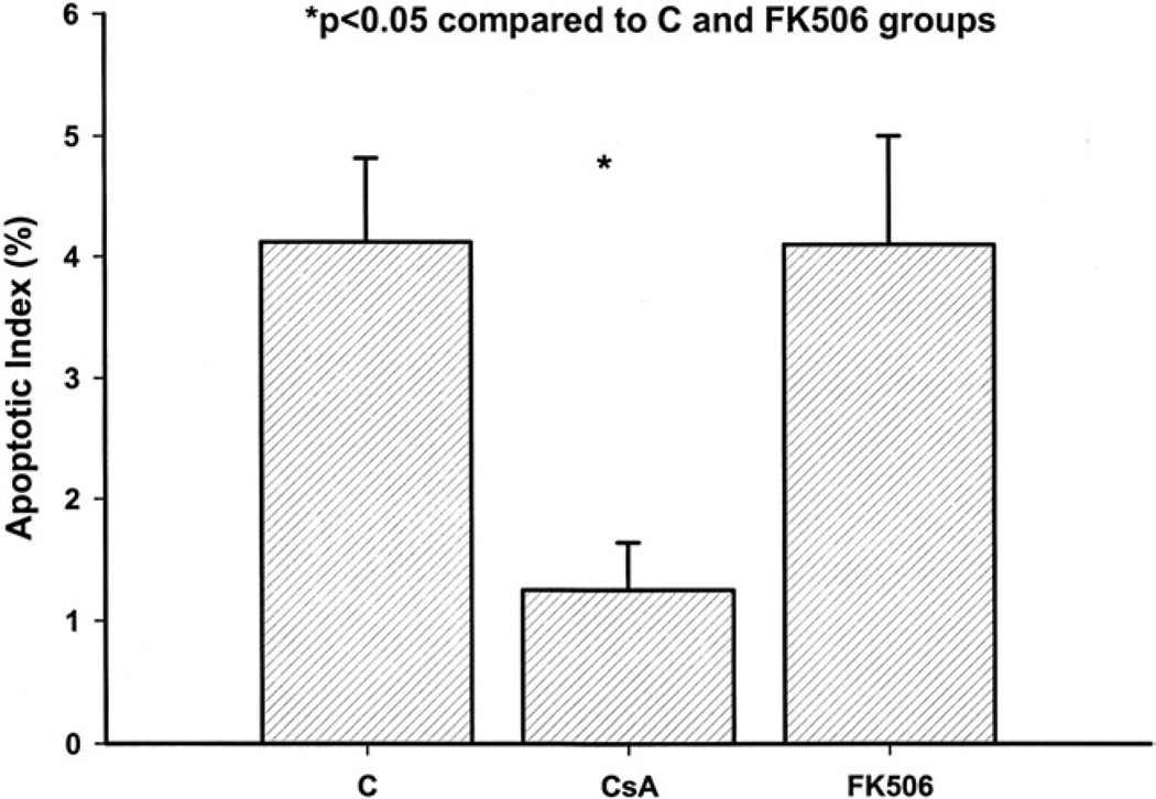
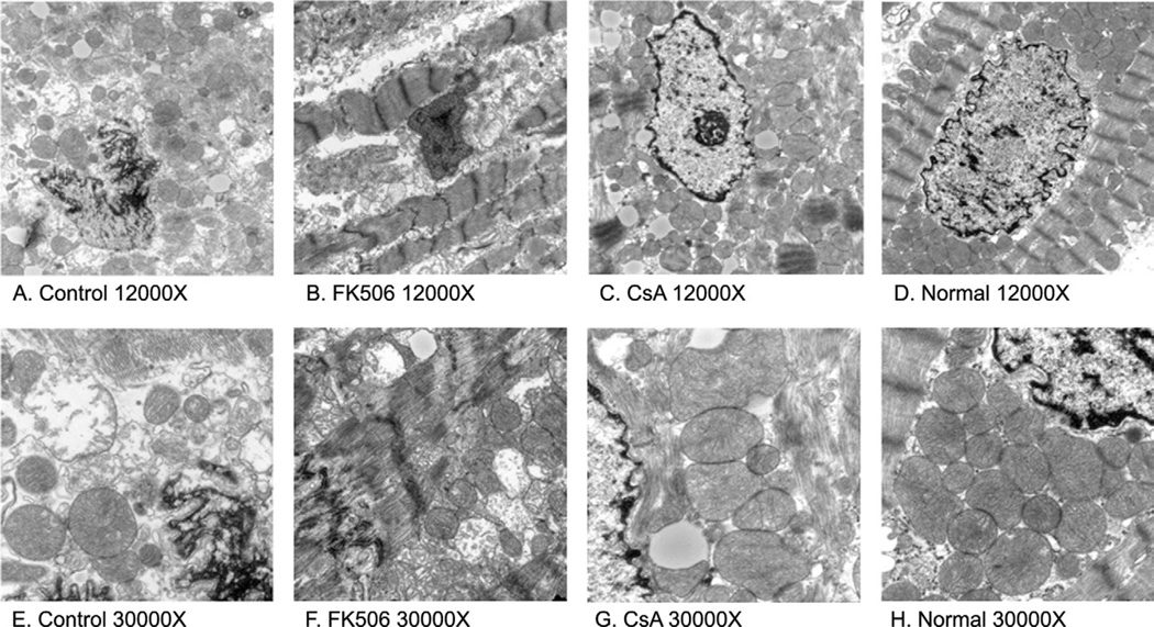
Methods: Thirty-seven rabbits were divided into three groups: control (n = 15), CsA (MPTP and calcineurin inhibitor, n = 12), or FK506 (calcineurin inhibitor, n = 10). Each group received a 1-hour infusion of either a saline vehicle, 25 mg/kg CsA or 1 mg/kg FK506. All animals underwent 30 minutes of regional ischemia and 3 hours of reperfusion. Myocardial infarct size was determined using Evans blue dye and triphenyltetrazolium chloride. In situ oligo ligation was used to assess apoptotic cell death. Transmission electron microscopy was used to quantitatively evaluate morphologic differences in the mitochondria between groups.
Results: Infarct size in the CsA group (39% +/- 3%) was significantly reduced compared with the control group (60% +/- 2%, p < 0.001) and FK506 group (55% +/- 3%, p = 0.001). Apoptotic cell death was also attenuated in the CsA group (1.2% +/- 0.5%) compared with the control group (4.3% +/- 0.8%, p = 0.01) and FK506 group (4.1% +/- 0.9%, p = 0.05). Transmission electron microscopy revealed a preservation of normal mitochondrial morphology and a reduction in the percentage of disrupted mitochondria in the CsA group (20% +/- 7%) compared with the control group (53% +/- 12%) and FK506 group (47% +/- 9%).
Conclusions: Cyclosporine A-induced MPTP inhibition preserves mitochondrial morphology after myocardial ischemia/reperfusion and limits myocyte necrosis and apoptosis. These effects are independent of calcineurin inhibition.
Figures
Comment in
-
Invited commentary.Ann Thorac Surg. 2008 Oct;86(4):1292. doi: 10.1016/j.athoracsur.2008.07.012. Ann Thorac Surg. 2008. PMID: 18805179 No abstract available.
Similar articles
-
In vivo fluorometric assessment of cyclosporine on mitochondrial function during myocardial ischemia and reperfusion.Ann Thorac Surg. 2010 May;89(5):1532-7. doi: 10.1016/j.athoracsur.2010.01.065. Ann Thorac Surg. 2010. PMID: 20417773 Free PMC article.
-
Inhibiting mitochondrial permeability transition pore opening: a new paradigm for myocardial preconditioning?Cardiovasc Res. 2002 Aug 15;55(3):534-43. doi: 10.1016/s0008-6363(02)00455-8. Cardiovasc Res. 2002. PMID: 12160950
-
Inhibition of GSK3beta by postconditioning is required to prevent opening of the mitochondrial permeability transition pore during reperfusion.Circulation. 2008 May 27;117(21):2761-8. doi: 10.1161/CIRCULATIONAHA.107.755066. Epub 2008 May 19. Circulation. 2008. PMID: 18490522
-
Targeting myocardial reperfusion injuries with cyclosporine in the CIRCUS Trial - pharmacological reasons for failure.Fundam Clin Pharmacol. 2016 Apr;30(2):191-3. doi: 10.1111/fcp.12177. Epub 2016 Jan 15. Fundam Clin Pharmacol. 2016. PMID: 26713664 Review.
-
[Endogenous cardioprotective mechanisms - what is it about and how does it work?].Kardiol Pol. 2011;69 Suppl 3:59-66. Kardiol Pol. 2011. PMID: 22125205 Review. Polish.
Cited by
-
Cyclosporin variably and inconsistently reduces infarct size in experimental models of reperfused myocardial infarction: a systematic review and meta-analysis.Br J Pharmacol. 2012 Apr;165(7):2034-43. doi: 10.1111/j.1476-5381.2011.01691.x. Br J Pharmacol. 2012. PMID: 21950961 Free PMC article.
-
Beneficial effects of Cyclosporine A in combination with Nortriptyline on germ cell-specific apoptosis, oxidative stress and epididymal sperm qualities following testicular ischemia/reperfusion in rats: a comparative study.BMC Pharmacol Toxicol. 2022 Aug 5;23(1):59. doi: 10.1186/s40360-022-00601-6. BMC Pharmacol Toxicol. 2022. PMID: 35932053 Free PMC article.
-
Therapeutic potential and recent advances on targeting mitochondrial dynamics in cardiac hypertrophy: A concise review.Mol Ther Nucleic Acids. 2021 Jun 24;25:416-443. doi: 10.1016/j.omtn.2021.06.006. eCollection 2021 Sep 3. Mol Ther Nucleic Acids. 2021. PMID: 34484866 Free PMC article. Review.
-
New Nanoparticle Formulation for Cyclosporin A: In Vitro Assessment.Pharmaceutics. 2021 Jan 12;13(1):91. doi: 10.3390/pharmaceutics13010091. Pharmaceutics. 2021. PMID: 33445646 Free PMC article.
-
In vivo fluorometric assessment of cyclosporine on mitochondrial function during myocardial ischemia and reperfusion.Ann Thorac Surg. 2010 May;89(5):1532-7. doi: 10.1016/j.athoracsur.2010.01.065. Ann Thorac Surg. 2010. PMID: 20417773 Free PMC article.
References
-
- Halestrap AP. Calcium, mitochondria and reperfusion injury: a pore way to die. Biochem Soc Trans. 2006;34:232–237. - PubMed
-
- Nazareth W, Yafei N, Crompton M. Inhibition of anoxia-induced injury in heart myocytes by cyclosporin. A. J Mol Cell Cardiol. 1991;23:1351–1354. - PubMed
-
- Weinbrenner C, Liu GS, Downey JM, Cohen MV. Cyclosporine A limits myocardial infarct size even when administered after onset of ischemia. Cardiovasc Res. 1998;38:678–684. - PubMed
-
- Griffiths EJ, Halestrap AP. Protection by cyclosporin A of ischemia/reperfusion-induced damage in isolated rat hearts. J Mol Cell Cardiol. 1993;25:1461–1469. - PubMed
-
- Hausenloy DJ, Maddock HL, Baxter GF, Yellon DM. Inhibiting mitochondrial permeability transition pore opening: a new paradigm for myocardial preconditioning? Cardiovasc Res. 2002;55:534–543. - PubMed
Publication types
MeSH terms
Substances
Grants and funding
LinkOut - more resources
Full Text Sources
Other Literature Sources
