

Perivascular cells increase expression of ciliary neurotrophic factor following partial denervation of the rat neurohypophysis
- PMID: 18805412
- PMCID: PMC2998289
- DOI: 10.1016/j.expneurol.2008.08.011
Perivascular cells increase expression of ciliary neurotrophic factor following partial denervation of the rat neurohypophysis
Abstract
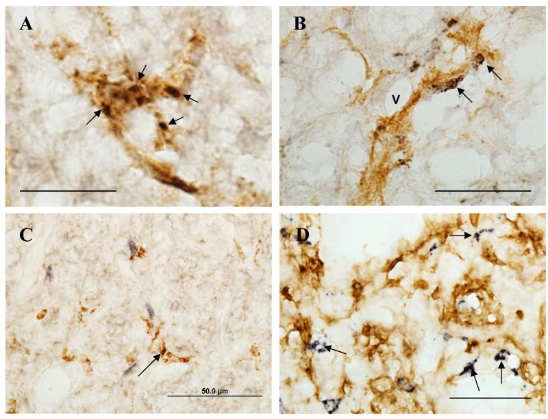
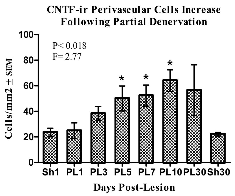
The expression of ciliary neurotrophic factor (CNTF) was investigated immunocytochemically during the axonal degeneration and collateral axonal sprouting response that follows partial denervation of the rat neurohypophysis. A significant increase in the number of CNTF-immunoreactive (CNTF-ir) cells was observed in the neurohypophysis of partially denervated animals compared to age-matched sham-operated controls by 5 days post-denervation, remaining elevated throughout the 30 day post-denervation period. Stereometric assessment of the numbers of CNTF-ir cells within the partially denervated neurohypophysis demonstrated a 36% increase by 3 days following denervation reaching 130% of control values by 10 days post-lesion. The cell numbers remained elevated throughout the 30 day post-lesion period suggesting that CNTF may play a role in the neurosecretory axonal sprouting process known to occur between 10 and 30 days post-denervation. Subsequent preparations pairing anti-CNTF with antibodies against ED1, CR3, p75 low affinity neurotrophin receptor (p75(LNGFR)), and S100beta, demonstrated that CNTF was exclusively localized in a phenotypically-distinct population of perivascular cells. The association of perivascular cells with phagocytic activity was confirmed by dual-label fluorescence microscopy showing the colocalization of P75(LNGFR)-ir and OX-42-ir in cells expressing the ED-1 antigen. No increase in CNTF-ir was observed in non-injured animals in which heightened levels of neurosecretory activity were induced physiologically. These results suggest that increased CNTF-ir occurs in response to conditions which induce high levels of phagocytic activity by perivascular cells in the axotomized neurohypophysis which is sustained throughout a period in which axonal sprouting is known to occur in the partially denervated neurohypophysis.
Figures

Similar articles
-
Upregulation of the p75 low-affinity neurotrophin receptor by phagocytically active perivascular active cells in the rat neural lobe.Cell Tissue Res. 2001 Jan;303(1):81-91. doi: 10.1007/s004410000295. Cell Tissue Res. 2001. PMID: 11236007
-
CNTF receptor alpha is expressed by magnocellular neurons and expression is upregulated in the rat supraoptic nucleus during axonal sprouting.Exp Neurol. 2009 Jan;215(1):135-41. doi: 10.1016/j.expneurol.2008.09.021. Epub 2008 Oct 11. Exp Neurol. 2009. PMID: 18973757 Free PMC article.
-
Neuronal activity and axonal sprouting differentially regulate CNTF and CNTF receptor complex in the rat supraoptic nucleus.Exp Neurol. 2012 Jan;233(1):243-52. doi: 10.1016/j.expneurol.2011.10.009. Epub 2011 Oct 19. Exp Neurol. 2012. PMID: 22037350 Free PMC article.
-
Adenovirus-mediated expression of ciliary neurotrophic factor (CNTF) rescues axotomized rat retinal ganglion cells but does not support axonal regeneration in vivo.Neurobiol Dis. 2000 Jun;7(3):212-23. doi: 10.1006/nbdi.2000.0285. Neurobiol Dis. 2000. PMID: 10860786
-
Ciliary neurotrophic factor is expressed in the magnocellular neurosecretory system of the rat in vivo: evidence for injury- and activity-induced upregulation.Exp Neurol. 2006 Jan;197(1):206-14. doi: 10.1016/j.expneurol.2005.09.009. Epub 2005 Oct 14. Exp Neurol. 2006. PMID: 16226750
Cited by
-
Effects of ciliary neurotrophic factor and leukemia inhibiting factor on oxytocin and vasopressin magnocellular neuron survival in rat and mouse hypothalamic organotypic cultures.J Neurosci Methods. 2009 Mar 30;178(1):128-33. doi: 10.1016/j.jneumeth.2008.12.004. Epub 2008 Dec 11. J Neurosci Methods. 2009. PMID: 19118574 Free PMC article.
-
Inhibition of the Jak-STAT pathway prevents CNTF-mediated survival of axotomized oxytocinergic magnocellular neurons in organotypic cultures of the rat supraoptic nucleus.Exp Neurol. 2013 Feb;240:75-87. doi: 10.1016/j.expneurol.2012.10.023. Epub 2012 Nov 1. Exp Neurol. 2013. PMID: 23123407 Free PMC article.
-
Absence of axonal sprouting following unilateral lesion in 125-day-old rat supraoptic nucleus may be due to age-dependent decrease in protein levels of ciliary neurotrophic factor receptor alpha.J Comp Neurol. 2019 Oct 1;527(14):2291-2301. doi: 10.1002/cne.24675. Epub 2019 Mar 25. J Comp Neurol. 2019. PMID: 30861131 Free PMC article.
References
-
- Adams JH, Daniel PM, Prichard MMl. Degeneration and regeneration of hypothalamic nerve fibers in the neurohypophysis after pituitary stalk section in the ferret. J Comp Neurol. 1969;135:121–144. - PubMed
-
- Antunes JL, Louis KM, Huang S, Zimmerman E. Section of the pituitary stalk in the rhesus monkey: Morphological and endocrine observations. Ann Neurol. 1980;8:308–316. - PubMed
-
- Beck E, Daniel PM, Prichard MM. Regeneration of hypothalamic nerve fibers in the goat. Neuroendo. 1969;5:161–182. - PubMed
-
- Billenstein DC, Leveque TF. The reorganization of the neurohypophysial stalk following hypophysectomy in the rat. Endocrinology. 1955;56:704–717. - PubMed
-
- Cocchia D, Miana N. Immunocytochemical localization of the brain-specific S-100 protein in the pituitary gland of adult rat. J of Neurocytol. 1980;9(6):771–782. - PubMed
Publication types
MeSH terms
Substances
Grants and funding
LinkOut - more resources
Full Text Sources
Research Materials
