. 2008 Dec;295(6):L988-97.
doi: 10.1152/ajplung.90202.2008. Epub 2008 Sep 19.

Integrin-linked kinase regulates smooth muscle differentiation marker gene expression in airway tissue

Affiliations

Integrin-linked kinase regulates smooth muscle differentiation marker gene expression in airway tissue

Yidi Wu et al. Am J Physiol Lung Cell Mol Physiol. 2008 Dec.

Abstract

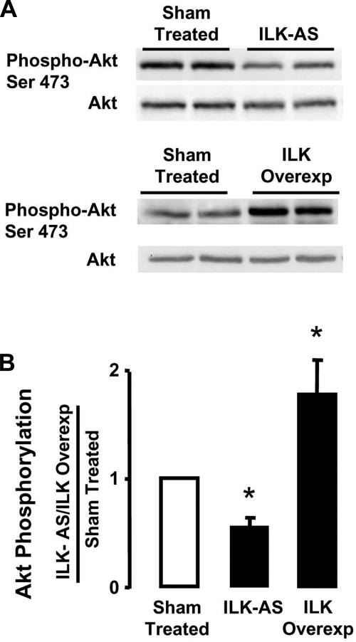
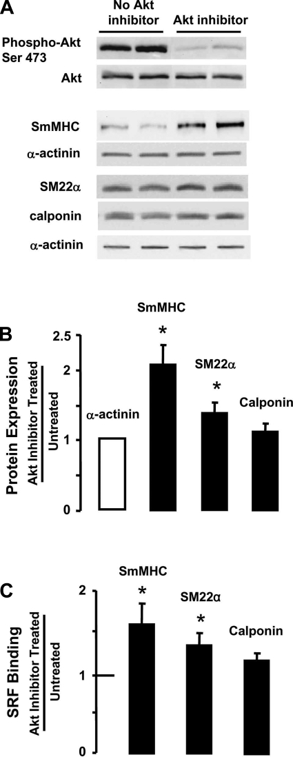
Phenotypic changes in airway smooth muscle occur with airway inflammation and asthma. These changes may be induced by alterations in the extracellular matrix that initiate signaling pathways mediated by integrin receptors. We hypothesized that integrin-linked kinase (ILK), a multidomain protein kinase that binds to the cytoplasmic tail of beta-integrins, may be an important mediator of signaling pathways that regulate the growth and differentiation state of airway smooth muscle. We disrupted signaling pathways mediated by ILK in intact differentiated tracheal muscle tissues by depleting ILK protein using ILK antisense. The depletion of ILK protein increased the expression of the smooth muscle differentiation marker genes myosin heavy chain (SmMHC), SM22alpha, and calponin and increased the expression of SmMHC protein. Conversely, the overexpression of ILK protein reduced the mRNA levels of SmMHC, SM22alpha, and calponin and SmMHC protein. Analysis by chromatin immunoprecipitation showed that the binding of the transcriptional regulator serum response factor (SRF) to the promoters of SmMHC, SM22alpha, and calponin genes was increased in ILK-depleted tissues and decreased in tissues overexpressing ILK. ILK depletion also increased the amount of SRF that localized within the nucleus. ILK depletion and overexpression, respectively, decreased and increased the activation of its downstream substrate protein kinase B (PKB/Akt). The pharmacological inhibition of Akt activity also increased SRF binding to the promoters of smooth muscle-specific genes and increased expression of smooth muscle proteins, suggesting that ILK may exert its effects by regulating the activity of Akt. We conclude that ILK is a critical regulator of airway smooth muscle differentiation. ILK may mediate signals from integrin receptors that control airway smooth muscle differentiation in response to alterations in the extracellular matrix.


Figures

Fig. 1.
Fig. 1.
Treatment with integrin-linked kinase (ILK) antisense depresses ILK protein expression in airway smooth muscle tissues. A: immunoblot illustrating the effects of ILK antisense treatment on ILK protein expression. ILK protein was depleted by treating smooth muscle tissues with ILK antisense (ILK-AS) for 24 h. Treatment with ILK sense (ILK-S) had no effect on the expression of ILK protein. Sham-treated muscles were subjected to identical procedures in the absence of oligonucleotides. B: mean expression levels of ILK protein in tissues treated with ILK-AS or ILK-S, normalized to protein levels in sham-treated tissues. ILK protein was significantly decreased in tissues treated with ILK antisense but was unaffected by treatment with ILK sense. Means ± SE (n = 6). *Significantly different from sham treated, P < 0.05.
Fig. 2.
Fig. 2.
Depletion of ILK increased mRNA levels for smooth muscle myosin heavy chain (SmMHC), SM22α, and calponin and SmMHC protein in tracheal smooth muscle tissues. A: mean expression of mRNA transcripts for smooth muscle-specific genes as measured by quantitative RT-PCR. Values for each experiment were the average of measurements from duplicate or triplicate muscle tissues for each treatment, normalized to values for sham-treated muscles. N represents the no. of experiments. Values are means ± SE. Depletion of ILK protein significantly increased the mRNA levels for SmMHC (n = 4), SM22α (n = 6), and calponin (n = 5), but it did not significantly affect mRNA levels of α-actinin (n = 3). *Significantly different from 1 (sham-treated), P < 0.05. B: immunoblot of proteins from extracts of sham-treated and ILK-AS treated muscle tissues. ILK depletion significantly increased SmMHC protein expression, but it did not significantly affect SM22α, calponin, or α-actinin expression in tracheal muscle tissues. C: mean results for the effect of ILK depletion on protein expression. SmMHC is significantly increased relative to sham-treated tissues (n = 6). The expression of SM22α (n = 6) and calponin (n = 6) was not significantly altered relative to sham-treated tissues. The expression level of each protein in AS-treated tissues is normalized to the level of α-actinin in the same sample. Values are means ± SE. *Significantly different from 1 (α-actinin), P < 0.05.
Fig. 3.
Fig. 3.
Depletion of ILK protein in tracheal muscle tissues by ILK antisense increases the amount of SRF in the nucleus. A: tracheal muscle cells were enzymatically dissociated from tissues that had been depleted of ILK with antisense or that were sham-treated. Cells were immunostained for serum response factor (SRF) (green) and counterstained for nuclear DNA with propidium iodide (PI) (red). Yellow color indicates colocalization. White lines illustrate line scans for quantitation of pixel intensity. More SRF was localized to the nucleus of muscle cells from ILK antisense-treated tissues than in cells from sham-treated tissues. B: method for the quantitative analysis of SRF localization. The pixel intensity of SRF was quantified using a series of cross-sectional line scans through the area of the nucleus (3 lines) and the cytoplasm (4 lines). The ratio of nuclear to cytoplasmic SRF was determined for each cell by calculating the average pixel intensity of the lines across the nucleus relative to the cytoplasm. C: ratio of nuclear to cytoplasmic SRF was significantly higher in ILK-depleted tissues compared with sham-treated tissues. Values are means ± SE. *Significant difference between cells from sham-treated (n = 47) and ILK-AS-treated (n = 43) tissues, P < 0.05. The ratio of nuclear/cytoplasmic SRF was significantly greater than 1 in both groups of cells. D: nuclear and cytoplasmic extracts were prepared from sham-treated smooth muscle tissues and tissues treated with ILK antisense and were subjected to immunoblotting for SRF protein. More SRF was observed in nuclear extracts from ILK antisense-treated tissues than from sham-treated tissues. Glyceraldehyde-3-phosphate dehydrogenase was used as cytoplasmic marker. E: mean results for the effect of ILK depletion on the nuclear SRF. The amount of nuclear SRF was significantly increased in ILK-depleted tissues compared with sham-treated tissues. Values are means ± SE (n = 3). *P < 0.01.
Fig. 4.
Fig. 4.
Depletion of ILK protein expression increased the binding of SRF to the promoters of SmMHC, SM22α, and calponin in tracheal smooth muscle tissues. SRF was immunoprecipitated from chromatin extracts from ILK-depleted smooth muscle tissues. SRF immunoprecipitates were subjected to real-time PCR to amplify SmMHC, SM22α, and calponin promoter DNA. α-Actinin promoter DNA was used as a negative control for SRF binding. Depletion of ILK protein significantly increased the amount of SRF bound to the SmMHC promoter relative to sham-treated tissues (2.24 ± 0.42). ILK depletion also significantly increased the amount of SRF bound to the SM22α (2.10 ± 0.40) and calponin (1.54 ± 0.26) promoters compared with sham-treated tissues. Binding of SRF to the α-actinin promoter was negligible in extracts from both ILK-AS-treated and sham-treated tissues. Results represent means from 4 separate experiments. Values are means ± SE (n = 4). *Significantly different from sham-treated tissues (P < 0.05).
Fig. 5.
Fig. 5.
Treatment of airway smooth muscle tissues with ILK plasmids caused an increase in ILK protein expression. A: immunoblot indicating the effects of transfection with plasmids encoding WT-ILK on ILK protein expression. Treatment of smooth muscle tissues with ILK-WT plasmids followed by 2 days incubation caused upregulation of ILK protein expression (ILK Overexp). B: mean expression level of ILK protein in tissues treated with ILK plasmids relative to sham-treated tissues. ILK protein was significantly increased in tissues transfected with ILK plasmids (n = 14). Values are means ± SE. *Significantly different from 1 (Sham treated), P < 0.05. C: expression of recombinant FLAG-ILK protein was confirmed by immunoprecipitation of recombinant ILK from muscle extracts using anti-Flag antibody (Ab) followed by immunoblot using ILK Ab.
Fig. 6.
Fig. 6.
Overexpression of ILK decreases gene expression and SRF binding to the promoters of SmMHC, SM22α, and calponin. A: mean expression of mRNA transcripts for smooth muscle-specific genes as measured by quantitative RT-PCR. mRNA expression levels are expressed relative to values for sham-treated tissues. “N” represents the no. of separate experiments for each treatment. Values for each experiment were the average of measurements from duplicate or triplicate muscle tissues for each treatment. Values are means ± SE. Overexpression of ILK significantly decreased mRNA levels for SmMHC (n = 5), SM22α (n = 8), and calponin (n = 4), but ILK overexpression did not significantly affect mRNA levels for α-actinin (n = 4). B: SRF was immunoprecipitated from chromatin extracts from ILK-depleted smooth muscle tissues. SRF immunoprecipitates were subjected to real-time PCR to amplify SmMHC, SM22α, and calponin promoter DNA. α-Actinin promoter DNA was used as a negative control for SRF binding. The overexpression of ILK protein significantly decreased the amount of SRF binding to the promoters of SmMHC (0.58 ± 0.06), SM22α (0.65 ± 0.07), and calponin (0.79 ± 0.05) relative to sham-treated tissues.
Fig. 7.
Fig. 7.
Expression of smooth muscle SmMHC was decreased in tissues overexpressing ILK. A: immunoblots from extracts of tissues treated with ILK plasmids or sham treated. Expression of SmMHC is decreased in tissues overexpressing ILK, whereas there is little effect on the expression of SM22α, calponin, and α-actinin. B: mean results for the effect of ILK overexpression on protein expression. SmMHC expression is significantly decreased relative to sham-treated tissues (n = 9). The expressions of SM22α (n = 4) and calponin (n = 4) were not significantly affected by ILK overexpression. Amount of each protein is normalized to that of α-actinin in the same sample. Values are means ± SE. *Significantly different from 1 (sham treated), P < 0.05.
Fig. 8.
Fig. 8.
ILK depletion and ILK overexpression regulates protein kinase B (Akt) phosphorylation in smooth muscle tissues. A: ILK antisense treatment for 24 h caused an inhibition of Akt phosphorylation at Ser473 in smooth muscle tissues, indicating Akt inactivation. In contrast, ILK overexpression increased Akt phosphorylation at Ser473, indicating Akt activation. B: mean values for effect of ILK depletion (n = 3) or overexpression (n = 4) on Akt phosphorylation at Ser473 (means ± SE). *Significantly different from sham-treated tissues, P < 0.05.
Fig. 9.
Fig. 9.
Akt inhibitor treatment increased expression of smooth muscle-specific proteins and increased SRF binding to the promoters of smooth muscle-specific genes. A: treatment of muscle tissues with 50 μM Akt inhibitor VIII significantly inhibits Akt phosphorylation at Ser473. Akt treatment substantially increased SmMHC protein expression, but had less effect on the expression of SM22α and calponin h1. B: mean effect of Akt inhibition on SmMHC (n = 8), SM22α (n = 4), and calponin (n = 4) expression. Values are means ± SE. *Values are significantly different from untreated tissues, P < 0.05. C: SRF was immunoprecipitated from chromatin extracts from Akt inhibitor-treated and untreated smooth muscle tissues. SRF immunoprecipitates were subjected to real-time PCR to amplify SmMHC, SM22α, and calponin promoter DNA. α-Actinin promoter DNA was used as a negative control for SRF binding. Akt inhibitor treatment significantly increased the amount of SRF binding to the promoters of SmMHC and SM22α relative to untreated tissues.

Similar articles

Cited by

References

    1. Attwell S, Mills J, Troussard A, Wu C, Dedhar S. Integration of cell attachment, cytoskeletal localization, and signaling by integrin-linked kinase (ILK), CH-ILKBP, and the tumor suppressor PTEN. Mol Biol Cell 14: 4813–4825, 2003. - PMC - PubMed
    1. Bentley JK, Hershenson MB. Airway smooth muscle growth in asthma: proliferation, hypertrophy, and migration. Proc Am Thorac Soc 5: 89–96, 2008. - PMC - PubMed
    1. Brakebusch C, Fassler R. The integrin-actin connection, an eternal love affair. EMBO J 22: 2324–2333, 2003. - PMC - PubMed
    1. Burkin DJ, Kaufman SJ. The alpha7beta1 integrin in muscle development and disease. Cell Tissue Res 296: 183–190, 1999. - PubMed
    1. Chamley-Campbell J, Campbell GR, Ross R. The smooth muscle cell in culture. Physiol Rev 59: 1–61, 1979. - PubMed

Publication types

MeSH terms

LinkOut - more resources