. 2008 Dec;190(24):8145-54.
doi: 10.1128/JB.00983-08. Epub 2008 Sep 19.

Characterization of a Streptococcus sp.-Veillonella sp. community micromanipulated from dental plaque

Affiliations

Characterization of a Streptococcus sp.-Veillonella sp. community micromanipulated from dental plaque

Natalia I Chalmers et al. J Bacteriol. 2008 Dec.

Abstract

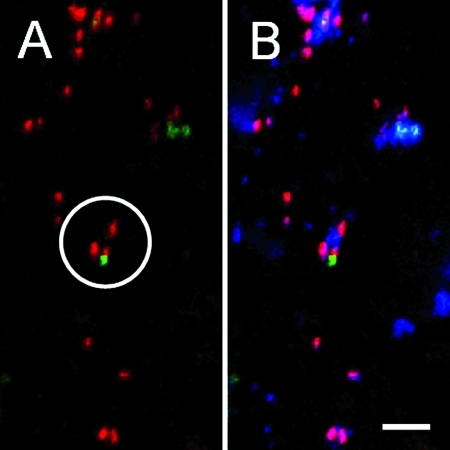
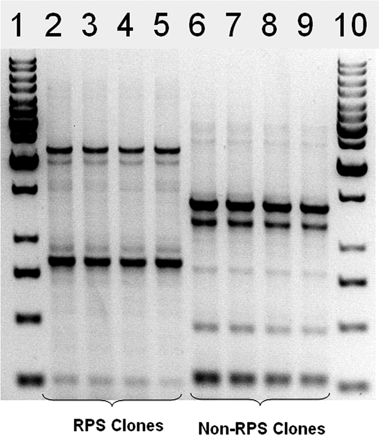
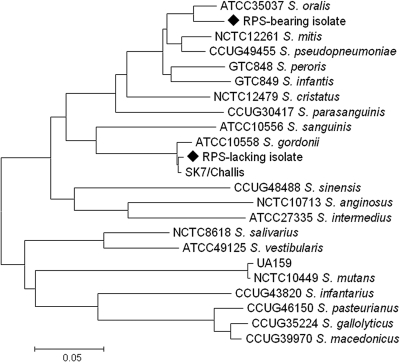
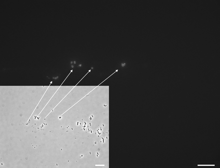
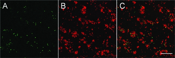
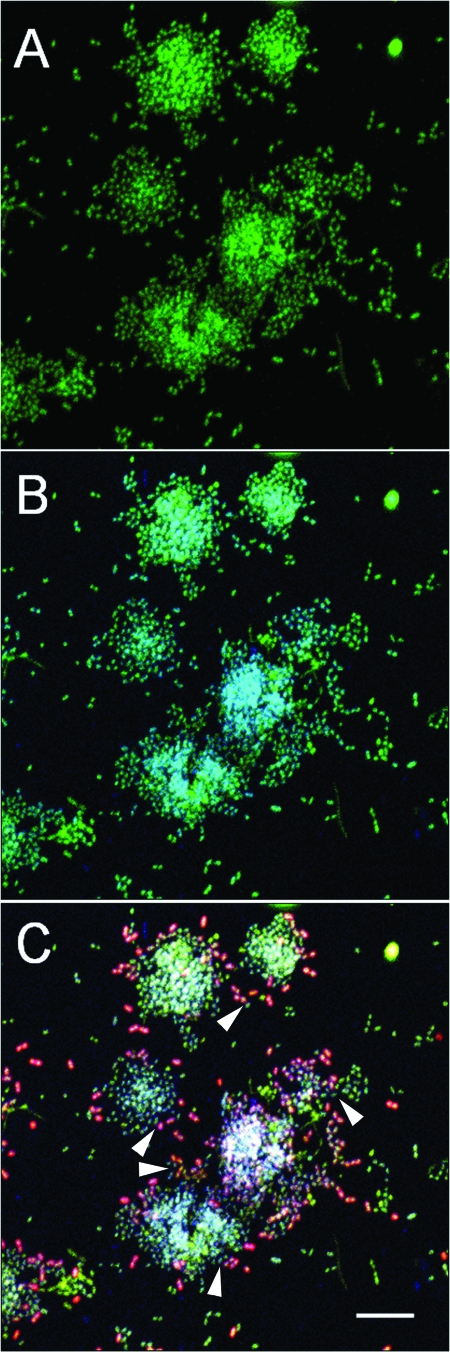
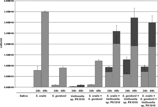
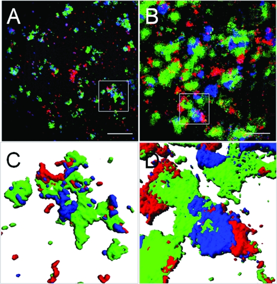
Streptococci and veillonellae occur in mixed-species colonies during formation of early dental plaque. One factor hypothesized to be important in assembly of these initial communities is coaggregation (cell-cell recognition by genetically distinct bacteria). Intrageneric coaggregation of streptococci occurs when a lectin-like adhesin on one streptococcal species recognizes a receptor polysaccharide (RPS) on the partner species. Veillonellae also coaggregate with streptococci. These genera interact metabolically; lactic acid produced by streptococci is a carbon source for veillonellae. To transpose these interactions from undisturbed dental plaque to an experimentally tractable in vitro biofilm model, a community consisting of RPS-bearing streptococci juxtaposed with veillonellae was targeted by quantum dot-based immunofluorescence and then micromanipulated off the enamel surface and cultured. Besides the expected antibody-reactive cell types, a non-antibody-reactive streptococcus invisible during micromanipulation was obtained. The streptococci were identified as Streptococcus oralis (RPS bearing) and Streptococcus gordonii (adhesin bearing). The veillonellae could not be cultivated; however, a veillonella 16S rRNA gene sequence was amplified from the original isolation mixture, and this sequence was identical to the sequence of the previously studied organism Veillonella sp. strain PK1910, an oral isolate in our culture collection. S. oralis coaggregated with S. gordonii by an RPS-dependent mechanism, and both streptococci coaggregated with PK1910, which was used as a surrogate during in vitro community reconstruction. The streptococci and strain PK1910 formed interdigitated three-species clusters when grown as a biofilm using saliva as the nutritional source. PK1910 grew only when streptococci were present. This study confirms that RPS-mediated intrageneric coaggregation occurs in the earliest stages of plaque formation by bringing bacteria together to create a functional community.


Figures

FIG. 1.
FIG. 1.
Confocal micrographs of 8-h dental plaque. (A) QD-based primary immunofluorescence revealing RPS-bearing streptococci reactive with QD655-conjugated anti-RPS (red) juxtaposed with veillonellae reactive with QD525-conjugated anti-R1 (green). A community representative of the cells selected for micromanipulation is circled. (B) Same field of view as that in panel A but with DAPI-stained cells (blue) also shown. The general nucleic acid stain DAPI revealed non-antibody-reactive cells, one of which was located in the representative community. DAPI was not used with micromanipulated samples. Bar, 10 μm.
FIG. 2.
FIG. 2.
REP-PCR patterns of four randomly selected RPS-bearing Streptococcus isolates (lanes 2 to 5) and four randomly selected RPS-negative Streptococcus isolates (lanes 6 to 9). Lanes 1 and 10 contained 1-kb molecular size markers.
FIG. 3.
FIG. 3.
Phylogenetic tree based on streptococcal sodA sequences. The neighbor-joining method was used to construct the tree. Filled diamonds indicate the two clinical isolates. Scale bar = 5% difference in nucleotide sequence. The type strains S. oralis ATCC 35037 and S. gordonii ATCC 10558 are included for reference.
FIG. 4.
FIG. 4.
Transmitted light micrograph (inset) of a wet mount of micromanipulated cells after outgrowth in an MSM broth culture and immunofluorescence microscopy (large image) of the same field of view showing cells labeled with anti-R1 antibody. The arrows indicate anti-R1-reactive cells in the two images. Note the non-antibody-reactive cells (presumed to be streptococci) in the transmitted light micrograph. Bars, 20 μm.
FIG. 5.
FIG. 5.
Confocal micrographs of immunofluorescence- and FISH-treated 8-h plaque on enamel showing (A) Veillonella cells reactive with the VEI488 FISH probe for veillonellae 16S rRNA (green), (B) RPS-bearing streptococci reactive with anti-S. oralis 34 RPS (red), and (C) an overlay of panels A and B showing juxtaposition of veillonellae and RPS-bearing streptococci. All images are maximum projection images. Bar, 40 μm.
FIG. 6.
FIG. 6.
Confocal micrographs of immunofluorescence- and FISH-treated 8-h plaque on enamel showing the distribution of RPS-bearing streptococci among other streptococci and nonstreptococcal bacteria. (A) All cells stained with the general nucleic acid stain acridine orange (green). (B) Streptococcus cells reactive with the 16S rRNA for streptococci appear blue-green through combination of acridine orange (green; shows all cells) with the streptococcal 16S rRNA probe (blue). (C) Streptococcus cells reactive with Alexa Fluor 546-conjugated anti-RPS. RPS-bearing Streptococcus cells are red with a white center. The bright white pixels in the center of large colonies result from colocalization of red, green, and blue. All images are maximum projection images. The arrowheads indicate RPS-negative streptococci (blue-green) that are close to RPS-bearing streptococci (red with white centers). Bar, 8 μm.
FIG. 7.
FIG. 7.
Q-PCR quantification of S. oralis, S. gordonii, and Veillonella sp. strain PK1910 in one-, two- and three-species biofilms at 24 and 48 h. For the two- and three-species biofilms, the number of streptococcal cells (S. oralis RPS-bearing isolate and S. gordonii RPS-negative isolate) is indicated by light gray bars, and the number of PK1910 cells is indicated by dark gray bars. The number of streptococcal cells increased between 24 and 48 h in the one-, two- and three-species biofilms. PK1910 did not form a single-species biofilm, but its biomass increased significantly in two- and three-species biofilms.
FIG. 8.
FIG. 8.
Representative confocal micrographs of 24-h (A and C) and 48-h (B and D) in vitro biofilms showing the intimate interaction between the RPS-bearing streptococcal isolate (S. oralis, labeled red by Alexa Fluor 546-conjugated anti-RPS), the RPS-negative streptococcal isolate (S. gordonii, labeled green by Alexa Fluor 488-conjugated anti-DL1), and the surrogate organism Veillonella sp. strain PK1910 (labeled blue by Alexa Fluor 633-conjugated anti-1910). (A and B) Distribution and juxtaposition of the three species on a peg surface after 24 h (A) and 48 h (B) of biofilm growth on saliva as the sole nutritional source. Significant growth of all species occurred at 48 h. (C and D) Three-dimensional volume renderings of the communities indicated by the squares in panels A and B, showing the interdigitation and spatial relationships of the three species. The arrowheads indicate interdigitation of the three species. Bars, 40 μm.

Comment in

Similar articles

Cited by

References

    1. Aas, J. A., B. J. Paster, L. N. Stokes, I. Olsen, and F. E. Dewhirst. 2005. Defining the normal bacterial flora of the oral cavity. J. Clin. Microbiol. 435721-5732. - PMC - PubMed
    1. Alam, S., S. R. Brailsford, R. A. Whiley, and D. Beighton. 1999. PCR-based methods for genotyping viridans group streptococci. J. Clin. Microbiol. 372772-2776. - PMC - PubMed
    1. Bruchez, M., Jr., M. Moronne, P. Gin, S. Weiss, and A. P. Alivisatos. 1998. Semiconductor nanocrystals as fluorescent biological labels. Science 2812013-2016. - PubMed
    1. Celesk, R. A., and J. London. 1980. Attachment of oral Cytophaga species to hydroxyapatite-containing surfaces. Infect. Immun. 29768-777. - PMC - PubMed
    1. Ceri, H., M. E. Olson, C. Stremick, R. R. Read, D. Morck, and A. Buret. 1999. The Calgary biofilm device: new technology for rapid determination of antibiotic susceptibilities of bacterial biofilms. J. Clin. Microbiol. 371771-1776. - PMC - PubMed

Publication types

MeSH terms

Associated data

LinkOut - more resources