

doi: 10.1128/JB.00839-08.
Epub 2008 Sep 19.
Assemblies of DivIVA mark sites for hyphal branching and can establish new zones of cell wall growth in Streptomyces coelicolor
Affiliations
- PMID: 18805980
- PMCID: PMC2576665
- DOI: 10.1128/JB.00839-08
Item in Clipboard
Assemblies of DivIVA mark sites for hyphal branching and can establish new zones of cell wall growth in Streptomyces coelicolor
J Bacteriol.
2008 Nov.
Abstract
Time-lapse imaging of Streptomyces hyphae revealed foci of the essential protein DivIVA at sites where lateral branches will emerge. Overexpression experiments showed that DivIVA foci can trigger establishment of new zones of cell wall assembly, suggesting a key role of DivIVA in directing peptidoglycan synthesis and cell shape in Streptomyces.
Figures
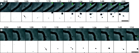
DivIVA marks sites of hyphal branching. Hyphae of strain K112 expressing divIVA-egfp were grown on agarose pads, and images were captured every 6 min. Time-lapse series of representative branching events are shown as overlays of fluorescence signal (green) on the phase contrast images (top) and as the fluorescence channel with the gray scale inverted and adjusted to clearly visualize the initially weak DivIVA-EGFP signals (bottom). The first time points when DivIVA-EGFP foci were detected at future branching sites are marked with arrows. The subsequent time points at which outgrowth of the branches can be seen are indicated by arrowheads. Time is shown in hours and minutes after starting time-lapse acquisition. Bars, 5 μm. The full time-lapse sequence from which the frames in panel A are derived is shown in Video S1 in the supplemental material.
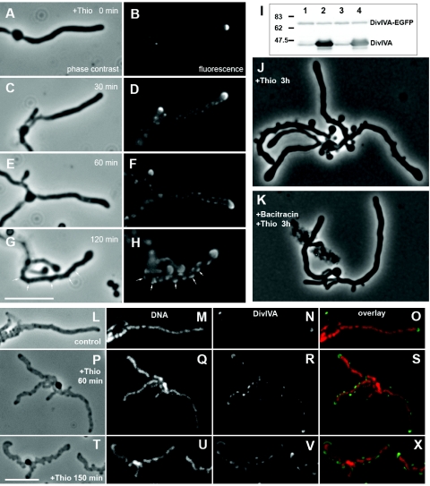
Ectopic overexpression of divIVA leads to redistribution of apical DivIVA, assembly of multiple lateral foci, and eventually initiation of cell wall assembly at sites coinciding with such foci. (A to H) Representative images of strain K121 (divIVA::pKF59[Φ(divIVA-egfp)Hyb] attBpSAM2::pKF58[tipAp-divIVA]) before and at intervals after induction of overexpression of tipAp-divIVA by addition of the inducer thiostrepton (Thio) to 10 μg/ml in YEME medium. Phase contrast images (A, C, E, and G) and fluorescence images of live cells show distribution of the DivIVA-EGFP protein (B, D, F, and H). Arrows indicate examples of the branch-like lateral outgrowths emerging at DivIVA foci. Immunoblotting with an anti-DivIVA antiserum (I) showed that the amount of DivIVA-EGFP did not change in response to induction of the tipAp promoter, while there was strong overproduction of normal DivIVA. Two independent cultures were each split in two halves, and thiostrepton (10 μg/ml) was added to one of the halves (lanes 2 and 4) while the other was uninduced for 2 h (lanes 1 and 3). A molecular weight standard is indicated. (J and K) Phase contrast images of cells from a similar experiment as described for panels A to H, except that 5 min prior to thiostrepton addition, bacitracin was added to block cell wall synthesis (K), or a mock addition of a corresponding amount of 0.1 M HCl was made (J). (L to X) Immunofluorescence microscopy images showing the relocalization of DivIVA in strain K114 (tipAp-divIVA) in response to overexpression induced by addition of thiostrepton (10 μg/ml) in YEME. Hyphae were attached to poly-l -lysine-coated slides and prepared for immunofluorescence essentially as described previously (27). DNA was stained with 7-aminoactinomycin D, and DivIVA was visualized using an anti-DivIVA antiserum and a secondary anti-rabbit immunoglobulin G antibody conjugated to Alexa Fluor 488 (Molecular Probes). Bars, 6 μm.
References
-
- Cabeen, M. T., and C. Jacobs-Wagner. 2005. Bacterial cell shape. Nat. Rev. Microbiol. 3601-610. - PubMed
-
- Chauhan, A., H. Lofton, E. Maloney, J. Moore, M. Fol, M. V. Madiraju, and M. Rajagopalan. 2006. Interference of Mycobacterium tuberculosis cell division by Rv2719c, a cell wall hydrolase. Mol. Microbiol. 62132-147. - PubMed
-
- Claessen, D., R. Emmins, L. W. Hamoen, R. A. Daniel, J. Errington, and D. H. Edwards. 2008. Control of the cell elongation-division cycle by shuttling of PBP1 protein in Bacillus subtilis. Mol. Microbiol. 681029-1046. - PubMed
-
- Daniel, R. A., and J. Errington. 2003. Control of cell morphogenesis in bacteria: two distinct ways to make a rod-shaped cell. Cell 113767-776. - PubMed
Publication types
MeSH terms
Substances
LinkOut - more resources
Full Text Sources
