. 2008 Nov;190(22):7464-78.
doi: 10.1128/JB.01075-08. Epub 2008 Sep 19.

Modification of Pseudomonas aeruginosa Pa5196 type IV Pilins at multiple sites with D-Araf by a novel GT-C family Arabinosyltransferase, TfpW

Affiliations

Modification of Pseudomonas aeruginosa Pa5196 type IV Pilins at multiple sites with D-Araf by a novel GT-C family Arabinosyltransferase, TfpW

Julianne V Kus et al. J Bacteriol. 2008 Nov.

Abstract

Pseudomonas aeruginosa Pa5196 produces type IV pilins modified with unusual alpha1,5-linked d-arabinofuranose (alpha1,5-D-Araf) glycans, identical to those in the lipoarabinomannan and arabinogalactan cell wall polymers from Mycobacterium spp. In this work, we identify a second strain of P. aeruginosa, PA7, capable of expressing arabinosylated pilins and use a combination of site-directed mutagenesis, electrospray ionization mass spectrometry (MS), and electron transfer dissociation MS to identify the exact sites and extent of pilin modification in strain Pa5196. Unlike previously characterized type IV pilins that are glycosylated at a single position, those from strain Pa5196 were modified at multiple sites, with modifications of alphabeta-loop residues Thr64 and Thr66 being important for normal pilus assembly. Trisaccharides of alpha1,5-D-Araf were the principal modifications at Thr64 and Thr66, with additional mono- and disaccharides identified on Ser residues within the antiparallel beta sheet region of the pilin. TfpW was hypothesized to encode the pilin glycosyltransferase based on its genetic linkage to the pilin, weak similarity to membrane-bound GT-C family glycosyltransferases (which include the Mycobacterium arabinosyltransferases EmbA/B/C), and the presence of characteristic motifs. Loss of TfpW or mutation of key residues within the signature GT-C glycosyltransferase motif completely abrogated pilin glycosylation, confirming its involvement in this process. A Pa5196 pilA mutant complemented with other Pseudomonas pilins containing potential sites of modification expressed nonglycosylated pilins, showing that TfpW's pilin substrate specificity is restricted. TfpW is the prototype of a new type IV pilin posttranslational modification system and the first reported gram-negative member of the GT-C glycosyltransferase family.


Figures

FIG. 1.
FIG. 1.
ClustalW amino acid alignment of mature pilins and predicted secondary structure. Pilin sequences from P. aeruginosa strains representing each pilin group and N. gonorrhoeae MS11 PilE are shown. Strains Pa5196 and PA7 represent pilin group IV, strain PA14 represents pilin group III, strain Pa281457 represents pilin group V, strain PAO1 represents pilin group II, and strain 1244 represents pilin group I. The T3 tryptic peptide of Pa5196 PilA is highlighted in pink, the T4 peptide in yellow and the T5 tryptic peptide in cyan. Candidate sites of glycosylation in PilA5196 are shown in boldface type; residues underlined were determined to be modified through SDM and/or ETD-MS. The boldface residues of the PA7 pilin are putative sites of glycosylation based on similarity to the Pa5196 pilin. The Ser63 residue of strain MS11 is glycosylated (22), as is the Ser148 residue of strain1244 (29); both are highlighted in green. The gray-shaded residues are Ser/Thr residues of pilin alleles I, II, III, and V corresponding to those in regions that are modified in PilA5196. The top line represents the predicted secondary (2°) structure of Pa5196 PilA based on the analysis by PHYRE, compared to the PAK PilA structure. The boldface E represents beta-strands, C represents coiled regions, and H represents α-helices. Gaps introduced to maximize alignment of sequences are indicated by dashes. Amino acids that are identical in all strains are indicated by asterisks below the sequence.
FIG. 2.
FIG. 2.
Pilins from strains Pa5196 and PA7 are modified with D-Araf and elicit antibodies that cross-react with mycobacterial lipoarabinomannan. (A) Strain PA7 produces functional pili when tested for twitching motility by agar subsurface assay, but it is less motile than strain PAO1 or Pa5196; for scale, the dish is 10 cm in diameter. See strain 5196NP in Fig. 3 for an example of a nonmotile strain. (B) SDS-PAGE and Western blot analyses of sheared surface pili demonstrate that Pa5196 and PA7 produce pilins with a larger mass than (nonglycosylated) PAO1, though PA7 produces fewer pili than Pa5196, corresponding with the reduced motility seen in panel A. Both Pa5196 and PA7 pilins react with anti-LAM and anti-PilA5196 antibodies, while the PAO1 pilins do not, confirming that PA7 pilins are also arabinosylated. The positions (in kilodaltons) of molecular mass markers (lane M) are shown to the left of the gel. (C) Immunization of rabbits with a sheared surface protein preparation from Pa5196 containing pilin and flagellin elicits antibodies that recognize cell wall material from Mycobacterium smegmatis (M.sm) of the same size range as anti-LAM sera. The flagellins of Pa5196 are not arabinosylated and do not react with anti-LAM sera. The positions of 15-kDa molecular mass markers (lane M) and of flagellin (F) and pilin (P) are shown to the left of the gel.
FIG. 3.
FIG. 3.
Twitching motility of PilA5196 site-directed mutants. All mutant pilins were able to complement twitching motility with the exception of the compound mutant T-5-A. The Thr66Ala and Thr64/Thr66Ala mutants produced slightly smaller, more intensely stained twitching zones, indicating increased adherence of cells to the plastic dish. The wild-type phenotype was restored when select Thr residues were replaced with Ser residues (Thr64/Thr66Ser). Each of the mutant pilins was expressed in the 5196NP (no pili) background from the pBADGr plasmid. T-3-A, Thr97/Thr99/Thr101Ala; T-4-A, Thr86/Thr97/Thr99/Thr101Ala; T-5-A, Thr64/Thr66/Thr97/Thr99/Thr101Ala.
FIG. 4.
FIG. 4.
SDS-PAGE and Western blots of surface T4P from site-directed mutants. (A) Coomassie blue-stained SDS-PAGE of sheared surface pilins expressed in the 5196NP (no pili) background. The levels of pili recovered from the Thr66Ala and Thr64/Thr66Ala mutants were reduced compared to those of other mutants. No pili could be recovered from the Thr64/Thr66/Thr97/Thr99/Thr101Ala mutant. WT, wild-type Pa5196 strain; NP, 5196NP (pilA mutant). The positions of molecular mass markers (lanes M) (in kilodaltons) and of flagellin (F) and pilin (P) are indicated to the left and right, respectively, of the gels. (B) Western blot with anti-PilA5196 antibody. Pilins from Thr64Ala, Thr66Ala, and Thr64/Thr66Ala mutants had reduced masses compared with the strain complemented with the wild-type pilA5196 gene. Thr66Ala and Thr64 Thr66Ala mutants also produced less surface pili than other mutants; to better visualize the pilin band, the samples are more concentrated compared with the others (as shown by the increased intensity of the flagellin band). (C) Western blot with anti-LAM antibody. All pilins reacted with the anti-LAM sera, although the Thr64/Thr66Ala mutant reacted weakly. Antibody reactivity is restored to the wild-type reactivity in the Thr64/Thr66Ser mutant.
FIG. 5.
FIG. 5.
Mass spectrometry of sheared intact pilins. Pili were isolated and subjected to ESI-MS as described in Materials and Methods to determine the extent of pilin glycosylation. (A) A characteristic pattern of evenly spaced peaks, each separated by 132 Da (the mass of a single d-Araf unit), is observed in the reconstructed molecular mass profile for the wild-type pilin expressed in the nonpiliated 5196NP background. The y axis shows the percent relative intensity [Rel. Int. (%)]. (B to D) Mutation of potential acceptor residue Thr64 (B) or Thr66 (C) to Ala causes the loss of approximately three or four d-Araf units each, while mutation of both sites (D) causes a cumulative loss of roughly six to eight d-Araf units. In addition, the glycoform distribution is narrower in the mutants (B to D) relative to the wild type (A), indicating a degree of glycan heterogeneity at these sites in the wild type.
FIG. 6.
FIG. 6.
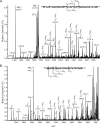
LC-MS analysis of the tryptic digestion products of pilin protein isolated from P. aeruginosa 5196NP plus PilA5196. (A) Mass spectrum of the 26- to 27-min HPLC fraction containing the T3-4 glycopeptide. The triply protonated ions of this glycopeptide are indicated and demonstrate the heterogeneity of the glycosylation associated with this peptide. The y axis shows the percent relative intensity [Rel. Int. (%)]. (B) Mass spectrum of the 21- to 22-min fraction containing the T5 glycopeptide. The doubly protonated glycopeptide ions are indicated in this instance.
FIG. 7.
FIG. 7.
ETD-MS analysis of glycosylated peptides from strain 5196NP plus pilA5196. (A) ETD-MS spectrum of the triply protonated ion at m/z 1424.7 corresponding to the T3-4 peptide modified with six d-Araf residues. The major c and z fragment ions are indicated in the spectrum and identify Thr64 and Thr66 as the sites of O glycosylation. Furthermore, each site is modified with three d-Araf residues. (B) ETD-MS spectrum of the doubly protonated ion at m/z 1634.2 corresponding to the T5 peptide modified with five d-Araf residues. This fragment ion spectrum indicates that all the serine residues in this peptide are partially modified with one or two d-Araf residues. Thr97, Thr99, and Thr101 are not glycosylated, and it is also very likely that Thr86 is not modified.
FIG. 8.
FIG. 8.
Topology model of TfpW. TMHMM (http://www.cbs.dtu.dk/services/TMHMM-2.0) and TMRPres2D (http://biophysics.biol.uoa.gr/TMRPres2D) were used to predict the putative transmembrane domains and topology of TfpW (646 residues). It has a short N-terminal cytoplasmic sequence, followed by 11 predicted membrane-spanning domains (numbered gray boxes) and a large (232-residue) C-terminal periplasmic domain containing predicted tetratricopeptide domains. The small black numbers at the top and bottom of the gray boxes correspond to residue numbers. The putative GT-C DDS motif is located in the first large periplasmic loop between transmembrane domains 1 and 2 and is followed by a characteristic hydrophobic region 45ILFL48.
FIG. 9.
FIG. 9.
TfpW influences twitching motility. A tfpW mutant displays an altered twitching phenotype compared to that of wild-type strain Pa5196; it produces a smaller, more densely stained twitching zone, suggesting that the mutant is more adherent to plastic than the wild type is. The wild-type twitching phenotype was restored when tfpW was expressed in trans from either pBADGr+5196pilA-tfpW or pBADGr+5196pilA-tfpW-tfpX, but not with constructs lacking tfpW. The P. aeruginosa 1244 oligosaccharide transferase PilO/TfpO was not able to complement the tfpW mutant. All recombinant strains are on the Pa5196 tfpW mutant background, with the exception of the wild-type control, Pa5196.
FIG. 10.
FIG. 10.
TfpW is a putative GT-C family glycosyltransferase. (A) Coomassie blue-stained SDS-polyacrylamide gel of sheared surface pili from P. aeruginosa Pa5196 and mutant strains. The tfpW mutant strain produces reduced amounts of surface pili as well as pilins of reduced mass. These lower-molecular-mass bands were confirmed to be PilA5196 by MS-MS (not shown). The pilins were restored to wild-type mass with the addition of tfpW in trans, but not by other genes in the pilin operon. The positions of molecular mass markers (lane M) (in kilodaltons) and of flagellin (F) and pilin (P) are shown to the left and right of the gel, respectively. (B) Western blot using anti-LAM antibody shows that the pilins from the tfpW mutant strain fail to react, while those strains complemented with tfpW in trans regain anti-LAM reactivity. (C) Mutation of the putative GT-C motif within TfpW results in loss of pilin glycosylation.
FIG. 11.
FIG. 11.
Models of type IV pilin glycosylation systems. Three systems involved in the glycosylation of type IV pilins have been identified: the Pgl system of Neisseria spp., the TfpO system of group I strains of P. aeruginosa, and the TfpW system of group IV strains of P. aeruginosa. The first two systems attach short hetero-oligosaccharides to a single site on the pilin, while the TfpW system attaches multiple single sugars or short homo-oligosaccharides (two to four units) to seven different acceptor sites on the protein. The Pgl system transfers a heterosaccharide synthesized by dedicated glycosyltransferases, while the TfpO system transfers a lipopolysaccharide O unit corresponding to the serotype of the host strain. The TfpW system transfers d-Araf which is synthesized by an as-yet unknown pathway. P, phosphate.

Similar articles

Cited by

References

    1. Aas, F. E., A. Vik, J. Vedde, M. Koomey, and W. Egge-Jacobsen. 2007. Neisseria gonorrhoeae O-linked pilin glycosylation: functional analyses define both the biosynthetic pathway and glycan structure. Mol. Microbiol. 65607-624. - PMC - PubMed
    1. Aas, F. E., W. Egge-Jacobsen, H. C. Winther-Larsen, C. Lovold, P. G. Hitchen, A. Dell, and M. Koomey. 2006. Neisseria gonorrhoeae type IV pili undergo multisite, hierarchical modifications with phosphoethanolamine and phosphocholine requiring an enzyme structurally related to lipopolysaccharide phosphoethanolamine transferases. J. Biol. Chem. 28127712-27723. - PubMed
    1. Alaimo, C., I. Catrein, L. Morf, C. L. Marolda, N. Callewaert, M. A. Valvano, M. F. Feldman, and M. Aebi. 2006. Two distinct but interchangeable mechanisms for flipping of lipid-linked oligosaccharides. EMBO J. 25967-976. - PMC - PubMed
    1. Altschul, S. F., W. Gish, W. Miller, E. W. Myers, and D. J. Lipman. 1990. Basic local alignment search tool. J. Mol. Biol. 215403-410. - PubMed
    1. Asikyan, M. L., J. V. Kus, and L. L. Burrows. 2008. Novel proteins that modulate type IV pilus retraction dynamics in Pseudomonas aeruginosa. J. Bacteriol. 1907022-7034. - PMC - PubMed

Publication types

MeSH terms

LinkOut - more resources